Structural and functional analysis of the C-terminal domain of Nup358/RanBP2 - PubMed (original) (raw)
Structural and functional analysis of the C-terminal domain of Nup358/RanBP2
Daniel H Lin et al. J Mol Biol. 2013.
Abstract
The nuclear pore complex is the sole mediator of bidirectional transport between the nucleus and cytoplasm. Nup358 is a metazoan-specific nucleoporin that localizes to the cytoplasmic filaments and provides several binding sites for the mobile nucleocytoplasmic transport machinery. Here we present the crystal structure of the C-terminal domain (CTD) of Nup358 at 1.75Å resolution. The structure reveals that the CTD adopts a cyclophilin-like fold with a non-canonical active-site configuration. We determined biochemically that the CTD possesses weak peptidyl-prolyl isomerase activity and show that the active-site cavity mediates a weak association with the human immunodeficiency virus-1 capsid protein, supporting its role in viral infection. Overall, the surface is evolutionarily conserved, suggesting that the CTD serves as a protein-protein interaction platform. However, we demonstrate that the CTD is dispensable for nuclear envelope localization of Nup358, suggesting that the CTD does not interact with other nucleoporins.
Copyright © 2013 Elsevier Ltd. All rights reserved.
Figures
Fig. 1
Domain organization and structure of Nup358CTD. (a) Domain organization of human Nup358. Domain boundaries are indicated by residue numbers. The bar above the domain structure denotes the crystallized fragment. I, II, III, and IV, Ran binding domains; NTD, N-terminal domain; CTD, C-terminal domain; E3, E3 ligase domain. (b) Cartoon representation of Nup358CTD with a view rotated 180° along the vertical axis shown on the right. (c) Cartoon representation of Nup358CTD rotated 90° along the horizontal axis from above. (d) Representative 2|Fo|-|Fc| electron density map contoured at 1.5 σ.
Fig. 2
Surface properties of Nup358CTD. (a) Surface representation of Nup358CTD, with the active site colored in red. (b) Surface representation colored according to sequence identity based on a multi-species sequence alignment (Fig. 2). The identity at each position is mapped onto the surface and is shaded in a color gradient from white (60 % less than 60 % identity) to red (100 % identity). (c) Surface representation colored according to electrostatic potential from -10 kBT/e (red) to +10 kBT/e (blue).
Fig. 3
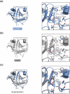
Comparison of Nup358CTD and Cyclophilin A active sites. (a) Detailed view of the Nup358CTD active site. (b) Detailed view of the Cyclophilin A active site (PDB Code 1M9C). (c) Overlay of the active sites from Nup358CTD and Cyclophilin A. Critical active site residues are shown in stick representation, and the Cα-traces are shown in coil representation, according to the coloring scheme in A. The orientation of all active sites is identical.
Fig. 4
Nup358CTD possesses peptidyl-prolyl isomerase activity. (a) Michaelis-Menten plot of the peptidyl-prolyl isomerization of Suc-Ala-Ala-Pro-Phe-2,4-difluoroanilide by Cyclophilin A, Nup358CTD, and Nup358CTD V3173W. (b) Michaelis-Menten plot of the peptidyl-prolyl isomerization of Suc-Ala-Ala-Pro-Phe-2,4-difluoroanilide by Nup358CTD and Nup358CTD V3173W. Note the different scale of the y-axis from panel (a). (c) Representative time-course traces of the peptidyl-prolyl isomerization of Suc-Ala-Ala-Pro-Phe-2,4-difluoroanilide by Cyclophilin A, Nup358CTD, Nup358CTD V3173W, and in the absence of an enzyme.
Fig. 5
Structural comparison of Nup358CTD to the Cyclophilin A•HIV-1CA complex. The structure of Nup358CTD overlaid on the structure of the Cyclophilin A•HIV-1CA complex (PDB code 1M9C). The right panel is a close-up view of the interaction with the HIV-1CA loop rotated 90° along the vertical axis from the left panel. Note the clash between the Nup358CTD Q3163 and the HIV-1CA proline-rich loop.
Fig. 6
Nup358CTD binds weakly to the HIV-1 capsid protein. (a-c) Size exclusion chromatography interaction analysis of HIV-1CA with (a) Cyclophilin A, (b) Nup358CTD, and (c) Nup358CTD V3173W. The analyzed fractions are indicated in gel-filtration profile by a grey bar. Notably, experiments were carried out with identical protein concentrations, the different peak height of the wild-type Nup358CTD is a result of a lower absorbance coefficient.
Fig. 7
Nup358CTD is dispensable for nuclear envelope localization. Nup358CTD and Nup358 fragments carrying a N-terminal HA-tag were transiently transfected into HEK293T cells and analyzed by fluorescence microscopy. HA-tagged Nup358 protein localization was detected with an α-HA antibody (green). The monoclonal α-mAb414 antibody (red) and DAPI (blue) were used as a reference for nuclear envelope and nucleus staining, respectively. The right panel shows the merged images.
Similar articles
- HIV-1 capsid undergoes coupled binding and isomerization by the nuclear pore protein NUP358.
Bichel K, Price AJ, Schaller T, Towers GJ, Freund SM, James LC. Bichel K, et al. Retrovirology. 2013 Jul 31;10:81. doi: 10.1186/1742-4690-10-81. Retrovirology. 2013. PMID: 23902822 Free PMC article. - Architecture of the cytoplasmic face of the nuclear pore.
Bley CJ, Nie S, Mobbs GW, Petrovic S, Gres AT, Liu X, Mukherjee S, Harvey S, Huber FM, Lin DH, Brown B, Tang AW, Rundlet EJ, Correia AR, Chen S, Regmi SG, Stevens TA, Jette CA, Dasso M, Patke A, Palazzo AF, Kossiakoff AA, Hoelz A. Bley CJ, et al. Science. 2022 Jun 10;376(6598):eabm9129. doi: 10.1126/science.abm9129. Epub 2022 Jun 10. Science. 2022. PMID: 35679405 Free PMC article. - Crystal structure of the N-terminal domain of Nup358/RanBP2.
Kassube SA, Stuwe T, Lin DH, Antonuk CD, Napetschnig J, Blobel G, Hoelz A. Kassube SA, et al. J Mol Biol. 2012 Nov 9;423(5):752-65. doi: 10.1016/j.jmb.2012.08.026. Epub 2012 Sep 7. J Mol Biol. 2012. PMID: 22959972 Free PMC article. - RANBP2 evolution and human disease.
Desgraupes S, Etienne L, Arhel NJ. Desgraupes S, et al. FEBS Lett. 2023 Oct;597(20):2519-2533. doi: 10.1002/1873-3468.14749. Epub 2023 Oct 15. FEBS Lett. 2023. PMID: 37795679 Review. - A model for cofactor use during HIV-1 reverse transcription and nuclear entry.
Hilditch L, Towers GJ. Hilditch L, et al. Curr Opin Virol. 2014 Feb;4(100):32-6. doi: 10.1016/j.coviro.2013.11.003. Epub 2014 Jan 14. Curr Opin Virol. 2014. PMID: 24525292 Free PMC article. Review.
Cited by
- Interactions of HIV-1 Capsid with Host Factors and Their Implications for Developing Novel Therapeutics.
Zhuang S, Torbett BE. Zhuang S, et al. Viruses. 2021 Mar 5;13(3):417. doi: 10.3390/v13030417. Viruses. 2021. PMID: 33807824 Free PMC article. Review. - Emerging role of cyclophilin A in HIV-1 infection: from producer cell to the target cell nucleus.
Padron A, Prakash P, Pandhare J, Luban J, Aiken C, Balasubramaniam M, Dash C. Padron A, et al. J Virol. 2023 Nov 30;97(11):e0073223. doi: 10.1128/jvi.00732-23. Epub 2023 Oct 16. J Virol. 2023. PMID: 37843371 Free PMC article. Review. - 8 Å structure of the outer rings of the Xenopus laevis nuclear pore complex obtained by cryo-EM and AI.
Tai L, Zhu Y, Ren H, Huang X, Zhang C, Sun F. Tai L, et al. Protein Cell. 2022 Oct;13(10):760-777. doi: 10.1007/s13238-021-00895-y. Epub 2022 Jan 11. Protein Cell. 2022. PMID: 35015240 Free PMC article. - The Structure of the Nuclear Pore Complex (An Update).
Lin DH, Hoelz A. Lin DH, et al. Annu Rev Biochem. 2019 Jun 20;88:725-783. doi: 10.1146/annurev-biochem-062917-011901. Epub 2019 Mar 18. Annu Rev Biochem. 2019. PMID: 30883195 Free PMC article. Review. - Down-modulation of nucleoporin RanBP2/Nup358 impaired chromosomal alignment and induced mitotic catastrophe.
Hashizume C, Kobayashi A, Wong RW. Hashizume C, et al. Cell Death Dis. 2013 Oct 10;4(10):e854. doi: 10.1038/cddis.2013.370. Cell Death Dis. 2013. PMID: 24113188 Free PMC article.
References
- Hoelz A, Debler EW, Blobel G. The structure of the nuclear pore complex. Annu. Rev. Biochem. 2011;80:613–643. - PubMed
- Wu J, Matunis MJ, Kraemer D, Blobel G, Coutavas E. Nup358, a cytoplasmically exposed nucleoporin with peptide repeats, Ran-GTP binding sites, zinc fingers, a cyclophilin A homologous domain, and a leucine-rich region. J. Biol. Chem. 1995;270:14209–14213. - PubMed
- Yokoyama N, et al. A giant nucleopore protein that binds Ran/TC4. Nature. 1995;376:184–188. - PubMed
- Vetter IR, Nowak C, Nishimoto T, Kuhlmann J, Wittinghofer A. Structure of a Ran-binding domain complexed with Ran bound to a GTP analogue: implications for nuclear transport. Nature. 1999;398:39–46. - PubMed
Publication types
MeSH terms
Substances
LinkOut - more resources
Full Text Sources
Other Literature Sources
Molecular Biology Databases
Miscellaneous


