Brain-wide pathway for waste clearance captured by contrast-enhanced MRI - PubMed (original) (raw)
. 2013 Mar;123(3):1299-309.
doi: 10.1172/JCI67677. Epub 2013 Feb 22.
Affiliations
- PMID: 23434588
- PMCID: PMC3582150
- DOI: 10.1172/JCI67677
Brain-wide pathway for waste clearance captured by contrast-enhanced MRI
Jeffrey J Iliff et al. J Clin Invest. 2013 Mar.
Abstract
The glymphatic system is a recently defined brain-wide paravascular pathway for cerebrospinal fluid (CSF) and interstitial fluid (ISF) exchange that facilitates efficient clearance of solutes and waste from the brain. CSF enters the brain along para-arterial channels to exchange with ISF, which is in turn cleared from the brain along para-venous pathways. Because soluble amyloid β clearance depends on glymphatic pathway function, we proposed that failure of this clearance system contributes to amyloid plaque deposition and Alzheimer's disease progression. Here we provide proof of concept that glymphatic pathway function can be measured using a clinically relevant imaging technique. Dynamic contrast-enhanced MRI was used to visualize CSF-ISF exchange across the rat brain following intrathecal paramagnetic contrast agent administration. Key features of glymphatic pathway function were confirmed, including visualization of para-arterial CSF influx and molecular size-dependent CSF-ISF exchange. Whole-brain imaging allowed the identification of two key influx nodes at the pituitary and pineal gland recesses, while dynamic MRI permitted the definition of simple kinetic parameters to characterize glymphatic CSF-ISF exchange and solute clearance from the brain. We propose that this MRI approach may provide the basis for a wholly new strategy to evaluate Alzheimer's disease susceptibility and progression in the live human brain.
Figures
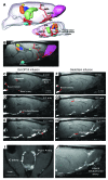
Figure 1. Paravascular influx of paramagnetic contrast.
(A) 3D visualization of key anatomical structures in the rat brain prior to administration of contrast. The anatomical structures include the pituitary (light blue), hippocampus (green), superior colliculus (orange), inferior colliculus (dark blue), pineal gland (yellow), and relevant arterial segments (red). The olfactory artery (OA), azygos of the anterior cerebral artery (azACA), azygos pericallosal artery (AzPA), the middle internal frontal artery (IFA), and the posterior lateral choroidal arterial complex were visualized. (B) A 2-dimensional T1-weighted MRI with the color-coded anatomical structures displayed. (C–E) The time series demonstrates early influx of the small molecular weight paramagnetic contrast agent Gd-DTPA (MW 938 Da). (C) The time at which the intrathecally infused Gd-DTPA appears in the cisterna magna is defined as 0 minutes, and (D and E) the earliest part of the influx process is demonstrated in the subsequent time frames and shows that Gd-DTPA enters the brain along paravascular pathways. (F–H) The dynamic time series of early influx of the large molecular weight paramagnetic contrast agent GadoSpin (MW 200 kDa) also shows that transport into the brain is paravascular. Note that it is evident that even though the 2 paramagnetic contrast agents differ in molecular weight, they pass through paravascular conduits at similar rates, supporting that CSF bulk flow governs this process. (I and J) Paravascular transport of the paramagnetic contrast agent is demonstrated particularly clearly at the level of the Circle of Willis along the internal carotid (IC) artery, posterior communicating (Pcom) arteries, and lateral orbitofrontal artery. Scale bar: 3 mm.
Figure 2. Gd-DTPA and GadoSpin transport along glymphatic pathways.
The average TACs for Gd-DTPA (blue circles) and GadoSpin (red circles) are shown for the (A) pituitary recess, (B) pineal recess, (C) cerebellum, (D) olfactory bulb, (E) aqueduct, and (F) pontine nucleus. The location of each of the ROIs is shown on the sagittal brain icon displayed in the top right corner of each of the graphs. (A and B) It is evident that contrast uptake in the pituitary recess and pineal recess is largely similar for Gd-DTPA and GadoSpin. This confirms that within the proximal portions of the glymphatic pathway, paramagnetic contrast agent transport is largely independent of molecular size. It is also clear that tissue uptake of Gd-DTPA is markedly higher than that observed for GadoSpin in the (C) cerebellum, (E) aqueduct, and (F) pontine nucleus. This confirms that the movement of contrast agent from the subarachnoid/proximal paravascular pathway into and through the brain interstitium is indeed dependent upon molecular size. Data are presented as mean ± SEM.
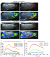
Figure 3. Cluster-based spatial distribution of Gd-DTPA and GadoSpin in the rat brain.
(A) T1-weighted sagittal midline section at the level of the aqueduct (Aq), with anatomical landmarks displayed prior to administration of GadoSpin. BA, basal artery; BC, basal cistern; Cb, cerebellum; GA, vein of Galen; Ob, olfactory bulb; Pin, pineal gland; Pit, pituitary gland. (B) Cluster analysis in GadoSpin rat. The paravascular cluster is overlaid on the corresponding MRI, demonstrating that the red and orange clusters match the spatial location of the basal cistern, pineal recess and pituitary recess, and tissue in the immediate vicinity of the major arteries. (C) Distribution of blue and green clusters from the same GadoSpin rat, demonstrating the more parenchymal location of these clusters. (D) All clusters displayed simultaneously and overlaid on the MRI. The red and orange clusters in the paravascular conduits are classified as zone 1, the green cluster is classified as zone 2, and the blue cluster is classified as zone 3. (E) T1-weighted MRI at the level of the aqueduct, with anatomical landmarks displayed from a rat prior to administration of Gd-DTPA. (F) Paravascular, zone 1 cluster displayed from the Gd-DTPA rat. (G) Green and blue cluster distribution in the Gd-DTPA rat. (H) All 4 clusters displayed. The red, green, and blue clusters are classified as zone 1, 2, and 3, respectively. Scale bar: 3 mm. The TACs for each of the 4 clusters for (I) a GadoSpin rat and (J) a Gd-DTPA rat are displayed in addition to the total number of voxels of each the clusters.
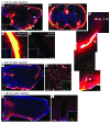
Figure 4. Fluorescence-based imaging of paravascular CSF-ISF exchange.
Small (TR-d3; MW 3 kDa) and large (FITC-d500; MW 500 kDa) molecular weight fluorescent tracers were injected intrathecally and imaged ex vivo by conventional and laser scanning confocal fluorescence microscopy. (A, B, F, and H) Whole-slice montages were generated, showing paravascular (arrowheads) CSF tracer influx into the brain at (A and B) 30 minutes, (F) 60 minutes, and (H) 180 minutes after injection. (B) Coronal slice counter labeled with vascular endothelial marker isolectin B4 (IB4) 30 minutes after injection. (C and D) High-power confocal imaging shows that CSF tracer enters the brain along penetrating arteries (arrowheads) (E) but not along draining veins. Large molecular weight FITC-d500 remains confined to the paravascular spaces, while small molecular weight TR-d3 moves readily into and through the surrounding interstitium. (F) 60 minutes after injection, small molecular weight TR-d3 is localized diffusely throughout the brain interstitium, (G) while large molecular weight FITC-d500 is apparent along terminal capillary bed. The inset depicts z-projection of image stack, demonstrating the extent of capillary labeling with FITC-d500. (H) At 180 minutes after injection, parenchymal tracer levels are reduced compared with 30 and 60 minutes after injection, (I) while intrathecal tracer persists along para-venous clearance pathways. Original magnification (scale bar measurements are shown parenthetically): ×40 (A, B, F, and H); ×200 (100 μm) (C); ×400 (50 μm) (D, E, G, and I and insets in D, E, and G); ×40 (inset, I).
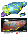
Figure 5. Brain-wide glymphatic pathways of CSF-ISF exchange, assessed by contrast-enhanced MRI in the rat.
After injection into the subarachnoid space of the cisterna magna, contrast agent follows specific paravascular pathways (yellow arrows) to enter the brain parenchyma and exchange with the interstitial compartments (orange arrows and fields). Acquisition of dynamic image series identified key CSF influx nodes at the pineal (Pin) and pituitary (Pit) recesses and allowed simple kinetic parameters to be derived that deflect the extent and rate of glymphatic CSF-ISF exchange throughout the whole brain. Scale bar: 3 mm.
Comment in
- Bathing the brain.
Strittmatter WJ. Strittmatter WJ. J Clin Invest. 2013 Mar;123(3):1013-5. doi: 10.1172/JCI68241. Epub 2013 Feb 22. J Clin Invest. 2013. PMID: 23434595 Free PMC article.
Similar articles
- Evaluating glymphatic pathway function utilizing clinically relevant intrathecal infusion of CSF tracer.
Yang L, Kress BT, Weber HJ, Thiyagarajan M, Wang B, Deane R, Benveniste H, Iliff JJ, Nedergaard M. Yang L, et al. J Transl Med. 2013 May 1;11:107. doi: 10.1186/1479-5876-11-107. J Transl Med. 2013. PMID: 23635358 Free PMC article. - Cerebrospinal and interstitial fluid transport via the glymphatic pathway modeled by optimal mass transport.
Ratner V, Gao Y, Lee H, Elkin R, Nedergaard M, Benveniste H, Tannenbaum A. Ratner V, et al. Neuroimage. 2017 May 15;152:530-537. doi: 10.1016/j.neuroimage.2017.03.021. Epub 2017 Mar 18. Neuroimage. 2017. PMID: 28323163 Free PMC article. - The role of brain barriers in fluid movement in the CNS: is there a 'glymphatic' system?
Abbott NJ, Pizzo ME, Preston JE, Janigro D, Thorne RG. Abbott NJ, et al. Acta Neuropathol. 2018 Mar;135(3):387-407. doi: 10.1007/s00401-018-1812-4. Epub 2018 Feb 10. Acta Neuropathol. 2018. PMID: 29428972 Review. - Current Concepts in Intracranial Interstitial Fluid Transport and the Glymphatic System: Part II-Imaging Techniques and Clinical Applications.
Klostranec JM, Vucevic D, Bhatia KD, Kortman HGJ, Krings T, Murphy KP, terBrugge KG, Mikulis DJ. Klostranec JM, et al. Radiology. 2021 Dec;301(3):516-532. doi: 10.1148/radiol.2021204088. Epub 2021 Oct 26. Radiology. 2021. PMID: 34698564 Review.
Cited by
- Fast whole brain MR imaging of dynamic susceptibility contrast changes in the cerebrospinal fluid (cDSC MRI).
Cao D, Kang N, Pillai JJ, Miao X, Paez A, Xu X, Xu J, Li X, Qin Q, Van Zijl PCM, Barker P, Hua J. Cao D, et al. Magn Reson Med. 2020 Dec;84(6):3256-3270. doi: 10.1002/mrm.28389. Epub 2020 Jul 3. Magn Reson Med. 2020. PMID: 32621291 Free PMC article. - Reduced Diffusivity along Perivascular Spaces on MR Imaging Associated with Younger Age of First Use and Cognitive Impairment in Recreational Marijuana Users.
Andica C, Kamagata K, Takabayashi K, Mahemuti Z, Hagiwara A, Aoki S. Andica C, et al. AJNR Am J Neuroradiol. 2024 Jul 8;45(7):912-919. doi: 10.3174/ajnr.A8215. AJNR Am J Neuroradiol. 2024. PMID: 38383055 - A new glaucoma hypothesis: a role of glymphatic system dysfunction.
Wostyn P, Van Dam D, Audenaert K, Killer HE, De Deyn PP, De Groot V. Wostyn P, et al. Fluids Barriers CNS. 2015 Jun 29;12:16. doi: 10.1186/s12987-015-0012-z. Fluids Barriers CNS. 2015. PMID: 26118970 Free PMC article. Review. - Contrast Enhancement of the Normal Infundibular Recess Using Heavily T2-weighted 3D FLAIR.
Osawa I, Kozawa E, Yamamoto Y, Tanaka S, Shiratori T, Kaizu A, Inoue K, Niitsu M. Osawa I, et al. Magn Reson Med Sci. 2022 Jul 1;21(3):469-476. doi: 10.2463/mrms.mp.2021-0021. Epub 2021 May 13. Magn Reson Med Sci. 2022. PMID: 33980787 Free PMC article. - Enlarged Perivascular Space and Index for Diffusivity Along the Perivascular Space as Emerging Neuroimaging Biomarkers of Neurological Diseases.
Zhang J, Liu S, Wu Y, Tang Z, Wu Y, Qi Y, Dong F, Wang Y. Zhang J, et al. Cell Mol Neurobiol. 2023 Dec 29;44(1):14. doi: 10.1007/s10571-023-01440-7. Cell Mol Neurobiol. 2023. PMID: 38158515 Review.
References
Publication types
MeSH terms
Substances
Grants and funding
- R01 NS078167/NS/NINDS NIH HHS/United States
- R01 NS078304/NS/NINDS NIH HHS/United States
- NS078167/NS/NINDS NIH HHS/United States
- R01 NS075177/NS/NINDS NIH HHS/United States
- NS078304/NS/NINDS NIH HHS/United States
LinkOut - more resources
Full Text Sources
Other Literature Sources
Medical


