The genomically mosaic brain: aneuploidy and more in neural diversity and disease - PubMed (original) (raw)
Review
The genomically mosaic brain: aneuploidy and more in neural diversity and disease
Diane M Bushman et al. Semin Cell Dev Biol. 2013 Apr.
Abstract
Genomically identical cells have long been assumed to comprise the human brain, with post-genomic mechanisms giving rise to its enormous diversity, complexity, and disease susceptibility. However, the identification of neural cells containing somatically generated mosaic aneuploidy - loss and/or gain of chromosomes from a euploid complement - and other genomic variations including LINE1 retrotransposons and regional patterns of DNA content variation (DCV), demonstrate that the brain is genomically heterogeneous. The precise phenotypes and functions produced by genomic mosaicism are not well understood, although the effects of constitutive aberrations, as observed in Down syndrome, implicate roles for defined mosaic genomes relevant to cellular survival, differentiation potential, stem cell biology, and brain organization. Here we discuss genomic mosaicism as a feature of the normal brain as well as a possible factor in the weak or complex genetic linkages observed for many of the most common forms of neurological and psychiatric diseases.
Copyright © 2013 Elsevier Ltd. All rights reserved.
Figures
Figure 1
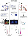
Schematic of genomic mosaicism analysis techniques. A, Cells from cycling populations may be arrested in metaphase for either chromosome spread enumeration by counting DAPI-stained chromosomes (bottom left), or full karyotype analysis by SKY (bottom right). B, Non-cycling or interphase cells are hybridized with chromosome-specific FISH probes (e.g., the chromosome 8 and 16 point probes shown here in red and green, respectively). Euploid cells, disomic for both chromosomes, would display 2 dots of each; here, both nuclei are disomic for chromosome 8, while the nucleus on the left is monosomic for chromosome 16 and the nucleus on the right is trisomic for chromosome 16. C, Isolated cells and nuclei are stained to saturation with dyes like the DNA-intercalating dye Propidium Iodide or DRAQ5 for flow cytometric analysis. The prominent peak of the resulting DNA content histogram contains cells in the G0/G1 phase of the cell cycle (2N DNA content); S phase (2
<n<4) and="" g2="" m="" (4n)="" phase="" are="" distinguishable="" on="" the="" linear="" scale="" of="" x-axis.="" <b="">D, Hetergeneous DNA content histogram from human frontal cortical nuclei (green, red and blue are separate individuals) stained with propidium iodide, showing broad bases and right-hand shoulders. Chicken erythrocyte nuclei (CEN) were included as an internal reference standard and control. E, Overlay of representative lymphocyte (green), cerebellar (red) and cortical (blue) histograms displaying an area of increased DCV unique to the cortical sample. (Adapted with permission from Peterson et al., 2012; Westra et al., 2010.)</n<4)>
Similar articles
- Genomic Mosaicism Formed by Somatic Variation in the Aging and Diseased Brain.
Costantino I, Nicodemus J, Chun J. Costantino I, et al. Genes (Basel). 2021 Jul 14;12(7):1071. doi: 10.3390/genes12071071. Genes (Basel). 2021. PMID: 34356087 Free PMC article. Review. - Neuronal DNA content variation (DCV) with regional and individual differences in the human brain.
Westra JW, Rivera RR, Bushman DM, Yung YC, Peterson SE, Barral S, Chun J. Westra JW, et al. J Comp Neurol. 2010 Oct 1;518(19):3981-4000. doi: 10.1002/cne.22436. J Comp Neurol. 2010. PMID: 20737596 Free PMC article. - Aneuploidy in the normal and diseased brain.
Kingsbury MA, Yung YC, Peterson SE, Westra JW, Chun J. Kingsbury MA, et al. Cell Mol Life Sci. 2006 Nov;63(22):2626-41. doi: 10.1007/s00018-006-6169-5. Cell Mol Life Sci. 2006. PMID: 16952055 Free PMC article. Review. - [Instability of chromosomes in human nerve cells (normal and with neuromental diseases)].
Iurov IuB, Vorsanova SG, Solov'ev IV, Iurov IIu. Iurov IuB, et al. Genetika. 2010 Oct;46(10):1352-5. Genetika. 2010. PMID: 21254554 Russian. - Genomic mosaicism in the developing and adult brain.
Rohrback S, Siddoway B, Liu CS, Chun J. Rohrback S, et al. Dev Neurobiol. 2018 Nov;78(11):1026-1048. doi: 10.1002/dneu.22626. Epub 2018 Aug 1. Dev Neurobiol. 2018. PMID: 30027562 Free PMC article. Review.
Cited by
- Quantitative FISHing: Implications for Chromosomal Analysis.
Vorsanova SG, Yurov YB, Iourov IY. Vorsanova SG, et al. Methods Mol Biol. 2024;2825:239-246. doi: 10.1007/978-1-0716-3946-7_13. Methods Mol Biol. 2024. PMID: 38913313 - FISHing for Chromosome Instability and Aneuploidy in the Alzheimer's Disease Brain.
Yurov YB, Vorsanova SG, Iourov IY. Yurov YB, et al. Methods Mol Biol. 2023;2561:191-204. doi: 10.1007/978-1-0716-2655-9_10. Methods Mol Biol. 2023. PMID: 36399271 - Somatic mosaicism in the diseased brain.
Iourov IY, Vorsanova SG, Kurinnaia OS, Kutsev SI, Yurov YB. Iourov IY, et al. Mol Cytogenet. 2022 Oct 21;15(1):45. doi: 10.1186/s13039-022-00624-y. Mol Cytogenet. 2022. PMID: 36266706 Free PMC article. Review. - Exploring the Origin and Physiological Significance of DNA Double Strand Breaks in the Developing Neuroretina.
Álvarez-Lindo N, Suárez T, de la Rosa EJ. Álvarez-Lindo N, et al. Int J Mol Sci. 2022 Jun 9;23(12):6449. doi: 10.3390/ijms23126449. Int J Mol Sci. 2022. PMID: 35742893 Free PMC article. Review. - The Role of Transposable Elements of the Human Genome in Neuronal Function and Pathology.
Chesnokova E, Beletskiy A, Kolosov P. Chesnokova E, et al. Int J Mol Sci. 2022 May 23;23(10):5847. doi: 10.3390/ijms23105847. Int J Mol Sci. 2022. PMID: 35628657 Free PMC article. Review.
References
- Alberman E, Mutton D, Morris JK. Cytological and epidemiological findings in trisomies 13, 18, and 21: England and Wales 2004-2009. Am J Med Genet A. 2012;158A:1145–50. - PubMed
- Kalousek DK, Howard-Peebles PN, Olson SB, Barrett IJ, Dorfmann A, Black SH, et al. Confirmation of CVS mosaicism in term placentae and high frequency of intrauterine growth retardation association with confined placental mosaicism. Prenatal Diagn. 1991;11:743–50. - PubMed
- Benn P, Hsu LY, Perlis T, Schonhaut A. Prenatal diagnosis of chromosome mosaicism. Prenatal Diagn. 1984;4:1–9. - PubMed
- Jinawath N, Zambrano R, Wohler E, Palmquist MK, Hoover-Fong J, Hamosh A, et al. Mosaic trisomy 13: understanding origin using SNP array. J Med Genet. 2011;48:323–6. - PubMed
Publication types
MeSH terms
LinkOut - more resources
Full Text Sources
Other Literature Sources
Medical
