Psoriatic arthritis - PubMed (original) (raw)
Psoriatic arthritis
Artur Jacek Sankowski et al. Pol J Radiol. 2013 Jan.
Abstract
Psoriatic arthritis (PsA) is a chronic inflammatory joint disease which develops in patients with psoriasis. It is characteristic that the rheumatoid factor in serum is absent. Etiology of the disease is still unclear but a number of genetic associations have been identified. Inheritance of the disease is multilevel and the role of environmental factors is emphasized. Immunology of PsA is also complex. Inflammation is caused by immunological reactions leading to release of kinins. Destructive changes in bones usually appear after a few months from the onset of clinical symptoms. Typically PsA involves joints of the axial skeleton with an asymmetrical pattern. The spectrum of symptoms include inflammatory changes in attachments of articular capsules, tendons, and ligaments to bone surface. The disease can have divers clinical course but usually manifests as oligoarthritis. Imaging plays an important role in the diagnosis of PsA. Classical radiography has been used for this purpose for over a hundred years. It allows to identify late stages of the disease, when bone tissue is affected. In the last 20 years many new imaging modalities, such as ultrasonography (US), computed tomography (CT) and magnetic resonance (MR), have been developed and became important diagnostic tools for evaluation of rheumatoid diseases. They enable the assessment and monitoring of early inflammatory changes. As a result, patients have earlier access to modern treatment and thus formation of destructive changes in joints can be markedly delayed or even avoided.
Keywords: genetics and immunology of psoriatic arthritis; imaging studies; psoriatic arthritis; spondyloarthropathies.
Figures
Figure 1.
X-ray of the heel: Destructive changes at the sight of calcaneal tendon attachment due to inflammation.
Figure 2.
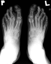
X-ray of feet: The destructive form of psoriatic arthritis (arthritis mutilans). Numerous destructive changes in matacarpophalangeal and intrerphalangeal joints.
Figure 3.
X-ray of hands: The destructive form of psoriatic arthritis (arthritis mutilans). Numerous destructive changes in joints of both hands. Ankylosis of the right wrist. Typical for PsA changes called “pencil-in-cup” involving metacarpophalangeal joints.
Figure 4.
Inflammatory changes in sacroiliac joints. Marked asymmetry with more prominent erosions on the left side.
Figure 5.
X-ray of sacroiliac joints and lumbar spine. Marked asymmetry of symptoms is visible.
Figure 6.
X-ray of intrerphalangeal joints of the hand. Minor erosive changes involving distal interphalangeal joints.
Figure 7.
Patient with PsA – X-ray of forefoot. Shows a form of the disease involving distal interphalangeal joints. Margin erosions and periostosis in DIP joint of the first finger of the left foot are visible.
Figure 8.
X-ray of hands: Ankylosis of the left wrist in a patient with the late stage of PsA.
Figure 9.
Hand X-ray examination: Superimposed degenerative and inflammatory changes in the course of psoriatic arthritis involving the interphalyngeal joints.
Figure 10.
The US examination in a grey scale, longitudinal plane. Investigation of the second matacarpophalangeal joint. Erosion and inflammatory changes presenting as a synovium hypertrophy.
Figure 11.
US examination in the same patient (Figure 7). Effusion, hypertrophy and hyperaemia of synovial membrane with erosions in interphalangeal joint of the left hallux.
Figure 12.
US examination Power Doppler of the proximal phalanx of the II finger, volar side. Inflammatory changes in the tendon sheath of the flexor muscle of the fingers. Joint effusion, synovial membrane hypertrophy and hyperaemia in PD.
Figure 13.
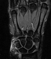
MR examination of the joints of the wrist. STIR images. The distal radio-ulnar joint effusion, less prominent in intercarpal joints.
Figure 14.
Whole body Scintigraphy (of skeleton) of a patient with PsA. Numerous joints of axial and peripheral skeleton involved in the process.
Figure 15.
Scintigraphy of hands and forearms. Radionuclide accumulation in the joints of the wrist, metacarpophalangeal and interphalangeal joints involved in the inflammatory process.
Similar articles
- Ultrasonography, magnetic resonance imaging, radiography, and clinical assessment of inflammatory and destructive changes in fingers and toes of patients with psoriatic arthritis.
Wiell C, Szkudlarek M, Hasselquist M, Møller JM, Vestergaard A, Nørregaard J, Terslev L, Østergaard M. Wiell C, et al. Arthritis Res Ther. 2007;9(6):R119. doi: 10.1186/ar2327. Arthritis Res Ther. 2007. PMID: 18001463 Free PMC article. - Psoriatic arthritis: imaging techniques.
Spadaro A, Lubrano E. Spadaro A, et al. Reumatismo. 2012 Jun 5;64(2):99-106. doi: 10.4081/reumatismo.2012.99. Reumatismo. 2012. PMID: 22690386 Review. - Recent Advances in Imaging for Diagnosis, Monitoring, and Prognosis of Psoriatic Arthritis.
Fassio A, Matzneller P, Idolazzi L. Fassio A, et al. Front Med (Lausanne). 2020 Oct 29;7:551684. doi: 10.3389/fmed.2020.551684. eCollection 2020. Front Med (Lausanne). 2020. PMID: 33195301 Free PMC article. Review. - [Imaging modalities in psoriatic arthritis].
Hermann KG, Ohrndorf S, Werner SG, Finzel S, Backhaus M. Hermann KG, et al. Z Rheumatol. 2013 Oct;72(8):771-8. doi: 10.1007/s00393-013-1188-8. Z Rheumatol. 2013. PMID: 24085530 Review. German.
Cited by
- A novel biomarker of MMP-cleaved prolargin is elevated in patients with psoriatic arthritis.
Sinkeviciute D, Skovlund Groen S, Sun S, Manon-Jensen T, Aspberg A, Önnerfjord P, Bay-Jensen AC, Kristensen S, Holm Nielsen S. Sinkeviciute D, et al. Sci Rep. 2020 Aug 11;10(1):13541. doi: 10.1038/s41598-020-70327-0. Sci Rep. 2020. PMID: 32782251 Free PMC article. - Do genetics contribute to TNF inhibitor response prediction in Psoriatic Arthritis?
Curry PDK, Morris AP, Barton A, Bluett J. Curry PDK, et al. Pharmacogenomics J. 2023 Jan;23(1):1-7. doi: 10.1038/s41397-022-00290-8. Epub 2022 Oct 15. Pharmacogenomics J. 2023. PMID: 36243888 Free PMC article. Review. - Time until onset of action when treating psoriatic arthritis: meta-analysis and novel approach of generating confidence intervals.
Pham PA, Dressler C, Eisert L, Nast A, Werner RN. Pham PA, et al. Rheumatol Int. 2019 Apr;39(4):605-618. doi: 10.1007/s00296-019-04244-5. Epub 2019 Jan 25. Rheumatol Int. 2019. PMID: 30684041 - Expression of MIF and TNFA in psoriatic arthritis: relationship with Th1/Th2/Th17 cytokine profiles and clinical variables.
Bautista-Herrera LA, De la Cruz-Mosso U, Morales-Zambrano R, Villanueva-Quintero GD, Hernández-Bello J, Ramírez-Dueñas MG, Martínez-López E, Brennan-Bourdon LM, Baños-Hernández CJ, Muñoz-Valle JF. Bautista-Herrera LA, et al. Clin Exp Med. 2018 May;18(2):229-235. doi: 10.1007/s10238-017-0475-0. Epub 2017 Sep 30. Clin Exp Med. 2018. PMID: 28965181 - Cartilage Degradation in Psoriatic Arthritis Is Associated With Increased Synovial Perfusion as Detected by Magnetic Resonance Imaging.
Abrar DB, Schleich C, Müller-Lutz A, Frenken M, Radke KL, Vordenbäumen S, Schneider M, Ostendorf B, Sewerin P. Abrar DB, et al. Front Med (Lausanne). 2020 Sep 25;7:539870. doi: 10.3389/fmed.2020.539870. eCollection 2020. Front Med (Lausanne). 2020. PMID: 33102496 Free PMC article.
References
- Moll JMH, Wright V. Psoriatic arthritis. Seminars in Arthritis and Rheumatism. 1973;3(1):55–78. - PubMed
- Hensler T, Christophers E. Psoriasis of early and late onset: characterization of two types of psoriasis vulgaris. J Am Acad Dermatol. 1985;13(3):450–56. - PubMed
- Allen MH, Ameen H, Veal C, et al. The Major Psoriasis Susceptibility Locus PSORS1 Is not a Risk Factor for Late-Onset Psoriasis. J Invest Dermatol. 2005;124:103–6. - PubMed
- Veale D, Rogers S, Fitzgerald O. Classification of Clinical Subsets in Psoriatic Arthritis. Br J Rheumatol. 1994;33:133–38. - PubMed
LinkOut - more resources
Full Text Sources
Other Literature Sources
Research Materials
Miscellaneous


