Landscape of the PARKIN-dependent ubiquitylome in response to mitochondrial depolarization - PubMed (original) (raw)
. 2013 Apr 18;496(7445):372-6.
doi: 10.1038/nature12043. Epub 2013 Mar 17.
Affiliations
- PMID: 23503661
- PMCID: PMC3641819
- DOI: 10.1038/nature12043
Landscape of the PARKIN-dependent ubiquitylome in response to mitochondrial depolarization
Shireen A Sarraf et al. Nature. 2013.
Abstract
The PARKIN ubiquitin ligase (also known as PARK2) and its regulatory kinase PINK1 (also known as PARK6), often mutated in familial early-onset Parkinson's disease, have central roles in mitochondrial homeostasis and mitophagy. Whereas PARKIN is recruited to the mitochondrial outer membrane (MOM) upon depolarization via PINK1 action and can ubiquitylate porin, mitofusin and Miro proteins on the MOM, the full repertoire of PARKIN substrates--the PARKIN-dependent ubiquitylome--remains poorly defined. Here we use quantitative diGly capture proteomics (diGly) to elucidate the ubiquitylation site specificity and topology of PARKIN-dependent target modification in response to mitochondrial depolarization. Hundreds of dynamically regulated ubiquitylation sites in dozens of proteins were identified, with strong enrichment for MOM proteins, indicating that PARKIN dramatically alters the ubiquitylation status of the mitochondrial proteome. Using complementary interaction proteomics, we found depolarization-dependent PARKIN association with numerous MOM targets, autophagy receptors, and the proteasome. Mutation of the PARKIN active site residue C431, which has been found mutated in Parkinson's disease patients, largely disrupts these associations. Structural and topological analysis revealed extensive conservation of PARKIN-dependent ubiquitylation sites on cytoplasmic domains in vertebrate and Drosophila melanogaster MOM proteins. These studies provide a resource for understanding how the PINK1-PARKIN pathway re-sculpts the proteome to support mitochondrial homeostasis.
Figures
Figure 1. QdiGLY proteomics for PARKIN-dependent ubiquitylation
a, Proteomics work flow. b, diGLY sites identified and quantified across 73 experiments. FDR, false discovery rate. c, log2(H:L) plots for quantified diGLY peptides for HCT116PARKIN (experiment 17) or HeLaPARKIN (experiment 57) cells (Table S2). d, Overlap of ubiquitylation sites in HeLaPARKIN biological triplicates (1h CCCP + Btz) (Table S1, S2). e, Ubiquitylation site and protein overlap between all HCT116PARKIN and HeLaPARKIN experiments treated with CCCP and Btz for 1h. f, log2(H:L) ratios for selected diGLY sites from HCT116PARKIN (Ex 17) and HeLaPARKIN (Ex 57) (1h CCCP + Btz).
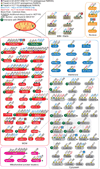
Figure 2. PARKIN-dependent ubiquitylation sites revealed by QdiGLY proteomics
Class 1 sites are in black font. Additional sites found in Class 1 proteins are in red (HCT116PARKIN) or blue font (HeLaPARKIN). Site overlap in HCT116 and/or SH-SY5Y: magenta, orange, and green octagons. Dotted lines: interacting proteins. Rectangles represent Class 2 substrates. Red or blue boxes refer to additional sites identified in either HCT116PARKIN and HeLaPARKIN cells (Table S2). * and ^, protein levels decrease or increase, respectively, upon depolarization.
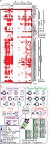
Figure 3. PARKIN associates with mitochondrial proteins and the proteasome in response to depolarization
a, Heat map of HCIPs (represented by APSMs) for HA-FLAG-PARKIN and mutants in response to depolarization in 293T cells, with or without Btz or BafA. Proteins indicated had WDN-scores ≥ 1.0, Z-score ≥ 5, and APSMs ≥ 2 unless otherwise noted (see METHODS).b, Summary of PARKIN-interacting proteins in 293T and HeLa and integration with diGLY sites.
Figure 4. Structural anatomy and conservation of PARKIN-dependent diGLY sites
Structures or domain schematics are shown for Class 1 depolarization and PARKIN-dependent diGLY sites (PDB codes, Table S5). Color-coded circles (a) indicate the conservation of Lys at the homologous position in M. musculus, D. rerio, and D. melanogaster (Table S5). For structures, regulated sites are shown in red space-filled models. b, Cytoplasmic proteins. RPN10, red circle. c, MOM proteins with structures. d, MOM proteins without structures.
Similar articles
- Defining roles of PARKIN and ubiquitin phosphorylation by PINK1 in mitochondrial quality control using a ubiquitin replacement strategy.
Ordureau A, Heo JM, Duda DM, Paulo JA, Olszewski JL, Yanishevski D, Rinehart J, Schulman BA, Harper JW. Ordureau A, et al. Proc Natl Acad Sci U S A. 2015 May 26;112(21):6637-42. doi: 10.1073/pnas.1506593112. Epub 2015 May 12. Proc Natl Acad Sci U S A. 2015. PMID: 25969509 Free PMC article. - PINK1 and Parkin target Miro for phosphorylation and degradation to arrest mitochondrial motility.
Wang X, Winter D, Ashrafi G, Schlehe J, Wong YL, Selkoe D, Rice S, Steen J, LaVoie MJ, Schwarz TL. Wang X, et al. Cell. 2011 Nov 11;147(4):893-906. doi: 10.1016/j.cell.2011.10.018. Cell. 2011. PMID: 22078885 Free PMC article. - Parkin recruitment to impaired mitochondria for nonselective ubiquitylation is facilitated by MITOL.
Koyano F, Yamano K, Kosako H, Tanaka K, Matsuda N. Koyano F, et al. J Biol Chem. 2019 Jun 28;294(26):10300-10314. doi: 10.1074/jbc.RA118.006302. Epub 2019 May 20. J Biol Chem. 2019. PMID: 31110043 Free PMC article. - N-degron-mediated degradation and regulation of mitochondrial PINK1 kinase.
Eldeeb MA, Ragheb MA. Eldeeb MA, et al. Curr Genet. 2020 Aug;66(4):693-701. doi: 10.1007/s00294-020-01062-2. Epub 2020 Mar 10. Curr Genet. 2020. PMID: 32157382 Review. - Targeting mitochondrial dysfunction: role for PINK1 and Parkin in mitochondrial quality control.
Narendra DP, Youle RJ. Narendra DP, et al. Antioxid Redox Signal. 2011 May 15;14(10):1929-38. doi: 10.1089/ars.2010.3799. Epub 2011 Mar 3. Antioxid Redox Signal. 2011. PMID: 21194381 Free PMC article. Review.
Cited by
- PINK1-PRKN mediated mitophagy: differences between in vitro and in vivo models.
Han R, Liu Y, Li S, Li XJ, Yang W. Han R, et al. Autophagy. 2023 May;19(5):1396-1405. doi: 10.1080/15548627.2022.2139080. Epub 2022 Nov 3. Autophagy. 2023. PMID: 36282767 Free PMC article. Review. - Interorganelle communication, aging, and neurodegeneration.
Petkovic M, O'Brien CE, Jan YN. Petkovic M, et al. Genes Dev. 2021 Apr 1;35(7-8):449-469. doi: 10.1101/gad.346759.120. Genes Dev. 2021. PMID: 33861720 Free PMC article. Review. - The E3 ubiquitin ligase parkin is recruited to the 26 S proteasome via the proteasomal ubiquitin receptor Rpn13.
Aguileta MA, Korac J, Durcan TM, Trempe JF, Haber M, Gehring K, Elsasser S, Waidmann O, Fon EA, Husnjak K. Aguileta MA, et al. J Biol Chem. 2015 Mar 20;290(12):7492-505. doi: 10.1074/jbc.M114.614925. Epub 2015 Feb 9. J Biol Chem. 2015. PMID: 25666615 Free PMC article. - Mitochondrial outer membrane integrity regulates a ubiquitin-dependent and NF-κB-mediated inflammatory response.
Vringer E, Heilig R, Riley JS, Black A, Cloix C, Skalka G, Montes-Gómez AE, Aguado A, Lilla S, Walczak H, Gyrd-Hansen M, Murphy DJ, Huang DT, Zanivan S, Tait SW. Vringer E, et al. EMBO J. 2024 Mar;43(6):904-930. doi: 10.1038/s44318-024-00044-1. Epub 2024 Feb 9. EMBO J. 2024. PMID: 38337057 Free PMC article. - Miro phosphorylation sites regulate Parkin recruitment and mitochondrial motility.
Shlevkov E, Kramer T, Schapansky J, LaVoie MJ, Schwarz TL. Shlevkov E, et al. Proc Natl Acad Sci U S A. 2016 Oct 11;113(41):E6097-E6106. doi: 10.1073/pnas.1612283113. Epub 2016 Sep 27. Proc Natl Acad Sci U S A. 2016. PMID: 27679849 Free PMC article.
References
- Glauser L, Sonnay S, Stafa K, Moore DJ. Parkin promotes the ubiquitination and degradation of the mitochondrial fusion factor mitofusin 1. J Neurochem. 2011;118:636–645. - PubMed
Publication types
MeSH terms
Substances
Grants and funding
- GM095567/GM/NIGMS NIH HHS/United States
- GM070565/GM/NIGMS NIH HHS/United States
- F32 CA139885/CA/NCI NIH HHS/United States
- R01 GM095567/GM/NIGMS NIH HHS/United States
- R01 GM070565/GM/NIGMS NIH HHS/United States
- CA139885/CA/NCI NIH HHS/United States
- R01 GM067945/GM/NIGMS NIH HHS/United States
- GM067945/GM/NIGMS NIH HHS/United States
LinkOut - more resources
Full Text Sources
Other Literature Sources
Molecular Biology Databases
Miscellaneous


