An enlarged parietal foramen in the late archaic Xujiayao 11 neurocranium from Northern China, and rare anomalies among Pleistocene Homo - PubMed (original) (raw)
An enlarged parietal foramen in the late archaic Xujiayao 11 neurocranium from Northern China, and rare anomalies among Pleistocene Homo
Xiu-Jie Wu et al. PLoS One. 2013.
Abstract
We report here a neurocranial abnormality previously undescribed in Pleistocene human fossils, an enlarged parietal foramen (EPF) in the early Late Pleistocene Xujiayao 11 parietal bones from the Xujiayao (Houjiayao) site, northern China. Xujiayao 11 is a pair of partial posteromedial parietal bones from an adult. It exhibits thick cranial vault bones, arachnoid granulations, a deviated posterior sagittal suture, and a unilateral (right) parietal lacuna with a posteriorly-directed and enlarged endocranial vascular sulcus. Differential diagnosis indicates that the perforation is a congenital defect, an enlarged parietal foramen, commonly associated with cerebral venous and cranial vault anomalies. It was not lethal given the individual's age-at-death, but it may have been associated with secondary neurological deficiencies. The fossil constitutes the oldest evidence in human evolution of this very rare condition (a single enlarged parietal foramen). In combination with developmental and degenerative abnormalities in other Pleistocene human remains, it suggests demographic and survival patterns among Pleistocene Homo that led to an elevated frequency of conditions unknown or rare among recent humans.
Conflict of interest statement
Competing Interests: The authors have declared that no competing interests exist.
Figures
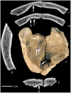
Figure 1. The Xujiayao 11 parietal bones. Exterior view (A).
CT horizontal section images showing the linear sagittal suture (B), the posterior left postmortem breaks (C), and the posterior right oblique sagittal suture (D). Anterior is above.
Figure 2. The Xujiayao 11 parietal bones. Interior view (A).
CT sagittal section image showing the thickness of the bone (B). CT coronal section images (C, D, E) showing the large Pacchionion depression (a), the two small granular foveolae (b, c) and the wide venous sulcus (d) in the bone. Anterior is above.
Figure 3. The right parietal perforation of Xujiayao 11.
Exocranial (A) and endocranial (B) details of the opening. The bone is oriented with the opening approximately horizontal, such that anterior is above-left in the exocranial view and above-right in the endocranial view. The rounded and beveled edge is evident in the external table (C). The vascular groove is evident on the inner table (D). The arrows delimit the preserved right rounded margins of the hole, to distinguish it from the left postmortem breakage of the margins.
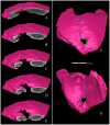
Figure 4. CT reconstruction of the Xujiyao 11 parietal bones with sequential coronal slices through the perforation.
The slices extend from the anterior edge of the opening (A) to close to the posterior margin (E). A 3D CT reconstruction of the specimen is shown in external (F) and internal (G) views, with the postmortem breakage filled in and the sagittal suture line provided.
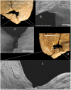
Figure 5. The Xujiayao 11 posteromedial parietal lacuna, with SEM details of the margins.
Exocranial (A) and endocranial (D) views of the opening with details of the anterolateral corner (B), the posterolateral sutural margin (C), and the posterolateral endocranial sulcus margin (E). Scale bars are 1 mm for B and 2 mm for C and E.
Similar articles
- Enlarged parietal foramina: a rare forensic autopsy finding.
Durão C, Carpinteiro D, Pedrosa F, Machado MP, Cunha E. Durão C, et al. Int J Legal Med. 2016 May;130(3):855-7. doi: 10.1007/s00414-015-1239-6. Epub 2015 Aug 2. Int J Legal Med. 2016. PMID: 26233611 Review. - Evolution of cranial capacity revisited: A view from the late Middle Pleistocene cranium from Xujiayao, China.
Wu XJ, Bae CJ, Friess M, Xing S, Athreya S, Liu W. Wu XJ, et al. J Hum Evol. 2022 Feb;163:103119. doi: 10.1016/j.jhevol.2021.103119. Epub 2022 Jan 10. J Hum Evol. 2022. PMID: 35026677 - An updated age for the Xujiayao hominin from the Nihewan Basin, North China: Implications for Middle Pleistocene human evolution in East Asia.
Ao H, Liu CR, Roberts AP, Zhang P, Xu X. Ao H, et al. J Hum Evol. 2017 May;106:54-65. doi: 10.1016/j.jhevol.2017.01.014. Epub 2017 Mar 17. J Hum Evol. 2017. PMID: 28434540 - Hominin teeth from the early Late Pleistocene site of Xujiayao, Northern China.
Xing S, Martinón-Torres M, Bermúdez de Castro JM, Wu X, Liu W. Xing S, et al. Am J Phys Anthropol. 2015 Feb;156(2):224-40. doi: 10.1002/ajpa.22641. Epub 2014 Oct 20. Am J Phys Anthropol. 2015. PMID: 25329008 - The origin and evolution of Homo sapiens.
Stringer C. Stringer C. Philos Trans R Soc Lond B Biol Sci. 2016 Jul 5;371(1698):20150237. doi: 10.1098/rstb.2015.0237. Philos Trans R Soc Lond B Biol Sci. 2016. PMID: 27298468 Free PMC article. Review.
Cited by
- Multiple occipital, parietal, temporal, and frontal foramina: a variant of enlarged parietal foramina in an infant.
Yılmaz E, Yetim A, Erol OB, Pekcan M, Yekeler E. Yılmaz E, et al. Balkan Med J. 2014 Dec;31(4):345-8. doi: 10.5152/balkanmedj.2014.14528. Epub 2014 Dec 1. Balkan Med J. 2014. PMID: 25667790 Free PMC article. - 26Al/10Be burial dating of Xujiayao-Houjiayao site in Nihewan Basin, northern China.
Tu H, Shen G, Li H, Xie F, Granger DE. Tu H, et al. PLoS One. 2015 Feb 23;10(2):e0118315. doi: 10.1371/journal.pone.0118315. eCollection 2015. PLoS One. 2015. PMID: 25706272 Free PMC article. - Middle pleistocene human remains from Tourville-la-Rivière (Normandy, France) and their archaeological context.
Faivre JP, Maureille B, Bayle P, Crevecoeur I, Duval M, Grün R, Bemilli C, Bonilauri S, Coutard S, Bessou M, Limondin-Lozouet N, Cottard A, Deshayes T, Douillard A, Henaff X, Pautret-Homerville C, Kinsley L, Trinkaus E. Faivre JP, et al. PLoS One. 2014 Oct 8;9(10):e104111. doi: 10.1371/journal.pone.0104111. eCollection 2014. PLoS One. 2014. PMID: 25295956 Free PMC article. - External auditory exostoses in the Xuchang and Xujiayao human remains: Patterns and implications among eastern Eurasian Middle and Late Pleistocene crania.
Trinkaus E, Wu XJ. Trinkaus E, et al. PLoS One. 2017 Dec 12;12(12):e0189390. doi: 10.1371/journal.pone.0189390. eCollection 2017. PLoS One. 2017. PMID: 29232394 Free PMC article. - Enlarged parietal foramina: a rare forensic autopsy finding.
Durão C, Carpinteiro D, Pedrosa F, Machado MP, Cunha E. Durão C, et al. Int J Legal Med. 2016 May;130(3):855-7. doi: 10.1007/s00414-015-1239-6. Epub 2015 Aug 2. Int J Legal Med. 2016. PMID: 26233611 Review.
References
- Hauser G, DeStefano GF (1989) Epigenetic variants of the human skull. Stuttgart: Schweizerbart. 301 p.
- Wu M (1980) Human fossils discovered at Xujiayao site in 1977. Vertebrata PalAsiatica 18: 227–238.
- Hatipoglu HG, Ozcan HN, Hatipoglu US, Yuksel E (2008) Age, sex and body mass index in relation to calvarial diploe thickness and craniometric data on MRI. Forensic Sci Intl 182: 46–51. - PubMed
- Chia LP, Wei C (1976) A Palaeolithic site at Hsü-Chia-Yao in Yangkao County, Shansi Province. Acta Archaeol Sinica 2: 97–114.
- Chia LP, Wei Q, Li CR (1979) Report on the excavation of Hsuchiayao man site in 1976. Vertebrata PalAsiatica 17: 277–293.
Publication types
MeSH terms
Supplementary concepts
Grants and funding
This work has been supported by the Chinese Academy of Sciences (KZZD-EW-03, XDA05130100) and the National Natural Science Foundation of China (41272034). The funders had no role in study design, data collection and analysis, decision to publish, or preparation of the manuscript.
LinkOut - more resources
Full Text Sources
Other Literature Sources


