Structural basis for termination of AIM2-mediated signaling by p202 - PubMed (original) (raw)
Structural basis for termination of AIM2-mediated signaling by p202
Heng Ru et al. Cell Res. 2013 Jun.
No abstract available
Figures
Figure 1
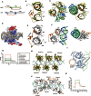
Structural basis for the termination of AIM2-mediated signaling by p202. (A) Schematic diagram of domain arrangement of mAIM2 and p202. Arrows indicate the length of the construct used for structural studies. (B) Cartoon representation of the structure of the HIN domain of mAIM2 bound with DNA. (C) Side view of the cartoon representation of the structure of the HIN domain of mAIM2 bound with dsDNA (left panel). Right panel shows the top view of the structure showing a helix from the linker inserted into the DNA spiral. DNA strand are shown in blue and green. (D) Electrostatic surface potential representation of the HIN domain of mAIM2 bound with dsDNA. 2Fo-Fc electron density map for DNA contoured at 1σ is shown. Positive potential is shown in blue, negative potential in red. DNA is shown as sticks. (E) Cartoon representation of the structure of the HINa domain of p202. Secondary structural elements are labeled according to the canonical OB-fold convention. (F) Side view of the structure of p202-HINa bound with dsDNA (left panel). Right panel shows the top view of the same structure showing loops protruding out from the β-barrel of OB-fold. Strands of DNA are shown in blue and green. (G) Surface plasmon resonance (SPR) assay of the DNA-binding affinity of wild-type p202 HINa domain and its mutants harboring clusters of mutations (M3, M6, M9, M10, M13, M16 and M20) around the DNA-binding region. Data shown represent the binding to AT-rich dsDNA (5′-TTATATATATATATATATAA-3′). (H) Cartoon representation of the mAIM2 HIN molecules bound to dsDNA. (I) Modes of dsDNA binding by the HIN domains of p202 and AIM2. Cα atoms of the HINa domain of p202 (grey) bound with DNA (green ribbon) are superimposed over Cα atoms of the HIN domain of murine AIM2 (blue) bound with DNA (orange ribbon). (J) Cartoon of the p202 HINa molecules bound with DNA. (K) The HINa domain of p202 (green) binds more DNA than mAIM2 HIN domain (red) when assayed under identical conditions using equal molar ratios of protein and DNA.
Similar articles
- Structural mechanism of DNA recognition by the p202 HINa domain: insights into the inhibition of Aim2-mediated inflammatory signalling.
Li H, Wang J, Wang J, Cao LS, Wang ZX, Wu JW. Li H, et al. Acta Crystallogr F Struct Biol Commun. 2014 Jan;70(Pt 1):21-9. doi: 10.1107/S2053230X1303135X. Epub 2013 Dec 24. Acta Crystallogr F Struct Biol Commun. 2014. PMID: 24419611 Free PMC article. - Molecular mechanism for p202-mediated specific inhibition of AIM2 inflammasome activation.
Yin Q, Sester DP, Tian Y, Hsiao YS, Lu A, Cridland JA, Sagulenko V, Thygesen SJ, Choubey D, Hornung V, Walz T, Stacey KJ, Wu H. Yin Q, et al. Cell Rep. 2013 Jul 25;4(2):327-39. doi: 10.1016/j.celrep.2013.06.024. Epub 2013 Jul 11. Cell Rep. 2013. PMID: 23850291 Free PMC article. - AIM2 inflammasome activation and regulation: A structural perspective.
Wang B, Yin Q. Wang B, et al. J Struct Biol. 2017 Dec;200(3):279-282. doi: 10.1016/j.jsb.2017.08.001. Epub 2017 Aug 13. J Struct Biol. 2017. PMID: 28813641 Free PMC article. Review. - Aim2 deficiency stimulates the expression of IFN-inducible Ifi202, a lupus susceptibility murine gene within the Nba2 autoimmune susceptibility locus.
Panchanathan R, Duan X, Shen H, Rathinam VA, Erickson LD, Fitzgerald KA, Choubey D. Panchanathan R, et al. J Immunol. 2010 Dec 15;185(12):7385-93. doi: 10.4049/jimmunol.1002468. Epub 2010 Nov 5. J Immunol. 2010. PMID: 21057088 Free PMC article. - AIM2 Inflammasome Assembly and Signaling.
Wang B, Tian Y, Yin Q. Wang B, et al. Adv Exp Med Biol. 2019;1172:143-155. doi: 10.1007/978-981-13-9367-9_7. Adv Exp Med Biol. 2019. PMID: 31628655 Review.
Cited by
- The Role of the AIM2 Gene in Obesity-Related Glucose and Lipid Metabolic Disorders: A Recent Update.
Zhang Y, Xuan X, Ye D, Liu D, Song Y, Gao F, Lu S. Zhang Y, et al. Diabetes Metab Syndr Obes. 2024 Oct 21;17:3903-3916. doi: 10.2147/DMSO.S488978. eCollection 2024. Diabetes Metab Syndr Obes. 2024. PMID: 39465122 Free PMC article. Review. - MNDA, a PYHIN factor involved in transcriptional regulation and apoptosis control in leukocytes.
Bottardi S, Layne T, Ramòn AC, Quansah N, Wurtele H, Affar EB, Milot E. Bottardi S, et al. Front Immunol. 2024 Apr 12;15:1395035. doi: 10.3389/fimmu.2024.1395035. eCollection 2024. Front Immunol. 2024. PMID: 38680493 Free PMC article. Review. - A 360° view of the inflammasome: Mechanisms of activation, cell death, and diseases.
Barnett KC, Li S, Liang K, Ting JP. Barnett KC, et al. Cell. 2023 May 25;186(11):2288-2312. doi: 10.1016/j.cell.2023.04.025. Cell. 2023. PMID: 37236155 Free PMC article. Review. - Macrophages in periodontitis: A dynamic shift between tissue destruction and repair.
Yin L, Li X, Hou J. Yin L, et al. Jpn Dent Sci Rev. 2022 Nov;58:336-347. doi: 10.1016/j.jdsr.2022.10.002. Epub 2022 Oct 28. Jpn Dent Sci Rev. 2022. PMID: 36340583 Free PMC article. Review. - Involvement of Inflammasome Components in Kidney Disease.
Aranda-Rivera AK, Srivastava A, Cruz-Gregorio A, Pedraza-Chaverri J, Mulay SR, Scholze A. Aranda-Rivera AK, et al. Antioxidants (Basel). 2022 Jan 27;11(2):246. doi: 10.3390/antiox11020246. Antioxidants (Basel). 2022. PMID: 35204131 Free PMC article. Review.
References
- Hornung V, Latz E. Nat Rev Immunol. 2010. pp. 123–130. - PubMed
- Roberts TL, Idris A, Dunn JA, et al. Science. 2009. pp. 1057–1060. - PubMed
Publication types
MeSH terms
Substances
LinkOut - more resources
Full Text Sources
Other Literature Sources
Molecular Biology Databases
