Age-associated changes in the ecological niche: implications for mesenchymal stem cell aging - PubMed (original) (raw)
Review
Age-associated changes in the ecological niche: implications for mesenchymal stem cell aging
Faizal Z Asumda. Stem Cell Res Ther. 2013.
Abstract
Adult stem cells are critical for organ-specific regeneration and self-renewal with advancing age. The prospect of being able to reverse tissue-specific post-injury sequelae by harvesting, culturing and transplanting a patient's own stem and progenitor cells is exciting. Mesenchymal stem cells have emerged as a reliable stem cell source for this treatment modality and are currently being tested in numerous ongoing clinical trials. Unfortunately, the fervor over mesenchymal stem cells is mitigated by several lines of evidence suggesting that their efficacy is limited by natural aging. This article discusses the mechanisms and manifestations of age-associated deficiencies in mesenchymal stem cell efficacy. A consideration of recent experimental findings suggests that the ecological niche might be responsible for mesenchymal stem cell aging.
Figures
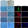
Figure 1
Representative micrographs depicting morphological change after 21 days of exposure to differentiation media. Representative micrographs show anti-FABP4, anti-osteocalcin, anti-aggrecan and anti-cardiac myosin heavy chain staining in differentiating bone marrow-derived mesenchymal stem cells (BM-MSCs). BM-MSCs derived from aged rats fail to undergo adipogenic, chondrogenic, osteogenic and cardiomyogenic differentiation. BM-MSCs derived from young rats transform into fat-forming adipocytes, cartilage-forming chondrocytes, bone-forming osteocytes and cardiomyogenic cells. BM-MSCs were isolated from ‘aged’ and ‘young’ Sprague Dawley rats (15 months and 4 months).
Figure 2
Diagrammatic illustration of potential factors that feed into the bone marrow-derived mesenchymal stem cell (BM-MSC) niche. Diminished BM-MSC function associated with natural aging may be due to deleterious changes at the niche level. Different factors that regulate and maintain the local BM-MSC microenvironment are depicted. Within the niche, BM-MSCs are responsive to metabolic factors and their products, such as oxidative stress and reactive oxygen species (ROS). Paracrine and signaling factors such as Notch, transforming growth factor (TGF)-β, mitogen-activated protein kinase (MAPK), Wnt and NF-ĸB are known to be age dysregulated in the stem cell niche. Physical and environmental factors such as space constraints, and cell-cell interactions between BM-MSCs and other stem and non-stem cells resident in the bone marrow, and between BM-MSCs and the local extracellular matrix may undergo age-associated changes. In response to these changes, BM-MSCs are likely to undergo molecular level changes, such as increased levels of pro-aging factors, DNA damage, telomerase attrition and transcription factor changes. The direct consequence of these changes is diminished BM-MSC function, self-renewal and differentiation capacity. ECM, extracellular matrix; HSC, hematopoietic stem cell.
Similar articles
- Abrogation of Age-Induced MicroRNA-195 Rejuvenates the Senescent Mesenchymal Stem Cells by Reactivating Telomerase.
Okada M, Kim HW, Matsu-ura K, Wang YG, Xu M, Ashraf M. Okada M, et al. Stem Cells. 2016 Jan;34(1):148-59. doi: 10.1002/stem.2211. Epub 2015 Oct 9. Stem Cells. 2016. PMID: 26390028 Free PMC article. - Telomerase protects werner syndrome lineage-specific stem cells from premature aging.
Cheung HH, Liu X, Canterel-Thouennon L, Li L, Edmonson C, Rennert OM. Cheung HH, et al. Stem Cell Reports. 2014 Mar 27;2(4):534-46. doi: 10.1016/j.stemcr.2014.02.006. eCollection 2014 Apr 8. Stem Cell Reports. 2014. PMID: 24749076 Free PMC article. - Therapeutic Potential for Regulation of the Nuclear Factor Kappa-B Transcription Factor p65 to Prevent Cellular Senescence and Activation of Pro-Inflammatory in Mesenchymal Stem Cells.
Mato-Basalo R, Morente-López M, Arntz OJ, van de Loo FAJ, Fafián-Labora J, Arufe MC. Mato-Basalo R, et al. Int J Mol Sci. 2021 Mar 25;22(7):3367. doi: 10.3390/ijms22073367. Int J Mol Sci. 2021. PMID: 33805981 Free PMC article. - Aging of tissue-resident adult stem/progenitor cells and their pathological consequences.
Mimeault M, Batra SK. Mimeault M, et al. Panminerva Med. 2009 Jun;51(2):57-79. Panminerva Med. 2009. PMID: 19776709 Review. - Targeting cellular senescence in kidney diseases and aging: A focus on mesenchymal stem cells and their paracrine factors.
Hejazian SM, Hejazian SS, Mostafavi SM, Hosseiniyan SM, Montazersaheb S, Ardalan M, Zununi Vahed S, Barzegari A. Hejazian SM, et al. Cell Commun Signal. 2024 Dec 18;22(1):609. doi: 10.1186/s12964-024-01968-1. Cell Commun Signal. 2024. PMID: 39696575 Free PMC article. Review.
Cited by
- Production of Inflammatory Mediators in Conditioned Medium of Adipose Tissue-Derived Mesenchymal Stem Cells (ATMSC)-Treated Fresh Frozen Plasma.
Laksmitawati DR, Widowati W, Noverina R, Ayuningtyas W, Kurniawan D, Kusuma HSW, Afifah E, Rinendyaputri R, Rilianawati R, Faried A, Susilarini NK. Laksmitawati DR, et al. Med Sci Monit Basic Res. 2022 Mar 23;28:e933726. doi: 10.12659/MSMBR.933726. Med Sci Monit Basic Res. 2022. PMID: 35318298 Free PMC article. - Allogeneic Mesenchymal Stem Cells Restore Endothelial Function in Heart Failure by Stimulating Endothelial Progenitor Cells.
Premer C, Blum A, Bellio MA, Schulman IH, Hurwitz BE, Parker M, Dermarkarian CR, DiFede DL, Balkan W, Khan A, Hare JM. Premer C, et al. EBioMedicine. 2015 Mar 28;2(5):467-75. doi: 10.1016/j.ebiom.2015.03.020. eCollection 2015 May. EBioMedicine. 2015. PMID: 26137590 Free PMC article. - Cryostorage of Mesenchymal Stem Cells and Biomedical Cell-Based Products.
Linkova DD, Rubtsova YP, Egorikhina MN. Linkova DD, et al. Cells. 2022 Aug 29;11(17):2691. doi: 10.3390/cells11172691. Cells. 2022. PMID: 36078098 Free PMC article. Review. - Focus on the Contribution of Oxidative Stress in Skin Aging.
Papaccio F, D Arino A, Caputo S, Bellei B. Papaccio F, et al. Antioxidants (Basel). 2022 Jun 6;11(6):1121. doi: 10.3390/antiox11061121. Antioxidants (Basel). 2022. PMID: 35740018 Free PMC article. Review. - Recapitulation of growth factor-enriched microenvironment via BMP receptor activating hydrogel.
Zhang Q, Liu Y, Li J, Wang J, Liu C. Zhang Q, et al. Bioact Mater. 2022 Jul 5;20:638-650. doi: 10.1016/j.bioactmat.2022.06.012. eCollection 2023 Feb. Bioact Mater. 2022. PMID: 35846838 Free PMC article.
References
- Traverse JH, Henry TD, Pepine CJ, Willerson JT, Zhao DX, Ellis SG, Forder JR, Anderson RD, Hatzopoulos AK, Penn MS, Perin EC, Chambers J, Baran KW, Raveendran G, Lambert C, Lerman A, Simon DI, Vaughan DE, Lai D, Gee AP, Taylor DA, Cogle CR, Thomas JD, Olson RE, Bowman S, Francescon J, Geither C, Handberg E, Kappenman C, Westbrook L. Effect of the use and timing of bone marrow mononuclear cell delivery on left ventricular function after acute myocardial infarction: the TIME randomized trial. JAMA. 2012;308:2380–2389. - PMC - PubMed
- Hare JM, Fishman JE, Gerstenblith G, DiFede Velazquez DL, Zambrano JP, Suncion VY, Tracy M, Ghersin E, Johnston PV, Brinker JA, Breton E, Davis-Sproul J, Schulman IH, Byrnes J, Mendizabal AM, Lowery MH, Rouy D, Altman P, Wong Po Foo C, Ruiz P, Amador A, Da Silva J, McNiece IK, Heldman A. Comparison of allogeneic vs autologous bone marrow-derived mesenchymal stem cells delivered by transendocardial injection in patients with ischemic cardiomyopathy: the POSEIDON randomized trial. JAMA. 2012;308:2369–2379. - PMC - PubMed
- Traverse JH, Henry TD, Vaughan DE, Ellis SG, Pepine CJ, Willerson JT, Zhao DX, Simpson LM, Penn MS, Byrne BJ, Perin EC, Gee AP, Hatzopoulos AK, McKenna DH, Forder JR, Taylor DA, Cogle CR, Baraniuk S, Olson RE, Jorgenson BC, Sayre SL, Vojvodic RW, Gordon DJ, Skarlatos SI, Moyè LA, Simari RD. Cardiovascular Cell Therapy Research Network. Late TIME: a phase-II, randomized, double-blinded, placebo-controlled, pilot trial evaluating the safety and effect of administration of bone marrow mononuclear cells 2 to 3 weeks after acute myocardial infarction. Tex Heart Inst J. 2010;37:412–420. - PMC - PubMed
- Weiss DJ, Casaburi R, Flannery R, Leroux-Williams M, Tashkin DP. A placebo-controlled randomized trial of mesenchymal stem cells in chronic obstructive pulmonary disease. Chest. 2012. Epub ahead of print.
Publication types
MeSH terms
Substances
LinkOut - more resources
Full Text Sources
Other Literature Sources

