Telomere length modulation in human astroglial brain tumors - PubMed (original) (raw)
Telomere length modulation in human astroglial brain tumors
Domenico La Torre et al. PLoS One. 2013.
Abstract
Background: Telomeres alteration during carcinogenesis and tumor progression has been described in several cancer types. Telomeres length is stabilized by telomerase (h-TERT) and controlled by several proteins that protect telomere integrity, such as the Telomere Repeat-binding Factor (TRF) 1 and 2 and the tankyrase-poli-ADP-ribose polymerase (TANKs-PARP) complex.
Objective: To investigate telomere dysfunction in astroglial brain tumors we analyzed telomeres length, telomerase activity and the expression of a panel of genes controlling the length and structure of telomeres in tissue samples obtained in vivo from astroglial brain tumors with different grade of malignancy.
Materials and methods: Eight Low Grade Astrocytomas (LGA), 11 Anaplastic Astrocytomas (AA) and 11 Glioblastoma Multiforme (GBM) samples were analyzed. Three samples of normal brain tissue (NBT) were used as controls. Telomeres length was assessed through Southern Blotting. Telomerase activity was evaluated by a telomere repeat amplification protocol (TRAP) assay. The expression levels of TRF1, TRF2, h-TERT and TANKs-PARP complex were determined through Immunoblotting and RT-PCR.
Results: LGA were featured by an up-regulation of TRF1 and 2 and by shorter telomeres. Conversely, AA and GBM were featured by a down-regulation of TRF1 and 2 and an up-regulation of both telomerase and TANKs-PARP complex.
Conclusions: In human astroglial brain tumours, up-regulation of TRF1 and TRF2 occurs in the early stages of carcinogenesis determining telomeres shortening and genomic instability. In a later stage, up-regulation of PARP-TANKs and telomerase activation may occur together with an ADP-ribosylation of TRF1, causing a reduced ability to bind telomeric DNA, telomeres elongation and tumor malignant progression.
Conflict of interest statement
Competing Interests: The authors have declared that no competing interests exist.
Figures
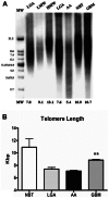
Figure 1. Terminal Restriction Fragment Analysis.
Representative autoradiogram of southern blot analysis. Telomere length was reduced in astroglial tumors as compared with NBT, but elongated telomere was frequently found in GBMs as compared with both AAs and LGGs (p<.001). A Bar Graph. Shows telomere length (x axis) in different grade of astrocytomas and controls (y axis). Error bars indicate standard deviation. ** indicates significance of p<.001. Abbreviations: Kbs: kilobase pair; NBT: normal brain tumor; LGA: Low grade Astrocytoma; AA: Anaplastic Astrocytoma; GBM: Glioblastoma Multiforme; LMW: Low molecular weight; HMW: High molecular weight; MW: Molecular Weight.
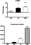
Figure 2. Telomerase activity and h-TERT mRNA expression.
Telomerase activity significantly correlates with h-TERT mRNA expression and WHO in astroglial brain tumors. The h-TERT expression levels in GBMs resulted statistically higher as compared with those in LGAs. The expressions in AAs differed significantly as compared with that in LGAs. A correlation was found between h-TERT and WHO grading (p<.001). Telomerase activity is expressed as relative telomerase activity (RTA) according to the formula showed in the methods. Error bars indicate standard deviation. * p<.05; ** p<.001. Abbreviations: NBT: normal brain tumor; LGA: Low grade Astrocytoma; AA: Anaplastic Astrocytoma; GBM: Glioblastoma Multiforme; h-TERT human telomerase reverse transcriptase.
Figure 3. Expression levels of Telomere-associated proteins among tumors with different grade of malignancy.
Quantitative mRNA expression levels of TRF1, TRF2, TANKs, and PARP-1 was determined by real-time RT-PCR analysis. Relative quantification for these genes was expressed as fold variation over control, and calculated by the ΔΔCt method, using control samples (actin) as calibrators. A TRF1 expression levels progressively decrease from LGAs to GBMs. The expression in LGAs was significantly higher than that in both GBMs (P = .007) and AAs (P = .009). B TRF2 expression levels in LGAs resulted statistically higher as compared with those in GBMs (P = .008), and showed a tendency toward a lower expression in AAs as compared with LGAs C Increased expression of TANKs resulted in AAs compared with LGAs and GBMs. D PARP1 mRNA expression levels resulted statistically higher in AAs as compared with those in LGAs. Moreover, PARP expression in GBMs showed a tendency toward the higher expression as compared with LGAs. Abbreviations: LGA: Low grade Astrocytoma; AA: Anaplastic Astrocytoma; GBM: Glioblastoma Multiforme; TRF1, Telomeric repeat-binding factors 1; TRF2, Telomeric repeat-binding factors 2; PARP1, Poly (ADP-ribose) polymerase 1.
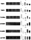
Figure 4. Western blot analysis of TRF1, TRF2, h-TERT, TANKs, and PARP-1.
Left: representative autoradiogram; right: graphs with quantitative data. Both TRF1 and TRF2 are over expressed in LGAs as compared with AAs and GBMs. h-TERT, TANKs, and PARP1 showed a tendency toward a higher expression in malignant astrocytomas (i.e. AAs and GBMs). Abbreviations: NBT: Normal Brain Tissue; LGA: Low grade Astrocytoma; AA: Anaplastic Astrocytoma; GBM: Glioblastoma Multiforme; TRF1, Telomeric repeat-binding factors 1; TRF2, Telomeric repeat-binding factors 2; h-TERT human telomerase reverse transcriptase; PARP1, Poly (ADP-ribose) polymerase 1.
Figure 5. Biomolecular profiles of expression for each tumor histotype.
Relative quantification of studied genes showed that expression levels of those genes are similar along with different histological grades. In LGA, an up-regulation of TRF1 occurs when telomerase has not yet been activated or is down regulated by TRF1. In AA, an up regulation of PARP-Tankirase complex and telomerase activation occurs. The ADP-ribosylation of TRFs, mediated by PARP1, diminish its ability to bind telomeric DNA, causing TRF1 down-regulation and allowing telomerase to elongate progressively telomeres. Finally, a down-regulation of TRF1 and TRF2, increasing telomerase activity, persistent over-expression of PARP-TANKs and elongated telomere are typical features in GBM. Black arrows indicates telomere length along with different stages of carcinogenesis and tumor progression in astroglial brain tumors. Abbreviations: LGA: Low grade Astrocytoma; AA: Anaplastic Astrocytoma; GBM: Glioblastoma Multiforme; TRF1, Telomeric repeat-binding factors 1; TRF2, Telomeric repeat-binding factors 2; h-TERT human telomerase reverse transcriptase; PARP1, Poly (ADP-ribose) polymerase 1.
Similar articles
- Expression of telomeric repeat binding factor 1 and 2 and TRF1-interacting nuclear protein 2 in human gastric carcinomas.
Matsutani N, Yokozaki H, Tahara E, Tahara H, Kuniyasu H, Haruma K, Chayama K, Yasui W, Tahara E. Matsutani N, et al. Int J Oncol. 2001 Sep;19(3):507-12. Int J Oncol. 2001. PMID: 11494028 - Role for the related poly(ADP-Ribose) polymerases tankyrase 1 and 2 at human telomeres.
Cook BD, Dynek JN, Chang W, Shostak G, Smith S. Cook BD, et al. Mol Cell Biol. 2002 Jan;22(1):332-42. doi: 10.1128/MCB.22.1.332-342.2002. Mol Cell Biol. 2002. PMID: 11739745 Free PMC article. - Tankyrase promotes telomere elongation in human cells.
Smith S, de Lange T. Smith S, et al. Curr Biol. 2000 Oct 19;10(20):1299-302. doi: 10.1016/s0960-9822(00)00752-1. Curr Biol. 2000. PMID: 11069113 - [The role of telomere-binding proteins in carcinogenesis].
Aragona M, Pontoriero A, Panetta S, La Torre I, La Torre F. Aragona M, et al. Minerva Med. 2000 Nov-Dec;91(11-12):299-304. Minerva Med. 2000. PMID: 11253711 Review. Italian. - Telomere repeat binding factors: keeping the ends in check.
Karlseder J. Karlseder J. Cancer Lett. 2003 May 15;194(2):189-97. doi: 10.1016/s0304-3835(02)00706-1. Cancer Lett. 2003. PMID: 12757977 Review.
Cited by
- Tankyrases/β-catenin Signaling Pathway as an Anti-proliferation and Anti-metastatic Target in Hepatocarcinoma Cell Lines.
Huang J, Qu Q, Guo Y, Xiang Y, Feng D. Huang J, et al. J Cancer. 2020 Jan 1;11(2):432-440. doi: 10.7150/jca.30976. eCollection 2020. J Cancer. 2020. PMID: 31897238 Free PMC article. - Non-canonical roles of canonical telomere binding proteins in cancers.
Akincilar SC, Chan CHT, Ng QF, Fidan K, Tergaonkar V. Akincilar SC, et al. Cell Mol Life Sci. 2021 May;78(9):4235-4257. doi: 10.1007/s00018-021-03783-0. Epub 2021 Feb 18. Cell Mol Life Sci. 2021. PMID: 33599797 Free PMC article. Review. - Pilocytic Astrocytoma-Derived Cells in Peripheral Blood: A Case Report.
Volpentesta G, Donato G, Ferraro E, Mignogna C, Radaelli R, Sabatini U, La Torre D, Malara N. Volpentesta G, et al. Front Oncol. 2021 Oct 27;11:737730. doi: 10.3389/fonc.2021.737730. eCollection 2021. Front Oncol. 2021. PMID: 34778052 Free PMC article. - Aging, Physical Exercise, Telomeres, and Sarcopenia: A Narrative Review.
Hernández-Álvarez D, Rosado-Pérez J, Gavia-García G, Arista-Ugalde TL, Aguiñiga-Sánchez I, Santiago-Osorio E, Mendoza-Núñez VM. Hernández-Álvarez D, et al. Biomedicines. 2023 Feb 17;11(2):598. doi: 10.3390/biomedicines11020598. Biomedicines. 2023. PMID: 36831134 Free PMC article. Review. - The neuro-protective role of telomerase via TERT/TERF-2 in the acute phase of spinal cord injury.
Chang DG, Kim JW, Kim HJ, Kim YH, Kim SI, Ha KY. Chang DG, et al. Eur Spine J. 2023 Jul;32(7):2431-2440. doi: 10.1007/s00586-023-07561-3. Epub 2023 May 10. Eur Spine J. 2023. PMID: 37165116
References
- Blackburn EH (1991) Structure and function of telomeres. Nature 350: 569–573. - PubMed
- Wright WE, Shay JW (1995) Time, telomeres and tumours: is cellular senescence more than an anticancer mechanism? Trends Cell Biol 5: 293–297. - PubMed
- Harley CB (1991) Telomere loss: mitotic clock or genetic time bomb? Mutat Res 256: 271–282. - PubMed
- Rudolph KL, Chang S, Lee HW, Blasco M, Gottlieb GJ, et al. (1999) Longevity, stress response, and cancer in aging telomerase-deficient mice. Cell 96: 701–712. - PubMed
- Harley CB, Futcher AB, Greider CW (1990) Telomeres shorten during ageing of human fibroblasts. Nature 345: 458–460. - PubMed
Publication types
MeSH terms
Substances
Grants and funding
This Paper was supported in part by Italian Ministry of University and Research; Grant number: prot. 2008979M8K. The funders had no role in study design, data collection and analysis, decision to publish, or preparation of the manuscript. No additional external funding was received for this study.
LinkOut - more resources
Full Text Sources
Other Literature Sources
Medical
Research Materials
Miscellaneous


