Traversing the Renal Pelvis during Percutaneous Nephrostomy Tube Placement ("Kidney Kabob") - PubMed (original) (raw)
Traversing the Renal Pelvis during Percutaneous Nephrostomy Tube Placement ("Kidney Kabob")
Mitchell Smith et al. Semin Intervent Radiol. 2012 Jun.
No abstract available
Figures
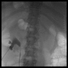
Figure 1
Initial nephrostogram, anteroposterior projection, after access of the collecting system under ultrasound guidance. Contrast fills the intrarenal collecting system and proximal ureter.
Figure 2
Noncontrast computed tomography scan of the abdomen and pelvis obtained after persistent low urine output from the percutaneous nephrostomy tube. Image demonstrates the nephrostomy tube traversing the renal parenchyma and coiling in the intra-abdominal fat anterior to the kidney.
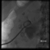
Figure 3
Percutaneous nephrostomy tube, anteroposterior projection, after nephrostomy tube replacement demonstrating tube and contrast within the renal pelvis.
Similar articles
- The results of ultrasound-guided percutaneous nephrostomy tube placement for obstructive uropathy: A single-centre 10-year experience.
Efesoy O, Saylam B, Bozlu M, Çayan S, Akbay E. Efesoy O, et al. Turk J Urol. 2018 Jul;44(4):329-334. doi: 10.5152/tud.2018.25205. Turk J Urol. 2018. PMID: 29799408 Free PMC article. - The use of percutaneous nephrostomy in patients with ureteric obstruction undergoing renal transplantation.
Ehrlichman RJ, Bettman M, Kirkman RL, Tilney NL. Ehrlichman RJ, et al. Surg Gynecol Obstet. 1986 Feb;162(2):121-5. Surg Gynecol Obstet. 1986. PMID: 3511554 - [Use of a gelatine-thrombin matrix for closure of the access tract without a nephrostomy tube in minimally invasive percutaneous nephrolitholapaxy].
Schilling D, Winter B, Merseburger AS, Anastasiadis AG, Walcher U, Stenzl A, Nagele U. Schilling D, et al. Urologe A. 2008 May;47(5):601-7. doi: 10.1007/s00120-008-1673-x. Urologe A. 2008. PMID: 18311555 German. - Do's and don't's of percutaneous nephrostomy.
Zagoria RJ, Dyer RB. Zagoria RJ, et al. Acad Radiol. 1999 Jun;6(6):370-7. doi: 10.1016/s1076-6332(99)80233-5. Acad Radiol. 1999. PMID: 10376069 Review. - Percutaneous nephrostomy with extensions of the technique: step by step.
Dyer RB, Regan JD, Kavanagh PV, Khatod EG, Chen MY, Zagoria RJ. Dyer RB, et al. Radiographics. 2002 May-Jun;22(3):503-25. doi: 10.1148/radiographics.22.3.g02ma19503. Radiographics. 2002. PMID: 12006684 Review.
References
- Goodwin W E, Casey W C, Woolf W. Percutaneous trocar (needle) nephrostomy in hydronephrosis. J Am Med Assoc. 1955;157(11):891–894. - PubMed
- Laggard L E, Fernstrom I. Percutaneous nephropyelostomy. Acta Radiol Diagn (Stockh) 1974;15(3):288–294. - PubMed
- Pedersen J F. Percutaneous nephrostomy guided by ultrasound. J Urol. 1974;112(2):157–159. - PubMed
- Farrell T A, Hicks M E. A review of radiologically guided percutaneous nephrostomies in 303 patients. J Vasc Interv Radiol. 1997;8(5):769–774. - PubMed
- Wah T M, Weston M J, Irving H C. Percutaneous nephrostomy insertion: outcome data from a prospective multi-operator study at a UK training centre. Clin Radiol. 2004;59(3):255–261. - PubMed


