Structural determinants for naturally evolving H5N1 hemagglutinin to switch its receptor specificity - PubMed (original) (raw)
Structural determinants for naturally evolving H5N1 hemagglutinin to switch its receptor specificity
Kannan Tharakaraman et al. Cell. 2013.
Abstract
Of the factors governing human-to-human transmission of the highly pathogenic avian-adapted H5N1 virus, the most critical is the acquisition of mutations on the viral hemagglutinin (HA) to "quantitatively switch" its binding from avian to human glycan receptors. Here, we describe a structural framework that outlines a necessary set of H5 HA receptor-binding site (RBS) features required for the H5 HA to quantitatively switch its preference to human receptors. We show here that the same RBS HA mutations that lead to aerosol transmission of A/Vietnam/1203/04 and A/Indonesia/5/05 viruses, when introduced in currently circulating H5N1, do not lead to a quantitative switch in receptor preference. We demonstrate that HAs from circulating clades require as few as a single base pair mutation to quantitatively switch their binding to human receptors. The mutations identified by this study can be used to monitor the emergence of strains having human-to-human transmission potential.
Copyright © 2013 Elsevier Inc. All rights reserved.
Figures
Figure 1. Comparing molecular features in the RBS of H5 and H2 HA
The RBS of H5 and its phylogenetically closest H2 HA is rendered as a cartoon and the key residues are labeled and their side chains are shown in stick representation. The human receptor in and avian receptor are shown with carbon atoms respectively colored in magenta and yellow in the stick representation at 50% transparency. The 4 key features are indicated based on coloring the carbon atom in different colors. The ‘130-loop’ (Feature 1) is shown in cyan. The ‘base’ of the RBS (Feature 2) including residue positions 136–138, 153, 221–228, is shown in gray. Sidechain of Arg in 224 position is shown at 50 % transparency. The ‘top’ of the RBS (Feature 3) including the ‘190-helix’ and residue positions 155, 156 and 219, is shown in green. The glycosylation sequon at 158 position (Feature 4) in H5 HA is shown in orange. The RBSN diagram is shown adjacent to the residue positions in that network. The circular nodes are colored according to their RBSN score (pink representing a low score to bright red representing a high score) and their connectivity to other nodes. The ‘*’ next to residue positions indicates that these positions belong to the adjacent HA1 domain in the HA trimer. The HA structure images were generated using Pymol (
). See also Figures S2, S3 and S4.
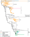
Figure 2. Phylogenetic tree of representative A/H5N1 HA1 protein sequences showing clustering of sequences based on antigenic clades
The sequences are color coded by the features observed in the HA1 domain. Scale bar represents 0.001 amino acid substitutions per site. Feature 1 has evolved in clade-2.2, -2.2.1 and -7 strains after 2006, while Feature 2 has evolved in clade-1 and -2.2.1 after 2007. Feature 3 has evolved in clade-2.2 and -7 strains. Some of the currently circulating clades (1, 2.1.3, 2.2, 2.2.1, 2.3.2, 2.3.4 and 7) have acquired mutations to match multiple RBS features and hence appear to be closer to human adaptation. Among these H5 HAs, the relative order of dominant circulating clades found to have acquired multiple features include clade-2.2.1, -2.2, -1 and -7. A subset of clade 2.2.1 and clade 7 has acquired amino acid changes in the RBS base towards matching Feature 1 and/or Feature 2. See also Table S1 and Figure S3.
Figure 3. Glycan receptor binding of wild type and mutant of H5 HAs HA
Dose-dependent direct binding of wild-type ckViet08, Eg09 and dkEgy10 are shown in A, C and E respectively and that of the V4.4, E4.3 and E5.1 mutants are shown in B, D, and F respectively. See also Table 1 and Table S1 for description of the mutants. Error bars were calculated based on normalized binding signals for glycan array assays done in triplicate for each HA sample. See also Figure S1 and Figure S5.
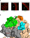
Figure 4. Physiological glycan receptor binding and summary of RBS features in current H5 HAs
A, left panel shows staining of human alveolar section by dkEgy09. The middle panel shows staining of human tracheal tissue section by the E5.1 mutant. The right panel shows staining of human tracheal tissue section by CA04. For all the tissue sections, the HA staining is shown in green against propidium iodide shown in red. Apical surface of trachea is indicated by white arrow B, Surface rendering of H5 RBS with the region corresponding to Features 1–4 are colored cyan, gray, green and orange in that order. The human receptor is shown as a stick. Feature 1 and 2 are colored dark red to indicate their ‘necessary’ requirement for human adaptation of H5 HA in the context of its current sequence evolution. See also Table S1 and Figure S3.
Similar articles
- Phenotypic Effects of Substitutions within the Receptor Binding Site of Highly Pathogenic Avian Influenza H5N1 Virus Observed during Human Infection.
Eggink D, Spronken M, van der Woude R, Buzink J, Broszeit F, McBride R, Pawestri HA, Setiawaty V, Paulson JC, Boons GJ, Fouchier RAM, Russell CA, de Jong MD, de Vries RP. Eggink D, et al. J Virol. 2020 Jun 16;94(13):e00195-20. doi: 10.1128/JVI.00195-20. Print 2020 Jun 16. J Virol. 2020. PMID: 32321815 Free PMC article. - Free energy simulations reveal a double mutant avian H5N1 virus hemagglutinin with altered receptor binding specificity.
Das P, Li J, Royyuru AK, Zhou R. Das P, et al. J Comput Chem. 2009 Aug;30(11):1654-63. doi: 10.1002/jcc.21274. J Comput Chem. 2009. PMID: 19399777 - Identification of amino acids in highly pathogenic avian influenza H5N1 virus hemagglutinin that determine avian influenza species specificity.
Li Z, Liu Z, Ma C, Zhang L, Su Y, Gao GF, Li Z, Cui L, He W. Li Z, et al. Arch Virol. 2011 Oct;156(10):1803-12. doi: 10.1007/s00705-011-1056-2. Epub 2011 Jul 9. Arch Virol. 2011. PMID: 21744000 Free PMC article. - [Host-adaptive mechanism of H5N1 avian influenza virus hemagglutininn].
Watanabe Y, Daidoji T, Nakaya T. Watanabe Y, et al. Uirusu. 2015;65(2):187-198. doi: 10.2222/jsv.65.187. Uirusu. 2015. PMID: 27760917 Review. Japanese. - H5N1 receptor specificity as a factor in pandemic risk.
Paulson JC, de Vries RP. Paulson JC, et al. Virus Res. 2013 Dec 5;178(1):99-113. doi: 10.1016/j.virusres.2013.02.015. Epub 2013 Apr 22. Virus Res. 2013. PMID: 23619279 Free PMC article. Review.
Cited by
- Fitness Determinants of Influenza A Viruses.
Griffin EF, Tompkins SM. Griffin EF, et al. Viruses. 2023 Sep 20;15(9):1959. doi: 10.3390/v15091959. Viruses. 2023. PMID: 37766365 Free PMC article. Review. - Determinant of receptor-preference switch in influenza hemagglutinin.
Ni F, Kondrashkina E, Wang Q. Ni F, et al. Virology. 2018 Jan 1;513:98-107. doi: 10.1016/j.virol.2017.10.010. Epub 2017 Oct 18. Virology. 2018. PMID: 29055255 Free PMC article. - Glycan-protein interactions in viral pathogenesis.
Raman R, Tharakaraman K, Sasisekharan V, Sasisekharan R. Raman R, et al. Curr Opin Struct Biol. 2016 Oct;40:153-162. doi: 10.1016/j.sbi.2016.10.003. Epub 2016 Oct 25. Curr Opin Struct Biol. 2016. PMID: 27792989 Free PMC article. Review. - Comparative review of respiratory diseases caused by coronaviruses and influenza A viruses during epidemic season.
Jiang C, Yao X, Zhao Y, Wu J, Huang P, Pan C, Liu S, Pan C. Jiang C, et al. Microbes Infect. 2020 Jul-Aug;22(6-7):236-244. doi: 10.1016/j.micinf.2020.05.005. Epub 2020 May 13. Microbes Infect. 2020. PMID: 32405236 Free PMC article. Review. - Evolutionary dynamics of highly pathogenic avian influenza A/H5N1 HA clades and vaccine implementation in Vietnam.
Le TH, Nguyen NT. Le TH, et al. Clin Exp Vaccine Res. 2014 Jul;3(2):117-27. doi: 10.7774/cevr.2014.3.2.117. Epub 2014 Jun 20. Clin Exp Vaccine Res. 2014. PMID: 25003084 Free PMC article. Review.
References
- Chandrasekaran A, Srinivasan A, Raman R, Viswanathan K, Raguram S, Tumpey TM, Sasisekharan V, Sasisekharan R. Glycan topology determines human adaptation of avian H5N1 virus hemagglutinin. Nat Biotechnol. 2008;26:107–113. - PubMed
- Gambaryan A, Tuzikov A, Pazynina G, Bovin N, Balish A, Klimov A. Evolution of the receptor binding phenotype of influenza A (H5) viruses. Virology. 2006;344:432–438. - PubMed
- Gamblin SJ, Haire LF, Russell RJ, Stevens DJ, Xiao B, Ha Y, Vasisht N, Steinhauer DA, Daniels RS, Elliot A, et al. The structure and receptor binding properties of the 1918 influenza hemagglutinin. Science. 2004;303:1838–1842. - PubMed
- Ge S, Wang Z. An overview of influenza A virus receptors. Crit Rev Microbiol. 2011;37:157–165. - PubMed
- Guan Y, Smith GJ, Webby R, Webster RG. Molecular epidemiology of H5N1 avian influenza. Rev Sci Tech. 2009;28:39–47. - PubMed
Publication types
MeSH terms
Substances
Grants and funding
- R01 GM057073/GM/NIGMS NIH HHS/United States
- R37 GM057073/GM/NIGMS NIH HHS/United States
- T32 ES007020/ES/NIEHS NIH HHS/United States
- R37GM057073-13/GM/NIGMS NIH HHS/United States
LinkOut - more resources
Full Text Sources
Other Literature Sources
Medical


