Molecular architecture of the uncleaved HIV-1 envelope glycoprotein trimer - PubMed (original) (raw)
Molecular architecture of the uncleaved HIV-1 envelope glycoprotein trimer
Youdong Mao et al. Proc Natl Acad Sci U S A. 2013.
Abstract
The human immunodeficiency virus type 1 (HIV-1) envelope glycoprotein (Env) trimer, a membrane-fusing machine, mediates virus entry into host cells and is the sole virus-specific target for neutralizing antibodies. Binding the receptors, CD4 and CCR5/CXCR4, triggers Env conformational changes from the metastable unliganded state to the fusion-active state. We used cryo-electron microscopy to obtain a 6-Å structure of the membrane-bound, heavily glycosylated HIV-1 Env trimer in its uncleaved and unliganded state. The spatial organization of secondary structure elements reveals that the unliganded conformations of both glycoprotein (gp)120 and gp41 subunits differ from those induced by receptor binding. The gp120 trimer association domains, which contribute to interprotomer contacts in the unliganded Env trimer, undergo rearrangement upon CD4 binding. In the unliganded Env, intersubunit interactions maintain the gp41 ectodomain helical bundles in a "spring-loaded" conformation distinct from the extended helical coils of the fusion-active state. Quaternary structure regulates the virus-neutralizing potency of antibodies targeting the conserved CD4-binding site on gp120. The Env trimer architecture provides mechanistic insights into the metastability of the unliganded state, receptor-induced conformational changes, and quaternary structure-based strategies for immune evasion.
Keywords: cryo-EM; membrane protein; retrovirus; spike; vaccine immunogen.
Conflict of interest statement
The authors declare no conflict of interest.
Figures
Fig. 1.
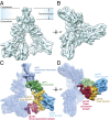
Architecture of the HIV-1 Env trimer. (A) Cryo-EM map of the HIV-1JR-FL Env trimer in a surface representation, viewed from a perspective parallel to the viral membrane. (B) Cryo-EM map of the HIV-1JR-FL Env trimer, viewed from the perspective of the target cell. (C and D) Domain organization of the Env protomer, revealed by segmentation of the density map. The gp120 domains are colored as follows: outer domain, blue; inner domain, orange; and TAD, red. The gp41 domains are colored as follows: ectodomain, green; and transmembrane region, cyan.
Fig. 2.
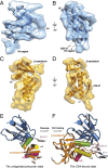
The unliganded gp120 subunit and CD4-induced changes. (A and B) Crystal structure of the outer domain of the gp120 core (17) was fitted into the cryo-EM map of the unliganded Env trimer and is shown from two perspectives parallel to the viral membrane. The approximate location of the V4 variable region, which was not resolved in the crystal structure, is indicated by a broken line. (C and D) Secondary structure organization of the inner domain of gp120 was approximated in the cryo-EM map of the unliganded Env trimer, which is viewed from two perspectives parallel to the viral membrane. (E and F) For comparison of the unliganded precursor state (E) and the CD4-bound state (F) of the gp120 core, the gp120 outer domains (blue) are aligned in the same orientation. The gp120 core in the unliganded precursor state is derived as described in A–D above, and the CD4-bound gp120 core structure is from an X-ray crystal structure (Protein Data Bank ID: 3JWD).
Fig. 3.
Architecture of the gp120 TAD. (A) Secondary structure elements in the gp120 TAD, viewed from a perspective perpendicular to the Env trimer axis. The TAD comprises an α-helix (red), a minibarrel structure (green), and a β-sheet-like element (yellow). (B) Three helical elements (red) pointing toward the central minibarrel structure (green), viewed from the center of mass of the trimer. (C) The TAD, viewed from the perspective of the target cell. The points at which the V1/V2 stem and V3 loop enter the TAD from the gp120 inner domain and outer domain, respectively, are indicated. The map segmentations of the gp120 TAD are shown as blue meshwork for a lower level of contour and as solid surfaces for slightly higher levels of contour. The potential α-helical elements shown as worm tubes are schematically illustrated and are not intended to represent definitive backbone traces.
Fig. 4.
gp41 subunit in the Env trimer. (A) Cryo-EM map of the three gp41 subunits in the Env trimer, viewed from an angle of ∼30° with respect to the viral membrane. (B) gp41 membrane-interactive region viewed from a perspective parallel to the viral membrane. (C) gp41 ectodomain, viewed from the perspective of the virus. The torus-like topology of the three gp41 subunits in the Env trimer is evident from this perspective. (D) gp41 ectodomain, viewed from a perspective parallel to the viral membrane. (E) Comparison of the gp41 ectodomain in the unliganded precursor state and the postfusion state with respect to the tertiary organization of helices. The perspective is identical to that shown in D, with a reduction in scale. For simplicity, only one of the three gp41 glycoprotein subunits in the Env trimer is depicted.
Fig. 5.
Env glycosylation. (A and B) Glycan-associated densities on the gp120 surface, viewed from two perspectives parallel to the viral membrane. The gp120 outer domain ribbon is colored blue and the asparagine residues associated with potential _N_-linked glycosylation sites are shown in Corey-Pauling-Koltun (CPK) representation and labeled. The blue meshwork shows the overall map segment associated with the gp120 outer domain. The yellow densities highlight the potential glycan-associated map segments in a surface representation. (C) Schematic showing the five glycan core residues associated with _N_-linked glycosylation. The trimannosyl core, which is common to high-mannose, complex, and hybrid oligosaccharides, is colored.
Fig. 6.
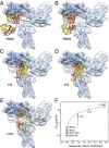
Effect of quaternary structure on virus neutralization by CD4BS antibodies. (A_–_E) Crystal structures of the gp120 core in complex with the CD4BS antibodies, VRC01 (A), VRC03 (B), b12 (C), b13 (D), and F105 (E) were superposed on the unliganded Env precursor map, with the Env trimer axis shown in A. The conformationally rigid gp120 outer domain was fitted to the Env trimer density map, and the outer domains in the crystal structures were aligned with the fitted outer domain (represented as a blue ribbon). The heavy and light chains of the CD4BS antibodies are colored red and yellow, respectively. The dashed red circle in C_–_E marks the approximate diameter of the space in the neighboring gp120 subunit that is overlapped by the antibodies, highlighting the quaternary steric hindrance that the antibodies experience when approaching their binding site on the gp120 outer domain. (F) Using the volume of the quaternary clash in the case of b12 as a normalization metric, the degree of quaternary steric hindrance encountered by each antibody as it accesses the Env trimer was quantified and was plotted against the geometric mean IC50 of the neutralization of multiple HIV-1 strains by the antibody (–, –45). The Protein Data Bank IDs of the antibodies in complex with gp120 core structures are 3NGB (A), 3SE8 (B), 2NY7 (C), 3IDX (D), and 3HI1 (E). The red line shows a linear regression of the relationship (rS: 0.9985, slope: 5.5, SE: 0.082).
Comment in
- Finding trimeric HIV-1 envelope glycoproteins in random noise.
van Heel M. van Heel M. Proc Natl Acad Sci U S A. 2013 Nov 5;110(45):E4175-7. doi: 10.1073/pnas.1314353110. Epub 2013 Oct 8. Proc Natl Acad Sci U S A. 2013. PMID: 24106301 Free PMC article. No abstract available. - Structure of trimeric HIV-1 envelope glycoproteins.
Subramaniam S. Subramaniam S. Proc Natl Acad Sci U S A. 2013 Nov 5;110(45):E4172-4. doi: 10.1073/pnas.1313802110. Epub 2013 Oct 8. Proc Natl Acad Sci U S A. 2013. PMID: 24106302 Free PMC article. No abstract available. - Avoiding the pitfalls of single particle cryo-electron microscopy: Einstein from noise.
Henderson R. Henderson R. Proc Natl Acad Sci U S A. 2013 Nov 5;110(45):18037-41. doi: 10.1073/pnas.1314449110. Epub 2013 Oct 8. Proc Natl Acad Sci U S A. 2013. PMID: 24106306 Free PMC article. - Reply to Subramaniam, van Heel, and Henderson: Validity of the cryo-electron microscopy structures of the HIV-1 envelope glycoprotein complex.
Mao Y, Castillo-Menendez LR, Sodroski JG. Mao Y, et al. Proc Natl Acad Sci U S A. 2013 Nov 5;110(45):E4178-82. doi: 10.1073/pnas.1316666110. Proc Natl Acad Sci U S A. 2013. PMID: 24350339 Free PMC article. No abstract available.
Similar articles
- Residues in the gp41 Ectodomain Regulate HIV-1 Envelope Glycoprotein Conformational Transitions Induced by gp120-Directed Inhibitors.
Pacheco B, Alsahafi N, Debbeche O, Prévost J, Ding S, Chapleau JP, Herschhorn A, Madani N, Princiotto A, Melillo B, Gu C, Zeng X, Mao Y, Smith AB 3rd, Sodroski J, Finzi A. Pacheco B, et al. J Virol. 2017 Feb 14;91(5):e02219-16. doi: 10.1128/JVI.02219-16. Print 2017 Mar 1. J Virol. 2017. PMID: 28003492 Free PMC article. - Effects of the I559P gp41 change on the conformation and function of the human immunodeficiency virus (HIV-1) membrane envelope glycoprotein trimer.
Alsahafi N, Debbeche O, Sodroski J, Finzi A. Alsahafi N, et al. PLoS One. 2015 Apr 7;10(4):e0122111. doi: 10.1371/journal.pone.0122111. eCollection 2015. PLoS One. 2015. PMID: 25849367 Free PMC article. - Cryo-EM structure of a CD4-bound open HIV-1 envelope trimer reveals structural rearrangements of the gp120 V1V2 loop.
Wang H, Cohen AA, Galimidi RP, Gristick HB, Jensen GJ, Bjorkman PJ. Wang H, et al. Proc Natl Acad Sci U S A. 2016 Nov 15;113(46):E7151-E7158. doi: 10.1073/pnas.1615939113. Epub 2016 Oct 31. Proc Natl Acad Sci U S A. 2016. PMID: 27799557 Free PMC article. - Quaternary Interaction of the HIV-1 Envelope Trimer with CD4 and Neutralizing Antibodies.
Liu Q, Zhang P, Lusso P. Liu Q, et al. Viruses. 2021 Jul 20;13(7):1405. doi: 10.3390/v13071405. Viruses. 2021. PMID: 34372611 Free PMC article. Review. - HIV-1 envelope glycoprotein structure.
Merk A, Subramaniam S. Merk A, et al. Curr Opin Struct Biol. 2013 Apr;23(2):268-76. doi: 10.1016/j.sbi.2013.03.007. Epub 2013 Apr 18. Curr Opin Struct Biol. 2013. PMID: 23602427 Free PMC article. Review.
Cited by
- New approaches to HIV vaccine development.
Haynes BF. Haynes BF. Curr Opin Immunol. 2015 Aug;35:39-47. doi: 10.1016/j.coi.2015.05.007. Epub 2015 Jun 8. Curr Opin Immunol. 2015. PMID: 26056742 Free PMC article. Review. - Crystal structure of a soluble cleaved HIV-1 envelope trimer.
Julien JP, Cupo A, Sok D, Stanfield RL, Lyumkis D, Deller MC, Klasse PJ, Burton DR, Sanders RW, Moore JP, Ward AB, Wilson IA. Julien JP, et al. Science. 2013 Dec 20;342(6165):1477-83. doi: 10.1126/science.1245625. Epub 2013 Oct 31. Science. 2013. PMID: 24179159 Free PMC article. - Antibody to gp41 MPER alters functional properties of HIV-1 Env without complete neutralization.
Kim AS, Leaman DP, Zwick MB. Kim AS, et al. PLoS Pathog. 2014 Jul 24;10(7):e1004271. doi: 10.1371/journal.ppat.1004271. eCollection 2014 Jul. PLoS Pathog. 2014. PMID: 25058619 Free PMC article. - Homology models of the HIV-1 attachment inhibitor BMS-626529 bound to gp120 suggest a unique mechanism of action.
Langley DR, Kimura SR, Sivaprakasam P, Zhou N, Dicker I, McAuliffe B, Wang T, Kadow JF, Meanwell NA, Krystal M. Langley DR, et al. Proteins. 2015 Feb;83(2):331-50. doi: 10.1002/prot.24726. Epub 2014 Dec 23. Proteins. 2015. PMID: 25401969 Free PMC article. - Complete dissociation of the HIV-1 gp41 ectodomain and membrane proximal regions upon phospholipid binding.
Roche J, Louis JM, Aniana A, Ghirlando R, Bax A. Roche J, et al. J Biomol NMR. 2015 Apr;61(3-4):235-48. doi: 10.1007/s10858-015-9900-4. Epub 2015 Jan 29. J Biomol NMR. 2015. PMID: 25631354 Free PMC article.
References
- Barré-Sinoussi F, et al. Isolation of a T-lymphotropic retrovirus from a patient at risk for acquired immune deficiency syndrome (AIDS) Science. 1983;220(4599):868–871. - PubMed
- Gallo RC, et al. Frequent detection and isolation of cytopathic retroviruses (HTLV-III) from patients with AIDS and at risk for AIDS. Science. 1984;224(4648):500–503. - PubMed
- Allan JS, et al. Major glycoprotein antigens that induce antibodies in AIDS patients are encoded by HTLV-III. Science. 1985;228(4703):1091–1094. - PubMed
- Robey WG, et al. Characterization of envelope and core structural gene products of HTLV-III with sera from AIDS patients. Science. 1985;228(4699):593–595. - PubMed
- Wyatt R, Sodroski J. The HIV-1 envelope glycoproteins: Fusogens, antigens, and immunogens. Science. 1998;280(5371):1884–1888. - PubMed
Publication types
MeSH terms
Substances
Grants and funding
- R37 AI024755/AI/NIAID NIH HHS/United States
- UM1 AI100645/AI/NIAID NIH HHS/United States
- U01 AI067854/AI/NIAID NIH HHS/United States
- AI67854/AI/NIAID NIH HHS/United States
- #257792/CAPMC/ CIHR/Canada
- U19 AI067854/AI/NIAID NIH HHS/United States
- R01 AI093256/AI/NIAID NIH HHS/United States
- AI93256/AI/NIAID NIH HHS/United States
- AI24755/AI/NIAID NIH HHS/United States
LinkOut - more resources
Full Text Sources
Other Literature Sources
Research Materials


