The haplotype-resolved genome and epigenome of the aneuploid HeLa cancer cell line - PubMed (original) (raw)
The haplotype-resolved genome and epigenome of the aneuploid HeLa cancer cell line
Andrew Adey et al. Nature. 2013.
Abstract
The HeLa cell line was established in 1951 from cervical cancer cells taken from a patient, Henrietta Lacks. This was the first successful attempt to immortalize human-derived cells in vitro. The robust growth and unrestricted distribution of HeLa cells resulted in its broad adoption--both intentionally and through widespread cross-contamination--and for the past 60 years it has served a role analogous to that of a model organism. The cumulative impact of the HeLa cell line on research is demonstrated by its occurrence in more than 74,000 PubMed abstracts (approximately 0.3%). The genomic architecture of HeLa remains largely unexplored beyond its karyotype, partly because like many cancers, its extensive aneuploidy renders such analyses challenging. We carried out haplotype-resolved whole-genome sequencing of the HeLa CCL-2 strain, examined point- and indel-mutation variations, mapped copy-number variations and loss of heterozygosity regions, and phased variants across full chromosome arms. We also investigated variation and copy-number profiles for HeLa S3 and eight additional strains. We find that HeLa is relatively stable in terms of point variation, with few new mutations accumulating after early passaging. Haplotype resolution facilitated reconstruction of an amplified, highly rearranged region of chromosome 8q24.21 at which integration of the human papilloma virus type 18 (HPV-18) genome occurred and that is likely to be the event that initiated tumorigenesis. We combined these maps with RNA-seq and ENCODE Project data sets to phase the HeLa epigenome. This revealed strong, haplotype-specific activation of the proto-oncogene MYC by the integrated HPV-18 genome approximately 500 kilobases upstream, and enabled global analyses of the relationship between gene dosage and expression. These data provide an extensively phased, high-quality reference genome for past and future experiments relying on HeLa, and demonstrate the value of haplotype resolution for characterizing cancer genomes and epigenomes.
Figures
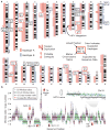
Figure 1. Haplotype-resolved copy number of the HeLa cancer cell line genome
a. Copy-number profile of HeLa split by haplotypes (red = A, blue = B). Links denote likely contiguity and tandem duplications. Boxes indicate marker chromosomes identified by copy number breakpoints (boxes colored by haplotype, or black for unknown); ‘S’ indicates links confirmed by mate-pair sequencing and italics indicate uncertain locations. b. Windowed copy number ratios for HeLa CCL-2 (green and purple, alternating chromosomes) and HeLa S3 (gray), with predicted integer copy number for S3 (black). Notable strain differences are indicated by red arrows (e.g., reduced copy over chromosome 18q). The window containing the HPV insertion and rearrangement is at elevated copy in both strains.
Figure 2. HeLa HPV integration locus
a. Chromosome 8 read depth flanking the HPV integration site (top, blue line), windowed copy-number ratios (purple points) and integer copy states (red bars, middle), and corresponding segments and breakpoints (circled numbers with genomic coordinates, bottom). b. Proposed HPV integration structure: per-segment copy number (upper left), non-rearranged haplotype B (CN=1, upper right), rearranged haplotype A with HPV insertion (CN=3, bottom) carrying ~3 and ~6 tandem copies of repeats ‘R1’ and ‘R2’ respectively. c. The partial HPV-18 genome and corresponding genes (gray and blue, top) with breakpoints highlighted by numbered circles. For reference, the entire HPV-18 genome is shown (bottom).
Figure 3. Gene expression by copy number and haplotype in HeLa S3
a. Transcript abundance (reads per kilobase per million, RPKM, for genes with an RPKM ≥ 1) is positively correlated with gene copy. b. Expression per copy (RPKM / gene copy-number) does not correlate with copy number. c. Fractional contribution of haplotype A to overall expression (RPKM averaged across megabase windows at phased sites) split by haplotype-resolved copy number. Open circles indicate expected fractions. d. Haplotype A-specific expression in HeLa S3 but not CCL-2 across S3-specific LOH on chr18q. e. Haplotype A fractional contribution to expression across the genome, color-coded by underlying haplotype-resolved copy number as in c (point size represents the log2 total RPKM, gray boxes indicate HeLa S3 LOH).
Figure 4. Haplotype-specific regulation near the HPV integration site
a. Long-range chromatin interactions between the HPV and MYC loci demonstrated by ChIA-PET with the RNA polymerase II signal (top) shown for HeLa S3 and an HPV− cell line (K562). Chromatin interactions (below) are highlighted with a green arrow. Bar graphs show read counts at phased, informative sites in MYC (red = A, blue = B). b. Transcript abundance in HeLa S3 across the MYC locus measured by RNA-Seq. Overall coverage is shown in gray (top) with phased, informative sites highlighted (green ticks; italic indicates non-reference allele). Haplotype contributions at each variant are shown at bottom, as in a.
Similar articles
- Haplotype-resolved and integrated genome analysis of the cancer cell line HepG2.
Zhou B, Ho SS, Greer SU, Spies N, Bell JM, Zhang X, Zhu X, Arthur JG, Byeon S, Pattni R, Saha I, Huang Y, Song G, Perrin D, Wong WH, Ji HP, Abyzov A, Urban AE. Zhou B, et al. Nucleic Acids Res. 2019 May 7;47(8):3846-3861. doi: 10.1093/nar/gkz169. Nucleic Acids Res. 2019. PMID: 30864654 Free PMC article. - Long-distance interaction of the integrated HPV fragment with MYC gene and 8q24.22 region upregulating the allele-specific MYC expression in HeLa cells.
Shen C, Liu Y, Shi S, Zhang R, Zhang T, Xu Q, Zhu P, Chen X, Lu F. Shen C, et al. Int J Cancer. 2017 Aug 1;141(3):540-548. doi: 10.1002/ijc.30763. Epub 2017 May 19. Int J Cancer. 2017. PMID: 28470669 Free PMC article. - Comprehensive, integrated, and phased whole-genome analysis of the primary ENCODE cell line K562.
Zhou B, Ho SS, Greer SU, Zhu X, Bell JM, Arthur JG, Spies N, Zhang X, Byeon S, Pattni R, Ben-Efraim N, Haney MS, Haraksingh RR, Song G, Ji HP, Perrin D, Wong WH, Abyzov A, Urban AE. Zhou B, et al. Genome Res. 2019 Mar;29(3):472-484. doi: 10.1101/gr.234948.118. Epub 2019 Feb 8. Genome Res. 2019. PMID: 30737237 Free PMC article. - Genome-based versus gene-based theory of cancer: possible implications for clinical practice.
Todorovic-Rakovic N. Todorovic-Rakovic N. J Biosci. 2011 Sep;36(4):719-24. doi: 10.1007/s12038-011-9099-9. J Biosci. 2011. PMID: 21857118 Review. - Browsing (Epi)genomes: a guide to data resources and epigenome browsers for stem cell researchers.
Karnik R, Meissner A. Karnik R, et al. Cell Stem Cell. 2013 Jul 3;13(1):14-21. doi: 10.1016/j.stem.2013.06.006. Cell Stem Cell. 2013. PMID: 23827707 Free PMC article. Review.
Cited by
- Building in vitro tools for livestock genomics: chromosomal variation within the PK15 cell line.
Johnsson M, Hickey JM, Jungnickel MK. Johnsson M, et al. BMC Genomics. 2024 Jan 11;25(1):49. doi: 10.1186/s12864-023-09931-z. BMC Genomics. 2024. PMID: 38200430 Free PMC article. - Comparative Transcriptomics and Proteomics of Cancer Cell Lines Cultivated by Physiological and Commercial Media.
Wang J, Peng W, Yu A, Fokar M, Mechref Y. Wang J, et al. Biomolecules. 2022 Oct 27;12(11):1575. doi: 10.3390/biom12111575. Biomolecules. 2022. PMID: 36358924 Free PMC article. - Super-enhancers in transcriptional regulation and genome organization.
Wang X, Cairns MJ, Yan J. Wang X, et al. Nucleic Acids Res. 2019 Dec 16;47(22):11481-11496. doi: 10.1093/nar/gkz1038. Nucleic Acids Res. 2019. PMID: 31724731 Free PMC article. - Haplotype-resolved and integrated genome analysis of the cancer cell line HepG2.
Zhou B, Ho SS, Greer SU, Spies N, Bell JM, Zhang X, Zhu X, Arthur JG, Byeon S, Pattni R, Saha I, Huang Y, Song G, Perrin D, Wong WH, Ji HP, Abyzov A, Urban AE. Zhou B, et al. Nucleic Acids Res. 2019 May 7;47(8):3846-3861. doi: 10.1093/nar/gkz169. Nucleic Acids Res. 2019. PMID: 30864654 Free PMC article. - Promoter of lncRNA Gene PVT1 Is a Tumor-Suppressor DNA Boundary Element.
Cho SW, Xu J, Sun R, Mumbach MR, Carter AC, Chen YG, Yost KE, Kim J, He J, Nevins SA, Chin SF, Caldas C, Liu SJ, Horlbeck MA, Lim DA, Weissman JS, Curtis C, Chang HY. Cho SW, et al. Cell. 2018 May 31;173(6):1398-1412.e22. doi: 10.1016/j.cell.2018.03.068. Epub 2018 May 3. Cell. 2018. PMID: 29731168 Free PMC article.
References
- Gey GO, Coffman WD, Kubicek MT. Tissue culture studies of the proliferative capacity of cervical carcinoma and normal epithelium. Cancer research. 1952;12:264–265.
- Gartler SM. Apparent Hela cell contamination of human heteroploid cell lines. Nature. 1968;217:750–751. - PubMed
- Skloot R. The immortal life of Henrietta Lacks. Crown Publishers; 2010.
- Macville M, et al. Comprehensive and definitive molecular cytogenetic characterization of HeLa cells by spectral karyotyping. Cancer Res. 1999;59:141–150. - PubMed
Publication types
MeSH terms
Grants and funding
- CA160080/CA/NCI NIH HHS/United States
- T32 HG000035/HG/NHGRI NIH HHS/United States
- F30 AG039173/AG/NIA NIH HHS/United States
- AG039173/AG/NIA NIH HHS/United States
- HG006283/HG/NHGRI NIH HHS/United States
- R01 HG006283/HG/NHGRI NIH HHS/United States
- R21 CA160080/CA/NCI NIH HHS/United States
- T32HG000035/HG/NHGRI NIH HHS/United States
- T32 GM007266/GM/NIGMS NIH HHS/United States
LinkOut - more resources
Full Text Sources
Other Literature Sources
Research Materials
Miscellaneous


