Therapeutic synergy between microRNA and siRNA in ovarian cancer treatment - PubMed (original) (raw)
. 2013 Nov;3(11):1302-15.
doi: 10.1158/2159-8290.CD-13-0159. Epub 2013 Sep 3.
Eun-Jung Jung # 3 4, Maitri Y Shah # 3 5, Chunhua Lu 1, Riccardo Spizzo 3, Masayoshi Shimizu 3, Hee Dong Han 1, Cristina Ivan 1 6, Simona Rossi 3 7, Xinna Zhang 1 6, Milena S Nicoloso 3, Sherry Y Wu 1, Maria Ines Almeida 3, Justin Bottsford-Miller 1, Chad V Pecot 8, Behrouz Zand 1, Koji Matsuo 1, Mian M Shahzad 1 9, Nicholas B Jennings 1, Cristian Rodriguez-Aguayo 3 6, Gabriel Lopez-Berestein 3 6 10, Anil K Sood 1 6 10, George A Calin 3 6
Affiliations
- PMID: 24002999
- PMCID: PMC3855315
- DOI: 10.1158/2159-8290.CD-13-0159
Therapeutic synergy between microRNA and siRNA in ovarian cancer treatment
Masato Nishimura et al. Cancer Discov. 2013 Nov.
Abstract
Development of improved RNA interference-based strategies is of utmost clinical importance. Although siRNA-mediated silencing of EphA2, an ovarian cancer oncogene, results in reduction of tumor growth, we present evidence that additional inhibition of EphA2 by a microRNA (miRNA) further "boosts" its antitumor effects. We identified miR-520d-3p as a tumor suppressor upstream of EphA2, whose expression correlated with favorable outcomes in two independent patient cohorts comprising 647 patients. Restoration of miR-520d-3p prominently decreased EphA2 protein levels, and suppressed tumor growth and migration/invasion both in vitro and in vivo. Dual inhibition of EphA2 in vivo using 1,2-dioleoyl-sn-glycero-3-phosphatidylcholine (DOPC) nanoliposomes loaded with miR-520d-3p and EphA2 siRNA showed synergistic antitumor efficiency and greater therapeutic efficacy than either monotherapy alone. This synergy is at least in part due to miR-520d-3p targeting EphB2, another Eph receptor. Our data emphasize the feasibility of combined miRNA-siRNA therapy, and will have broad implications for innovative gene silencing therapies for cancer and other diseases.
Significance: This study addresses a new concept of RNA inhibition therapy by combining miRNA and siRNA in nanoliposomal particles to target oncogenic pathways altered in ovarian cancer. Combined targeting of the Eph pathway using EphA2-targeting siRNA and the tumor suppressor miR-520d-3p exhibits remarkable therapeutic synergy and enhanced tumor suppression in vitro and in vivo compared with either monotherapy alone.
©2013 AACR.
Figures
Figure 1. miR-520d-3p is an independent positive prognostic factor in OC
(a) Analysis of variance (ANOVA) statistics identifying miR-520d-3p to be important predictor of overall survival (alive vs. deceased) and response to therapy (complete response vs. progressive disease), and cox proportional hazard model showing hazard ratio of miR-520d-3p using the 2009 TCGA database (n = 186). (b, c) Kaplan-Meier curves representing the percent overall survival in patients with OC based on miR-520d-3p median expression levels in TCGA 2009 database (n = 186) (b) and in MDACC cohort (n = 91) (c). (d, e, f) Kaplan-Meier curves representing the percent overall survival of 556 OC patients from TCGA 2012 dataset based on miR-520d-3p median expression alone (d) or EphA2 median expression alone (e) or after combined EphA2 and miR-520d-3p expression levels (f). The patients were grouped into percentiles according to median mRNA/miRNA expression. The Log-rank test was employed to determine the significance between mRNA/miRNA expression and overall survival. The colored numbers (red or blue) below the curves represent patients at risk at the specified time points.
Figure 2. EphA2 is a direct and functional target of miR-520d-3p
(a) Scatter plot showing negative correlation between EphA2 mRNA (normalized to 18S) and miR-520d-3p (normalized to U6) in MDACC patient set using Spearman’s correlation analysis (R < −0.248; P = 0.02). (b) Quantification of EphA2 and miR-520d-3p immunostaining from four patients showing negative correlation in OC tumors. (c) Representative images of the immunostaining in panel (b). (d) qRT-PCR analysis showing transient overexpression of miR-520d-3p in ES2 and SKOV3ip1 cells (upper panel) results in downregulation of EphA2 mRNA after 48 and 72 hours (lower panel). (e) Immunoblotting of EphA2 and GAPDH in ES2 and SKOV3ip1 cells transfected with miR-520d-3p (100nM or 200nM) or a scrambled control. (f) Representative diagram of the conserved binding site of miR-520d-3p in the 3’-UTR of EphA2 mRNA. (g) Luciferase activity of a reporter construct fused to wild-type or mutant EphA2 3′-UTR in ES2 and SKOV3ip1 cells with ectopic miR-520d-3p expression. Control, cells transfected with a scrambled miRNA control. Data are average of three independent experiments. Statistical significance was determined by unpaired, two-tailed Student’s t test. * P ≤ 0.05, ** P ≤ 0.01, *** P ≤ 0.001 Data are mean ± s.d.
Figure 3. miR-520d-3p expression inhibits migration, invasion and tumor growth
(a, b) Representative images (at 100×) showing effect of miR-520d-3p stable overexpression on migration (a) and invasion (b) of HeyA8 and SKOV3ip1 cells using Transwell migration and Matrigel invasion assays (Left panel). Absorbance was measured at 590nm after 24 hours. The data from one representative experiment are shown in right panel. Experiment was performed in triplicates at three independent times. (c, d) Total tumor weight (c) and number of metastatic tumor nodules (d) in mice (n = 10 per group) with intra peritoneal injection of miR-520d-3p-transduced or control-transduced or parental untreated HeyA8 (33 days) or SKOV3ip1 (46 days) cells after implantation. (e) Representative images of CD31 staining (at 100×) to identify endothelial cells in untreated, control miR-transfected and miR-520d-3p-transfected HeyA8 and SKOV3ip1 tumors. Quantification of CD31 staining is shown in right panel. A lumen with positive CD31 staining was counted as a single microvessel. Data are average of three independent experiments. (f) Immunoblotting for EphA2 and GAPDH in control or EphA2-transfected HeyA8 empty-E3 or miR-520d-3p-overexpressing-M10 clones. (g) Representative images (at 40×) showing migration of untreated control or EphA2-overexpressing HeyA8 empty-E3 or miR-520d-3p-overexpressing-M10 clones. Quantification of migratory cells counted is shown in right panel. Experiment was repeated in duplicates at three independent times. Statistical significance was determined by unpaired, two-tailed Student’s t test when compared to empty clones for all analyses. * P ≤ 0.05, ** P ≤ 0.01, *** P ≤ 0.001 Data are mean ± s.d.
Figure 4. Combination of miR-520d-3p and siRNA-EphA2 treatment shows enhanced EphA2 inhibition and anti-tumor efficiency in vitro
(a) Immunoblotting of EphA2 and GAPDH in HeyA8 and SKOV3ip1 cells after treatment with miR-520d-3p or different EphA2-targeting siRNAs or a combination of both (1 – 6). (b, c) Representative images showing effect of different combination treatments (1 – 4) on SKOV3ip1 and HeyA8 migration (b) and invasion (c) using Transwell migration assay (left panel). Cells were counted in 10 random fields per well at 40× after 6 hrs for migration and 24 hrs for invasion and the percent migratory or percent invasive cells were calculated compared to control treatment. A representative experiment is shown in right panel. Experiment was performed in duplicates at three independent times. (d) Representative images showing effect of rescue treatment with anti-miR-520d-3p in different combinations (1 – 6) on SKOV3ip1 migration using Transwell migration assay (left panel). Absorbance was measured at 590nm after 24 hours and the percent migratory cells were calculated compared to control treatment. The data from one representative experiment are shown in right panel. Experiment was performed in triplicates at three independent times. Statistical significance was determined by unpaired, two-tailed Student’s t test when compared to empty clones for all analyses. * P ≤ 0.05, ** P ≤ 0.01, *** P ≤ 0.001 Data are mean ± s.d.
Figure 5. Co-treatment with miR-520d-3p and siRNA-EphA2 shows potent synergy and improved therapeutic efficiency in vivo
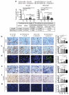
(a) Total tumor weight after various combination treatments (1 – 4) of HeyA8 (left panel) and SKOV3ip1 (right panel) tumors. The lower panel shows calculation to demonstrate synergism as described in Materials and Methods. (b, c, d) Effect of combined miR-520d-3p + siEphA2-1 treatment on angiogenesis, proliferation and apoptosis in SKOV3ip1 cells. Representative images of CD31 (b) Ki67 (c) and TUNEL (d) immunostaining following various combination treatments (1 – 4) are shown (images were acquired at 100×). Quantification of immunostaining in (b, c, d) are shown in the right panels. (e, f, g) Effect of combined miR-520d-3p + siEphA2-1 treatment on angiogenesis, proliferation and apoptosis in HeyA8 cells. Representative images of CD31 (e), Ki67 (f) and TUNEL (g) immunostaining following various combination treatments (1 – 4) are shown (images were acquired at 100×). Quantification of immunostaining in (e, f, g) are shown in the right panels. Statistical significance was determined by unpaired, two-tailed Student’s t test when compared to empty clones for all analyses. * P ≤ 0.05, ** P ≤ 0.01, *** P ≤ 0.001 Data are mean ± s.d.
Figure 6. EphB2 is a direct and functional target of miR-520d-3p, and a prognostic factor for patients with OC
(a) Luciferase activity of a reporter construct fused to wild-type or mutant EphB2 3′-UTR in MCF7 cells with ectopic miR-520d-3p expression (upper panel). Control, cells transfected with a scrambled control. Data are average of four independent experiments. Representative diagram of miR-520d-3p binding site on EphB2 mRNA (lower panel). (b) Immunoblotting of EphB2 and GAPDH in ES2 and SKOV3ip1 cells transfected with miR-520d-3p or a scrambled control. (c) Immunoblotting of EphB2 and GAPDH in miR-520d-3p overexpressing SKOV3ip1 stable clones. (d) Representative images of the immunostaining for EphA2 and miR-520d-3p from four patients showing negative correlation in OC tumors. (e) Immunoblotting of EphA2, EphB2 and GAPDH in SKOV3ip1 cells after various combination treatments (1 – 4). (f) Immunoblotting of EphB2 and GAPDH in SKOV3ip1 cells after treatment with miR-520d-3p or different EphA2-targeting siRNAs or a combination of both (1 – 6). (g, h, i) Kaplan-Meier curves representing the percent overall survival of 556 patients from TCGA 2012 dataset based on EphB2 median expression (f), combined EphB2 and miR-520d-3p expression levels (g) or combined EphA2, EphB2 and miR-520d-3p expression levels (h). The colored numbers (red or blue) below the curves represent patients at risk at the specified time points. Statistical significance was determined by unpaired, two-tailed Student’s t test. * P ≤ 0.05, ** P ≤ 0.01, *** P ≤ 0.001 Data are mean ± s.d.
Comment in
- Small RNAs deliver a blow to ovarian cancer.
Kasinski A, Slack FJ. Kasinski A, et al. Cancer Discov. 2013 Nov;3(11):1220-1. doi: 10.1158/2159-8290.CD-13-0667. Cancer Discov. 2013. PMID: 24203953 Free PMC article.
Similar articles
- Therapeutic EphA2 gene targeting in vivo using neutral liposomal small interfering RNA delivery.
Landen CN Jr, Chavez-Reyes A, Bucana C, Schmandt R, Deavers MT, Lopez-Berestein G, Sood AK. Landen CN Jr, et al. Cancer Res. 2005 Aug 1;65(15):6910-8. doi: 10.1158/0008-5472.CAN-05-0530. Cancer Res. 2005. PMID: 16061675 - MicroRNA 520d-3p inhibits gastric cancer cell proliferation, migration, and invasion by downregulating EphA2 expression.
Li R, Yuan W, Mei W, Yang K, Chen Z. Li R, et al. Mol Cell Biochem. 2014 Nov;396(1-2):295-305. doi: 10.1007/s11010-014-2164-6. Epub 2014 Jul 26. Mol Cell Biochem. 2014. PMID: 25063221 - Dual targeting of EphA2 and FAK in ovarian carcinoma.
Shahzad MM, Lu C, Lee JW, Stone RL, Mitra R, Mangala LS, Lu Y, Baggerly KA, Danes CG, Nick AM, Halder J, Kim HS, Vivas-Mejia P, Landen CN, Lopez-Berestein G, Coleman RL, Sood AK. Shahzad MM, et al. Cancer Biol Ther. 2009 Jun;8(11):1027-34. doi: 10.4161/cbt.8.11.8523. Epub 2009 Jun 24. Cancer Biol Ther. 2009. PMID: 19395869 Free PMC article. - EphA2 as a target for ovarian cancer therapy.
Landen CN, Kinch MS, Sood AK. Landen CN, et al. Expert Opin Ther Targets. 2005 Dec;9(6):1179-87. doi: 10.1517/14728222.9.6.1179. Expert Opin Ther Targets. 2005. PMID: 16300469 Review. - Preclinical and clinical development of siRNA-based therapeutics.
Ozcan G, Ozpolat B, Coleman RL, Sood AK, Lopez-Berestein G. Ozcan G, et al. Adv Drug Deliv Rev. 2015 Jun 29;87:108-19. doi: 10.1016/j.addr.2015.01.007. Epub 2015 Feb 7. Adv Drug Deliv Rev. 2015. PMID: 25666164 Free PMC article. Review.
Cited by
- MicroRNAs as therapeutic targets in human cancers.
Shah MY, Calin GA. Shah MY, et al. Wiley Interdiscip Rev RNA. 2014 Jul-Aug;5(4):537-48. doi: 10.1002/wrna.1229. Epub 2014 Mar 28. Wiley Interdiscip Rev RNA. 2014. PMID: 24687772 Free PMC article. Review. - Recent Advances in miRNA Delivery Systems.
Dasgupta I, Chatterjee A. Dasgupta I, et al. Methods Protoc. 2021 Jan 20;4(1):10. doi: 10.3390/mps4010010. Methods Protoc. 2021. PMID: 33498244 Free PMC article. - MiR-26b inhibits hepatocellular carcinoma cell proliferation, migration, and invasion by targeting EphA2.
Li H, Sun Q, Han B, Yu X, Hu B, Hu S. Li H, et al. Int J Clin Exp Pathol. 2015 May 1;8(5):4782-90. eCollection 2015. Int J Clin Exp Pathol. 2015. PMID: 26191168 Free PMC article. - SP1-mediated microRNA-520d-5p suppresses tumor growth and metastasis in colorectal cancer by targeting CTHRC1.
Yan L, Yu J, Tan F, Ye GT, Shen ZY, Liu H, Zhang Y, Wang JF, Zhu XJ, Li GX. Yan L, et al. Am J Cancer Res. 2015 Mar 15;5(4):1447-59. eCollection 2015. Am J Cancer Res. 2015. PMID: 26101709 Free PMC article. - Targeting oncomiRNAs and mimicking tumor suppressor miRNAs: Νew trends in the development of miRNA therapeutic strategies in oncology (Review).
Gambari R, Brognara E, Spandidos DA, Fabbri E. Gambari R, et al. Int J Oncol. 2016 Jul;49(1):5-32. doi: 10.3892/ijo.2016.3503. Epub 2016 May 4. Int J Oncol. 2016. PMID: 27175518 Free PMC article. Review.
References
- Bartel DP. MicroRNAs: genomics, biogenesis, mechanism, and function. Cell. 2004;116:281–97. - PubMed
- Calin GA, Croce CM. MicroRNA signatures in human cancers. Nature Reviews Cancer. 2006;6:857–66. - PubMed
- American Cancer Society . Cancer Facts & Figures 2012. American Cancer Society; Atlanta:
Publication types
MeSH terms
Substances
Grants and funding
- CA128797/CA/NCI NIH HHS/United States
- P30 CA016672/CA/NCI NIH HHS/United States
- UH2 TR000943-01/TR/NCATS NIH HHS/United States
- CA135444/CA/NCI NIH HHS/United States
- P50 CA098258/CA/NCI NIH HHS/United States
- R01 CA135444/CA/NCI NIH HHS/United States
- P50 CA083639/CA/NCI NIH HHS/United States
- R01 CA128797/CA/NCI NIH HHS/United States
- CA 109298/CA/NCI NIH HHS/United States
- CA151668/CA/NCI NIH HHS/United States
- T32 CA009666/CA/NCI NIH HHS/United States
- CA16672/CA/NCI NIH HHS/United States
- UH2 TR000943/TR/NCATS NIH HHS/United States
- R01 CA109298/CA/NCI NIH HHS/United States
- RC2GM092599/GM/NIGMS NIH HHS/United States
- U54 CA151668/CA/NCI NIH HHS/United States
- RC2 GM092599/GM/NIGMS NIH HHS/United States
LinkOut - more resources
Full Text Sources
Other Literature Sources
Medical
Molecular Biology Databases
Miscellaneous


