Type-specific cell line models for type-specific ovarian cancer research - PubMed (original) (raw)
. 2013 Sep 4;8(9):e72162.
doi: 10.1371/journal.pone.0072162. eCollection 2013.
Kimberly C Wiegand, Nataliya Melnyk, Christine Chow, Clara Salamanca, Leah M Prentice, Janine Senz, Winnie Yang, Monique A Spillman, Dawn R Cochrane, Karey Shumansky, Sohrab P Shah, Steve E Kalloger, David G Huntsman
Affiliations
- PMID: 24023729
- PMCID: PMC3762837
- DOI: 10.1371/journal.pone.0072162
Type-specific cell line models for type-specific ovarian cancer research
Michael S Anglesio et al. PLoS One. 2013.
Erratum in
- PLoS One. 2013;8(10). doi:10.1371/annotation/856f0890-9d85-4719-8e54-c27530ac94f4
- Correction: Type-Specific Cell Line Models for Type-Specific Ovarian Cancer Research.
Anglesio MS, Wiegand KC, Melnyk N, Chow C, Salamanca C, Prentice LM, Senz J, Yang W, Spillman MA, Cochrane DR, Shumansky K, Shah SP, Kalloger SE, Huntsman DG. Anglesio MS, et al. PLoS One. 2013 Sep 27;8(9):10.1371/annotation/ffcaf179-872f-470b-8bb6-f06d8ba6d03a. doi: 10.1371/annotation/ffcaf179-872f-470b-8bb6-f06d8ba6d03a. eCollection 2013. PLoS One. 2013. PMID: 24116245 Free PMC article.
Abstract
Background: OVARIAN CARCINOMAS CONSIST OF AT LEAST FIVE DISTINCT DISEASES: high-grade serous, low-grade serous, clear cell, endometrioid, and mucinous. Biomarker and molecular characterization may represent a more biologically relevant basis for grouping and treating this family of tumors, rather than site of origin. Molecular characteristics have become the new standard for clinical pathology, however development of tailored type-specific therapies is hampered by a failure of basic research to recognize that model systems used to study these diseases must also be stratified. Unrelated model systems do offer value for study of biochemical processes but specific cellular context needs to be applied to assess relevant therapeutic strategies.
Methods: We have focused on the identification of clear cell carcinoma cell line models. A panel of 32 "ovarian cancer" cell lines has been classified into histotypes using a combination of mutation profiles, IHC mutation-surrogates, and a validated immunohistochemical model. All cell lines were identity verified using STR analysis.
Results: Many described ovarian clear cell lines have characteristic mutations (including ARID1A and PIK3CA) and an overall molecular/immuno-profile typical of primary tumors. Mutations in TP53 were present in the majority of high-grade serous cell lines. Advanced genomic analysis of bona-fide clear cell carcinoma cell lines also support copy number changes in typical biomarkers such at MET and HNF1B and a lack of any recurrent expressed re-arrangements.
Conclusions: As with primary ovarian tumors, mutation status of cancer genes like ARID1A and TP53 and a general immuno-profile serve well for establishing histotype of ovarian cancer cell We describe specific biomarkers and molecular features to re-classify generic "ovarian carcinoma" cell lines into type specific categories. Our data supports the use of prototype clear cell lines, such as TOV21G and JHOC-5, and questions the use of SKOV3 and A2780 as models of high-grade serous carcinoma.
Conflict of interest statement
Competing Interests: The authors have declared that no competing interests exist.
Figures
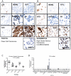
Figure 1. Prediction of histotype was in part based on the COSP algorithm using 9 IHC markers .
(A–B) representative IHC from a typical high-grade serous ovarian carcinoma cell line, Kuramochi, and a clear cell carcinoma cell line, TOV21G. In addition to the 9-marker COSP panel, IHC for ARID1A (BAF250a) is also shown as a mutation surrogate. (C) TFF3 mRNA expression from 60 ovarian cancer samples (12 of each histotype). As noted previously high expression is most prevalent in MUC, followed by ENOCa and LGSC , . Expression in our pilot cohort suggests the highest levels of TFF3 in MUC, which was significantly higher than all other groups (Tukey's adjusted p<0.01); no other pairwise comparisons had p<0.05. (D) TFF3 mRNA detected in ovarian cancer cell lines was used in place of an IHC score as the secreted TFF3 was considered a poor biomarker for cell culture conditions. Any cell line with measurable TFF3 mRNA above the NanoString detection threshold (see methods) was considered positive (score of 1 for use in the COSP algorithm).
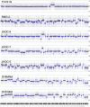
Figure 2. Genome-wide copy number profiles of bona-fide ovarian CCC cell lines.
A large range of copy number changes are seen including typical Chr8 gains and Chr17 gains surrounding the CCC biomarker HNF1B gene, see also Table 3.
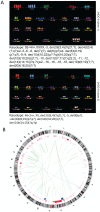
Figure 3. Genomic structure of CCC cell line JHOC-9. (A) 24 color FISH analysis suggested the presence of two dominant clones; one near-diploid and one near-tetraploid in the JHOC-9 CCC cell line.
A number of translocations and rearrangements can be seen in each representative clone. The complex karyotype of each dominant clone is noted below the corresponding 24-colour FISH results. Not all derivative chromosomes were identifiable resulting in a large number of ambiguous translocations and fragments (denoted by question marks in the karyotype notations). (B) Circos plot of RNAseq data and deFuse analysis depicting expressed genomic rearrangements in the JHOC-9 cell line. Translocations seen in the 24-color FISH profile are also visible as expressed transcripts including t(8;19) observed in both 2N and 4N dominant clones. No recurrent translocations were seen across our series (see also Table S3).
Similar articles
- TP53 mutations are common in all subtypes of epithelial ovarian cancer and occur concomitantly with KRAS mutations in the mucinous type.
Rechsteiner M, Zimmermann AK, Wild PJ, Caduff R, von Teichman A, Fink D, Moch H, Noske A. Rechsteiner M, et al. Exp Mol Pathol. 2013 Oct;95(2):235-41. doi: 10.1016/j.yexmp.2013.08.004. Epub 2013 Aug 18. Exp Mol Pathol. 2013. PMID: 23965232 - Molecular pathogenesis and extraovarian origin of epithelial ovarian cancer--shifting the paradigm.
Kurman RJ, Shih IeM. Kurman RJ, et al. Hum Pathol. 2011 Jul;42(7):918-31. doi: 10.1016/j.humpath.2011.03.003. Hum Pathol. 2011. PMID: 21683865 Free PMC article. Review. - Establishment and Characterization of the Novel High-Grade Serous Ovarian Cancer Cell Line OVPA8.
Tudrej P, Olbryt M, Zembala-Nożyńska E, Kujawa KA, Cortez AJ, Fiszer-Kierzkowska A, Pigłowski W, Nikiel B, Głowala-Kosińska M, Bartkowska-Chrobok A, Smagur A, Fidyk W, Lisowska KM. Tudrej P, et al. Int J Mol Sci. 2018 Jul 17;19(7):2080. doi: 10.3390/ijms19072080. Int J Mol Sci. 2018. PMID: 30018258 Free PMC article. - Morphologic, Immunophenotypic, and Molecular Features of Epithelial Ovarian Cancer.
Ramalingam P. Ramalingam P. Oncology (Williston Park). 2016 Feb;30(2):166-76. Oncology (Williston Park). 2016. PMID: 26892153 Review. - [Significance and expression of PAX8, PAX2, p53 and RAS in ovary and fallopian tubes to origin of ovarian high grade serous carcinoma].
Mao YN, Zeng LX, Li YH, Liu YZ, Wu JY, Li L, Wang Q. Mao YN, et al. Zhonghua Fu Chan Ke Za Zhi. 2017 Oct 25;52(10):687-696. doi: 10.3760/cma.j.issn.0529-567X.2017.10.008. Zhonghua Fu Chan Ke Za Zhi. 2017. PMID: 29060967 Chinese.
Cited by
- Integrative proteomic profiling of ovarian cancer cell lines reveals precursor cell associated proteins and functional status.
Coscia F, Watters KM, Curtis M, Eckert MA, Chiang CY, Tyanova S, Montag A, Lastra RR, Lengyel E, Mann M. Coscia F, et al. Nat Commun. 2016 Aug 26;7:12645. doi: 10.1038/ncomms12645. Nat Commun. 2016. PMID: 27561551 Free PMC article. - In vivo tumor growth of high-grade serous ovarian cancer cell lines.
Mitra AK, Davis DA, Tomar S, Roy L, Gurler H, Xie J, Lantvit DD, Cardenas H, Fang F, Liu Y, Loughran E, Yang J, Sharon Stack M, Emerson RE, Cowden Dahl KD, V Barbolina M, Nephew KP, Matei D, Burdette JE. Mitra AK, et al. Gynecol Oncol. 2015 Aug;138(2):372-7. doi: 10.1016/j.ygyno.2015.05.040. Epub 2015 Jun 5. Gynecol Oncol. 2015. PMID: 26050922 Free PMC article. - Lipid droplets: a candidate new research field for epithelial ovarian cancer.
Koizume S, Takahashi T, Miyagi Y. Koizume S, et al. Front Pharmacol. 2024 Jul 1;15:1437161. doi: 10.3389/fphar.2024.1437161. eCollection 2024. Front Pharmacol. 2024. PMID: 39011508 Free PMC article. - COL11A1 confers chemoresistance on ovarian cancer cells through the activation of Akt/c/EBPβ pathway and PDK1 stabilization.
Wu YH, Chang TH, Huang YF, Chen CC, Chou CY. Wu YH, et al. Oncotarget. 2015 Sep 15;6(27):23748-63. doi: 10.18632/oncotarget.4250. Oncotarget. 2015. PMID: 26087191 Free PMC article. - Differential epithelial and stromal LGR5 expression in ovarian carcinogenesis.
Kim H, Lee DH, Park E, Myung JK, Park JH, Kim DI, Kim SI, Lee M, Kim Y, Park CM, Hyun CL, Maeng YH, Lee C, Jang B. Kim H, et al. Sci Rep. 2022 Jul 1;12(1):11200. doi: 10.1038/s41598-022-15234-2. Sci Rep. 2022. PMID: 35778589 Free PMC article.
References
- Auersperg N (2011) The origin of ovarian carcinomas: a unifying hypothesis. Int J Gynecol Pathol 30: 12–21. - PubMed
- Kalloger SE, Kobel M, Leung S, Mehl E, Gao D, et al. (2011) Calculator for ovarian carcinoma subtype prediction. Mod Pathol 24: 512–521. - PubMed
- Soslow RA (2008) Histologic subtypes of ovarian carcinoma: an overview. Int J Gynecol Pathol 27: 161–174. - PubMed
Publication types
MeSH terms
Substances
Grants and funding
Support for this project was provided to the Ovarian Cancer Research Team of BC (OVCARE; http://www.ovcare.ca) through the BC Cancer Foundation, The VGH and UBC Hospitals Foundation and the Canadian Institutes for Health Research (CIHR) Emerging Team Grant: Personalized siRNA-Based Nanomedicines (FRN: 111627). Funders had no role in study design, data collection and analysis, decision to publish, or preparation of the manuscript.
LinkOut - more resources
Full Text Sources
Other Literature Sources
Medical
Molecular Biology Databases
Research Materials
Miscellaneous


