The role of primary cilia in the development and disease of the retina - PubMed (original) (raw)
Review
. 2014 Jan 1;10(1):69-85.
doi: 10.4161/org.26710. Epub 2013 Oct 25.
Affiliations
- PMID: 24162842
- PMCID: PMC4049897
- DOI: 10.4161/org.26710
Review
The role of primary cilia in the development and disease of the retina
Gabrielle Wheway et al. Organogenesis. 2014.
Abstract
The normal development and function of photoreceptors is essential for eye health and visual acuity in vertebrates. Mutations in genes encoding proteins involved in photoreceptor development and function are associated with a suite of inherited retinal dystrophies, often as part of complex multi-organ syndromic conditions. In this review, we focus on the role of the photoreceptor outer segment, a highly modified and specialized primary cilium, in retinal health and disease. We discuss the many defects in the structure and function of the photoreceptor primary cilium that can cause a class of inherited conditions known as ciliopathies, often characterized by retinal dystrophy and degeneration, and highlight the recent insights into disease mechanisms.
Keywords: ciliopathy; inherted retinal conditions; intraflagellar transport; photoreceptor development; primary cilia; retina.
Figures
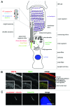
Figure 1. Schematic representation of a rod photoreceptor cell and localization of ciliary proteins.
(A) The schematic represents the rod photoreceptor cell outer segment, connecting cilium, inner segment, outer fiber, cell body, inner fiber and synaptic terminus. A number of key components of the ciliary apparatus are color coded and indicated. The IFT complex A (blue) and complex B (red) are represented in the magnified inset. A retinal pigmentary epithelial (RPE) cell is shown in gray at the top. (B) Confocal microscopy images of an immunofluorescent stained P20 mouse retinal cryosection showing the stratified layers of the retina. Cilium transition zone and basal body protein MKS1 is stained in green, and a novel interactant of MKS1, RNF34, is stained in red. These proteins localize to the base of the connecting cilium, as shown by the arrowheads in the enlarged insets. (C) Confocal microscopy image of a human adult retinal pigment epithelium (ARPE19) cell overexpressing enhanced-GFP-tagged lebercilin and immunostained with an antibody against acetylated α tubulin, which marks the axoneme of the cilium. Lebercilin can be seen in a punctuate pattern along the axoneme. Scale bar = 10μm.
Similar articles
- On the Wrong Track: Alterations of Ciliary Transport in Inherited Retinal Dystrophies.
Sánchez-Bellver L, Toulis V, Marfany G. Sánchez-Bellver L, et al. Front Cell Dev Biol. 2021 Mar 5;9:623734. doi: 10.3389/fcell.2021.623734. eCollection 2021. Front Cell Dev Biol. 2021. PMID: 33748110 Free PMC article. Review. - Non-syndromic retinal ciliopathies: translating gene discovery into therapy.
Estrada-Cuzcano A, Roepman R, Cremers FP, den Hollander AI, Mans DA. Estrada-Cuzcano A, et al. Hum Mol Genet. 2012 Oct 15;21(R1):R111-24. doi: 10.1093/hmg/dds298. Epub 2012 Jul 26. Hum Mol Genet. 2012. PMID: 22843501 Review. - Reserpine maintains photoreceptor survival in retinal ciliopathy by resolving proteostasis imbalance and ciliogenesis defects.
Chen HY, Swaroop M, Papal S, Mondal AK, Song HB, Campello L, Tawa GJ, Regent F, Shimada H, Nagashima K, de Val N, Jacobson SG, Zheng W, Swaroop A. Chen HY, et al. Elife. 2023 Mar 28;12:e83205. doi: 10.7554/eLife.83205. Elife. 2023. PMID: 36975211 Free PMC article. - By the Tips of Your Cilia: Ciliogenesis in the Retina and the Ubiquitin-Proteasome System.
Toulis V, Marfany G. Toulis V, et al. Adv Exp Med Biol. 2020;1233:303-310. doi: 10.1007/978-3-030-38266-7_13. Adv Exp Med Biol. 2020. PMID: 32274763 Review. - Targeting the photoreceptor cilium for the treatment of retinal diseases.
Ran J, Zhou J. Ran J, et al. Acta Pharmacol Sin. 2020 Nov;41(11):1410-1415. doi: 10.1038/s41401-020-0486-3. Epub 2020 Aug 4. Acta Pharmacol Sin. 2020. PMID: 32753732 Free PMC article. Review.
Cited by
- SANS (USH1G) Molecularly Links the Human Usher Syndrome Protein Network to the Intraflagellar Transport Module by Direct Binding to IFT-B Proteins.
Sorusch N, Yildirim A, Knapp B, Janson J, Fleck W, Scharf C, Wolfrum U. Sorusch N, et al. Front Cell Dev Biol. 2019 Oct 4;7:216. doi: 10.3389/fcell.2019.00216. eCollection 2019. Front Cell Dev Biol. 2019. PMID: 31637240 Free PMC article. - Bex1 is essential for ciliogenesis and harbours biomolecular condensate-forming capacity.
Hibino E, Ichiyama Y, Tsukamura A, Senju Y, Morimune T, Ohji M, Maruo Y, Nishimura M, Mori M. Hibino E, et al. BMC Biol. 2022 Feb 10;20(1):42. doi: 10.1186/s12915-022-01246-x. BMC Biol. 2022. PMID: 35144600 Free PMC article. - 661W Photoreceptor Cell Line as a Cell Model for Studying Retinal Ciliopathies.
Wheway G, Nazlamova L, Turner D, Cross S. Wheway G, et al. Front Genet. 2019 Apr 5;10:308. doi: 10.3389/fgene.2019.00308. eCollection 2019. Front Genet. 2019. PMID: 31024622 Free PMC article. - A Splice Variant of Bardet-Biedl Syndrome 5 (BBS5) Protein that Is Selectively Expressed in Retina.
Bolch SN, Dugger DR, Chong T, McDowell JH, Smith WC. Bolch SN, et al. PLoS One. 2016 Feb 11;11(2):e0148773. doi: 10.1371/journal.pone.0148773. eCollection 2016. PLoS One. 2016. PMID: 26867008 Free PMC article. - Insights into Ciliary Genes and Evolution from Multi-Level Phylogenetic Profiling.
Nevers Y, Prasad MK, Poidevin L, Chennen K, Allot A, Kress A, Ripp R, Thompson JD, Dollfus H, Poch O, Lecompte O. Nevers Y, et al. Mol Biol Evol. 2017 Aug 1;34(8):2016-2034. doi: 10.1093/molbev/msx146. Mol Biol Evol. 2017. PMID: 28460059 Free PMC article.
References
Publication types
MeSH terms
LinkOut - more resources
Full Text Sources
Other Literature Sources
Medical
