Structure-based design of a fusion glycoprotein vaccine for respiratory syncytial virus - PubMed (original) (raw)
. 2013 Nov 1;342(6158):592-8.
doi: 10.1126/science.1243283.
Man Chen, M Gordon Joyce, Mallika Sastry, Guillaume B E Stewart-Jones, Yongping Yang, Baoshan Zhang, Lei Chen, Sanjay Srivatsan, Anqi Zheng, Tongqing Zhou, Kevin W Graepel, Azad Kumar, Syed Moin, Jeffrey C Boyington, Gwo-Yu Chuang, Cinque Soto, Ulrich Baxa, Arjen Q Bakker, Hergen Spits, Tim Beaumont, Zizheng Zheng, Ningshao Xia, Sung-Youl Ko, John-Paul Todd, Srinivas Rao, Barney S Graham, Peter D Kwong
Affiliations
- PMID: 24179220
- PMCID: PMC4461862
- DOI: 10.1126/science.1243283
Structure-based design of a fusion glycoprotein vaccine for respiratory syncytial virus
Jason S McLellan et al. Science. 2013.
Erratum in
- Science. 2013 Nov 22;342(6161):931
Abstract
Respiratory syncytial virus (RSV) is the leading cause of hospitalization for children under 5 years of age. We sought to engineer a viral antigen that provides greater protection than currently available vaccines and focused on antigenic site Ø, a metastable site specific to the prefusion state of the RSV fusion (F) glycoprotein, as this site is targeted by extremely potent RSV-neutralizing antibodies. Structure-based design yielded stabilized versions of RSV F that maintained antigenic site Ø when exposed to extremes of pH, osmolality, and temperature. Six RSV F crystal structures provided atomic-level data on how introduced cysteine residues and filled hydrophobic cavities improved stability. Immunization with site Ø-stabilized variants of RSV F in mice and macaques elicited levels of RSV-specific neutralizing activity many times the protective threshold.
Figures
Fig. 1. Design of soluble site Ø-stabilized RSV F trimers
Over 100 variants of RSV F containing the T4 fibritin-trimerization domain (foldon) were designed to provide greater stability to antigenic site Ø (table S1). Shown here is the structure of the RSV F trimer in its D25-bound conformation with modeled C-terminal appended foldon. The trimer is displayed with two of the three F1F2protomers in molecular surface representation (colored tan and pink), and the third F1F2 protomer in ribbon representation. The ribbon is colored gray in regions where it is relatively fixed between pre- and postfusion, while the N- and C-terminal residues that move more than 5 Å between pre- and postfusion conformations are colored blue and green, respectively. Mutations compatible with RSV F expression and initial D25 recognition (table S1), but insufficiently stable to allow purification of RSV F as a homogenous trimer (Table 1), are labeled and shown in stick representation (colored black). Insets show enlargements of stabilizing mutations in stick representation (colored red) for DS, Cav1 and TriC variants, all of which sufficiently stabilize antigenic site Ø to allow purification as a homogeneous trimer (Table 1).
Fig. 2. Crystal structures of RSV F trimers, engineered to preserve antigenic site Ø
(A-C) Six structures for RSV F variants are shown, labeled by stabilizing mutation (DS, Cav1, DS-Cav1, and DS-Cav1-TriC) and the crystal lattice (cubic or tetragonal). (A) RSV F trimers are displayed in Ca-worm representation, colored according to atomic mobility factor, with regions of higher flexibility in warmer colors (red) and regions of lower flexibility in cooler colors (blue). Missing regions are shown as dotted lines. These occur at the C-terminal membrane-proximal region, where the foldon motif is not seen, except in the DS-Cav1-TriC structure (far right). In the DS structure, two loops in the head region are also disordered. (B) Antigenic site Ø of a RSV F protomer is displayed in ribbon diagram, with the structure of D25-bound RSV F in gray and different variants colored yellow (DS), light and dark blue (Cav1), light and dark green (DS-Cav1), and black (DS-Cav1-TriC). Residues corresponding to antigenic site Ø are highlighted in black on the image at far left, and secondary structural elements are shown on the second image from left. Stabilizing mutations are labeled and shown in stick representation (colored red). Perpendicular view presented in figure S8. (C) Atomic-level details are shown in stick representation, colored the same as in Fig. 2, with regions of RSV F that change conformation between prefusion and postfusion conformation in red and blue, and those that remain constant in gray. Stabilizing carbon atoms for stabilizing mutations are highlighted in red. In Cav1 (pH5.5) and in DS-Cav1 (pH5.5) novel features were observed involving the interaction of the C-terminus of the F2 peptide with a sulfate ion and the fusion peptide. In the DS-Cav1-TriC structure, the D486H-E487Q-F488W-D489H mutations interact with the two neighboring protomers (colored tan and pink) around the trimer axis.
Fig. 3. Immunogenicity of engineered RSV F trimers
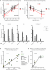
RSV F proteins engineered to stably display antigenic site Ø elicit neutralizing titers significantly higher than those elicited by postfusion F. (A) Neutralization titers of sera from mice immunized with 10 μg of RSV F (left). Postfusion F, as well as RSV F bound by antibodies AM22 or D25, were administered at 20 μg of the RSV F-antibody complex per mouse (right). Titers from each mouse are shown as individual black dots, and geometric means are indicated by red horizontal lines. (B) Neutralization titers of sera from rhesus macaques immunized with 50 μg of RSV F protein variants. Titers from each macaque are shown as individual black dots, and geometric means are indicated by red horizontal lines. Protective threshold (
) is indicated by a dotted line, and _p_-values are provided for postfusion versus DS-Cav1 as assessed by two-tailed T-test.
Fig. 4. Informatics of site Ø-stabilized RSV F immunogens
(A) Physical stability of site Ø versus immunogenicity. Physical stability (horizontal axis), as determined by the average of seven measurements of D25-retained activity in Table 1, is compared to elicited RSV-protective titers from Fig. 3 (vertical axis). (B) Structural mimicry of site Ø versus immunogenicity. Structural mimicry (horizontal axis) is the root-mean-square deviation of atom distances between different unbound RSV F structures (Fig. 2) and the D25-bound RSV F structure for all atoms within 10 Å of D25. This is compared to elicited RSV-protective titers from Fig. 3 (vertical axis). (C) Antigenic analysis of sera from immunized macaques. Binding of sera to sensor-tip immobilized DS-Cav1 (left), DS (middle), or postfusion F (right) was measured directly (black bars) or after incubation with excess postfusion F (light grey bars), DS (grey bars), or DS-Cav1 (dark grey bars). The mean responses of four macaque sera are shown, with standard deviations as error bars. Additional analysis of immunized sera is shown in fig. S12. (D) Elicited binding responses relative to surface areas and shared or unique portions of immunogens. Surface areas were calculated (fig. S11 and table S3) and compared to binding responses for the 36 response measurements in (C). (E) Correlation of immunogenicity and antigenic specificity of immunized macaque sera. The mean EC50 neutralization titers (week 6) of the four macaque sera in each group are plotted against the ratio of prefusion-specific/postfusion F-specific binding responses in (C) (
).
Comment in
- Vaccines. Structural biology triumph offers hope against a childhood killer.
Cohen J. Cohen J. Science. 2013 Nov 1;342(6158):546-7. doi: 10.1126/science.342.6158.546-a. Science. 2013. PMID: 24179197 No abstract available. - Viral diseases: Zeroing in on RSV vaccine design.
Crunkhorn S. Crunkhorn S. Nat Rev Drug Discov. 2014 Jan;13(1):17. doi: 10.1038/nrd4207. Epub 2013 Dec 13. Nat Rev Drug Discov. 2014. PMID: 24336502 No abstract available. - Structural Neuroimaging in Polysubstance Users.
Meyerhoff DJ. Meyerhoff DJ. Curr Opin Behav Sci. 2017 Feb;13:13-18. doi: 10.1016/j.cobeha.2016.07.006. Curr Opin Behav Sci. 2017. PMID: 28094824 Free PMC article.
Similar articles
- Design and characterization of a fusion glycoprotein vaccine for Respiratory Syncytial Virus with improved stability.
Zhang L, Durr E, Galli JD, Cosmi S, Cejas PJ, Luo B, Touch S, Parmet P, Fridman A, Espeseth AS, Bett AJ. Zhang L, et al. Vaccine. 2018 Dec 18;36(52):8119-8130. doi: 10.1016/j.vaccine.2018.10.032. Epub 2018 Oct 16. Vaccine. 2018. PMID: 30340881 - Alternative Virus-Like Particle-Associated Prefusion F Proteins as Maternal Vaccines for Respiratory Syncytial Virus.
Blanco JCG, Fernando LR, Zhang W, Kamali A, Boukhvalova MS, McGinnes-Cullen L, Morrison TG. Blanco JCG, et al. J Virol. 2019 Nov 13;93(23):e00914-19. doi: 10.1128/JVI.00914-19. Print 2019 Dec 1. J Virol. 2019. PMID: 31511382 Free PMC article. - Novel Respiratory Syncytial Virus-Like Particle Vaccine Composed of the Postfusion and Prefusion Conformations of the F Glycoprotein.
Cimica V, Boigard H, Bhatia B, Fallon JT, Alimova A, Gottlieb P, Galarza JM. Cimica V, et al. Clin Vaccine Immunol. 2016 Jun 6;23(6):451-9. doi: 10.1128/CVI.00720-15. Print 2016 Jun. Clin Vaccine Immunol. 2016. PMID: 27030590 Free PMC article. - Clinical Potential of Prefusion RSV F-specific Antibodies.
Rossey I, McLellan JS, Saelens X, Schepens B. Rossey I, et al. Trends Microbiol. 2018 Mar;26(3):209-219. doi: 10.1016/j.tim.2017.09.009. Epub 2017 Oct 17. Trends Microbiol. 2018. PMID: 29054341 Review. - Human respiratory syncytial virus: pathogenesis, immune responses, and current vaccine approaches.
Taleb SA, Al Thani AA, Al Ansari K, Yassine HM. Taleb SA, et al. Eur J Clin Microbiol Infect Dis. 2018 Oct;37(10):1817-1827. doi: 10.1007/s10096-018-3289-4. Epub 2018 Jun 6. Eur J Clin Microbiol Infect Dis. 2018. PMID: 29876771 Review.
Cited by
- Virology-The next fifty years.
Holmes EC, Krammer F, Goodrum FD. Holmes EC, et al. Cell. 2024 Sep 19;187(19):5128-5145. doi: 10.1016/j.cell.2024.07.025. Cell. 2024. PMID: 39303682 - Vaccine effect of recombinant single-chain hemagglutinin protein as an antigen.
Kawai A, Yamamoto Y, Yoshioka Y. Kawai A, et al. Heliyon. 2020 Jun 27;6(6):e04301. doi: 10.1016/j.heliyon.2020.e04301. eCollection 2020 Jun. Heliyon. 2020. PMID: 32637694 Free PMC article. - Stability Characterization of a Vaccine Antigen Based on the Respiratory Syncytial Virus Fusion Glycoprotein.
Flynn JA, Durr E, Swoyer R, Cejas PJ, Horton MS, Galli JD, Cosmi SA, Espeseth AS, Bett AJ, Zhang L. Flynn JA, et al. PLoS One. 2016 Oct 20;11(10):e0164789. doi: 10.1371/journal.pone.0164789. eCollection 2016. PLoS One. 2016. PMID: 27764150 Free PMC article. - Protein-Protein Interactions of Viroporins in Coronaviruses and Paramyxoviruses: New Targets for Antivirals?
Torres J, Surya W, Li Y, Liu DX. Torres J, et al. Viruses. 2015 Jun 4;7(6):2858-83. doi: 10.3390/v7062750. Viruses. 2015. PMID: 26053927 Free PMC article. Review. - Hepatitis C virus Broadly Neutralizing Monoclonal Antibodies Isolated 25 Years after Spontaneous Clearance.
Merat SJ, Molenkamp R, Wagner K, Koekkoek SM, van de Berg D, Yasuda E, Böhne M, Claassen YB, Grady BP, Prins M, Bakker AQ, de Jong MD, Spits H, Schinkel J, Beaumont T. Merat SJ, et al. PLoS One. 2016 Oct 24;11(10):e0165047. doi: 10.1371/journal.pone.0165047. eCollection 2016. PLoS One. 2016. PMID: 27776169 Free PMC article.
References
- Johnson S, et al. Development of a humanized monoclonal antibody (MEDI-493) with potent in vitro and in vivo activity against respiratory syncytial virus. J. Infect. Dis. 1997;176:1215–1224. - PubMed
- The IMpact-RSV Study Group Palivizumab, a humanized respiratory syncytial virus monoclonal antibody, reduces hospitalization from respiratory syncytial virus infection in high-risk infants. Pediatrics. 1998;102:531–537. - PubMed
Publication types
MeSH terms
Substances
LinkOut - more resources
Full Text Sources
Other Literature Sources
Medical


