Hepatitis-C-virus-like internal ribosome entry sites displace eIF3 to gain access to the 40S subunit - PubMed (original) (raw)
. 2013 Nov 28;503(7477):539-43.
doi: 10.1038/nature12658. Epub 2013 Nov 3.
Affiliations
- PMID: 24185006
- PMCID: PMC4106463
- DOI: 10.1038/nature12658
Hepatitis-C-virus-like internal ribosome entry sites displace eIF3 to gain access to the 40S subunit
Yaser Hashem et al. Nature. 2013.
Abstract
Hepatitis C virus (HCV) and classical swine fever virus (CSFV) messenger RNAs contain related (HCV-like) internal ribosome entry sites (IRESs) that promote 5'-end independent initiation of translation, requiring only a subset of the eukaryotic initiation factors (eIFs) needed for canonical initiation on cellular mRNAs. Initiation on HCV-like IRESs relies on their specific interaction with the 40S subunit, which places the initiation codon into the P site, where it directly base-pairs with eIF2-bound initiator methionyl transfer RNA to form a 48S initiation complex. However, all HCV-like IRESs also specifically interact with eIF3 (refs 2, 5-7, 9-12), but the role of this interaction in IRES-mediated initiation has remained unknown. During canonical initiation, eIF3 binds to the 40S subunit as a component of the 43S pre-initiation complex, and comparison of the ribosomal positions of eIF3 and the HCV IRES revealed that they overlap, so that their rearrangement would be required for formation of ribosomal complexes containing both components. Here we present a cryo-electron microscopy reconstruction of a 40S ribosomal complex containing eIF3 and the CSFV IRES. Remarkably, although the position and interactions of the CSFV IRES with the 40S subunit in this complex are similar to those of the HCV IRES in the 40S-IRES binary complex, eIF3 is completely displaced from its ribosomal position in the 43S complex, and instead interacts through its ribosome-binding surface exclusively with the apical region of domain III of the IRES. Our results suggest a role for the specific interaction of HCV-like IRESs with eIF3 in preventing ribosomal association of eIF3, which could serve two purposes: relieving the competition between the IRES and eIF3 for a common binding site on the 40S subunit, and reducing formation of 43S complexes, thereby favouring translation of viral mRNAs.
Figures
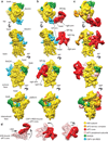
Figure 1. Cryo-EM structures of the CSFV ΔII-IRES•40S•DHX29 complex alone and bound to eIF3 compared to the structure of the DHX29-bound 43S preinitiation complex
(a) CSFV ΔII-IRES•40S•DHX29 complex (class 2, Extended Data Fig. 3). (b) CSFV ΔII-IRES•40S•DHX29 complex bound to eIF3 (class 4, Extended Data Fig. 3). (c) 43S preinitiation complex. (, from top to bottom) Complexes are viewed from the top, the back, the solvent and the intersubunit faces, respectively. In panels a–c, the 40S subunit is displayed in yellow, DXH29 in green, the eIF3 structural core in red and the CSFV ΔIIIRES in cyan. (d) Comparison between different positions and orientations of eIF3 in the 43S complex and in the CSFV ΔII-IRES•40S complexes, as indicated.
Figure 2. Different orientations of eIF3 and subdomain IIIb in the CSFV ΔII IRES•40S•DHX29 complex
(a) Solvent-side view of eIF3 in the two most divergent orientations, as it appears in classes 4 (solid red surface) and 6 (transparent pink surface) of the CSFV ΔII-IRES•40S•DHX29•eIF3 complex, bound to the CSFV ΔII-IRES (cyan) on the 40S subunit (yellow). (b, Left). Top view of eIF3 in the two most divergent orientations, bound to the CSFV ΔII-IRES on the 40S subunit. (b, Right) blowup focused on domain IIIb of the CSFV IRES, showing the extent of its reorientation. The brackets display the magnitude of the movement of eIF3 and of IRES domain IIIb in the two most divergent orientations.
Figure 3. Structure and atomic model of the CSFV ΔII-IRES bound to the 40S subunit
(a) Atomic model of the CSFV ΔII-IRES fitted into its density map (blue mesh), seen from the solvent (Left) and back (Middle) sides. Right panel displays a blowup on the CSFV ΔII-IRES atomic model (ribbon), colored variably to highlight its different subdomains. (b) Ribosomal proteins contacting the CSFV ΔII-IRES when bound to the 40S subunit, seen from the back (Left and Middle) and the front (Right).
Figure 4. Binding of eIF3 to subdomain IIIb of the CSFV IRES and effects on translation of the eIF3/IRES interaction
(a) eIF3 binding site on the CSFV IRES. The residues potentially interacting with eIF3 from domain IIIb in the CSFV IRES are highlighted by blue spheres and labeled (bottom). (b) Toeprinting analysis of 48S complexes assembled on wild-type HCV (nt. 1–349)–CAT mRNA (“wt HCV IRES”) and ΔIIIb-HCV IRES or ΔIIIc-HCV IRES derivatives lacking either domain IIIb (nt. 172–227) or domain IIIc (nt. 229–238) with 40S subunits, Met-tRNAiMet, eIF2 and eIF3 as indicated. Primer extension was arrested at nts. 342–345 by stably bound 40S subunits and at nts. 355–359 by 48S complexes, as indicated. Lanes C, T, A and G show the cDNA sequence corresponding to wt HCV IRES mRNA. The position of the initiation codon AUG373 is indicated on the left. (c) Inhibition of 43S complex formation by subdomain IIIabc of HCV and CSFV IRESs, assayed by sucrose density gradient centrifugation (SDG). The protein composition of ribosomal peak fractions was analyzed by SDS-PAGE and fluorescent SYPRO staining. (d) Inhibition of 48S complex formation on β-globin mRNA by IIIabc subdomains and by complete HCV and CSFV IRESs containing a 4nt. deletion in helix III2, assayed by toe-printing. Lanes C, T, A and G show the cDNA sequence corresponding to β-globin mRNA. The position of the initiation codon is indicated on the left. Each gel reported in the figure is representative of results obtained from three technical replicates.
Comment in
- HCV-like IRESs sequester eIF3: advantage virus.
Khan D, Bhat P, Das S. Khan D, et al. Trends Microbiol. 2014 Feb;22(2):57-8. doi: 10.1016/j.tim.2013.12.009. Epub 2013 Dec 31. Trends Microbiol. 2014. PMID: 24387882
Similar articles
- Specific interaction of eukaryotic translation initiation factor 3 with the 5' nontranslated regions of hepatitis C virus and classical swine fever virus RNAs.
Sizova DV, Kolupaeva VG, Pestova TV, Shatsky IN, Hellen CU. Sizova DV, et al. J Virol. 1998 Jun;72(6):4775-82. doi: 10.1128/JVI.72.6.4775-4782.1998. J Virol. 1998. PMID: 9573242 Free PMC article. - HCV-like IRESs sequester eIF3: advantage virus.
Khan D, Bhat P, Das S. Khan D, et al. Trends Microbiol. 2014 Feb;22(2):57-8. doi: 10.1016/j.tim.2013.12.009. Epub 2013 Dec 31. Trends Microbiol. 2014. PMID: 24387882 - Coordinated assembly of human translation initiation complexes by the hepatitis C virus internal ribosome entry site RNA.
Ji H, Fraser CS, Yu Y, Leary J, Doudna JA. Ji H, et al. Proc Natl Acad Sci U S A. 2004 Dec 7;101(49):16990-5. doi: 10.1073/pnas.0407402101. Epub 2004 Nov 24. Proc Natl Acad Sci U S A. 2004. PMID: 15563596 Free PMC article. - Hepatitis C Virus Translation Regulation.
Niepmann M, Gerresheim GK. Niepmann M, et al. Int J Mol Sci. 2020 Mar 27;21(7):2328. doi: 10.3390/ijms21072328. Int J Mol Sci. 2020. PMID: 32230899 Free PMC article. Review. - IRES-induced conformational changes in the ribosome and the mechanism of translation initiation by internal ribosomal entry.
Hellen CU. Hellen CU. Biochim Biophys Acta. 2009 Sep-Oct;1789(9-10):558-70. doi: 10.1016/j.bbagrm.2009.06.001. Epub 2009 Jun 17. Biochim Biophys Acta. 2009. PMID: 19539793 Free PMC article. Review.
Cited by
- Characterization of host factors associated with the internal ribosomal entry sites of foot-and-mouth disease and classical swine fever viruses.
Ide Y, Kitab B, Ito N, Okamoto R, Tamura Y, Matsui T, Sakoda Y, Tsukiyama-Kohara K. Ide Y, et al. Sci Rep. 2022 Apr 25;12(1):6709. doi: 10.1038/s41598-022-10437-z. Sci Rep. 2022. PMID: 35468926 Free PMC article. - Eukaryotic initiation factor (eIF) 3 mediates Barley Yellow Dwarf Viral mRNA 3'-5' UTR interactions and 40S ribosomal subunit binding to facilitate cap-independent translation.
Bhardwaj U, Powell P, Goss DJ. Bhardwaj U, et al. Nucleic Acids Res. 2019 Jul 9;47(12):6225-6235. doi: 10.1093/nar/gkz448. Nucleic Acids Res. 2019. PMID: 31114905 Free PMC article. - Kinetics of CrPV and HCV IRES-mediated eukaryotic translation using single-molecule fluorescence microscopy.
Bugaud O, Barbier N, Chommy H, Fiszman N, Le Gall A, Dulin D, Saguy M, Westbrook N, Perronet K, Namy O. Bugaud O, et al. RNA. 2017 Nov;23(11):1626-1635. doi: 10.1261/rna.061523.117. Epub 2017 Aug 2. RNA. 2017. PMID: 28768714 Free PMC article. - LOOP IIId of the HCV IRES is essential for the structural rearrangement of the 40S-HCV IRES complex.
Angulo J, Ulryck N, Deforges J, Chamond N, Lopez-Lastra M, Masquida B, Sargueil B. Angulo J, et al. Nucleic Acids Res. 2016 Feb 18;44(3):1309-25. doi: 10.1093/nar/gkv1325. Epub 2015 Nov 30. Nucleic Acids Res. 2016. PMID: 26626152 Free PMC article. - eIF3 targets cell-proliferation messenger RNAs for translational activation or repression.
Lee AS, Kranzusch PJ, Cate JH. Lee AS, et al. Nature. 2015 Jun 4;522(7554):111-4. doi: 10.1038/nature14267. Epub 2015 Apr 6. Nature. 2015. PMID: 25849773 Free PMC article.
References
Publication types
MeSH terms
Substances
Grants and funding
- R01 GM059660/GM/NIGMS NIH HHS/United States
- R01 GM029169/GM/NIGMS NIH HHS/United States
- R01 AI51340/AI/NIAID NIH HHS/United States
- R01GM29169/GM/NIGMS NIH HHS/United States
- R01 GM59660/GM/NIGMS NIH HHS/United States
- R56 AI051340/AI/NIAID NIH HHS/United States
- HHMI_/Howard Hughes Medical Institute/United States
LinkOut - more resources
Full Text Sources
Other Literature Sources


