The intestinal microbiota modulates the anticancer immune effects of cyclophosphamide - PubMed (original) (raw)
. 2013 Nov 22;342(6161):971-6.
doi: 10.1126/science.1240537.
Fabiana Saccheri, Grégoire Mignot, Takahiro Yamazaki, Romain Daillère, Dalil Hannani, David P Enot, Christina Pfirschke, Camilla Engblom, Mikael J Pittet, Andreas Schlitzer, Florent Ginhoux, Lionel Apetoh, Elisabeth Chachaty, Paul-Louis Woerther, Gérard Eberl, Marion Bérard, Chantal Ecobichon, Dominique Clermont, Chantal Bizet, Valérie Gaboriau-Routhiau, Nadine Cerf-Bensussan, Paule Opolon, Nadia Yessaad, Eric Vivier, Bernhard Ryffel, Charles O Elson, Joël Doré, Guido Kroemer, Patricia Lepage, Ivo Gomperts Boneca, François Ghiringhelli, Laurence Zitvogel
Affiliations
- PMID: 24264990
- PMCID: PMC4048947
- DOI: 10.1126/science.1240537
The intestinal microbiota modulates the anticancer immune effects of cyclophosphamide
Sophie Viaud et al. Science. 2013.
Abstract
Cyclophosphamide is one of several clinically important cancer drugs whose therapeutic efficacy is due in part to their ability to stimulate antitumor immune responses. Studying mouse models, we demonstrate that cyclophosphamide alters the composition of microbiota in the small intestine and induces the translocation of selected species of Gram-positive bacteria into secondary lymphoid organs. There, these bacteria stimulate the generation of a specific subset of "pathogenic" T helper 17 (pT(H)17) cells and memory T(H)1 immune responses. Tumor-bearing mice that were germ-free or that had been treated with antibiotics to kill Gram-positive bacteria showed a reduction in pT(H)17 responses, and their tumors were resistant to cyclophosphamide. Adoptive transfer of pT(H)17 cells partially restored the antitumor efficacy of cyclophosphamide. These results suggest that the gut microbiota help shape the anticancer immune response.
Figures
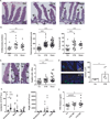
Fig. 1. Cyclophosphamide disrupts gut mucosal integrity
(A–B). Hematoxilin-eosin staining of the small intestine epithelium at 48h post-NaCl (Co) or CTX or doxorubicin (Doxo) therapy in C57BL/6 naïve mice (A). The numbers of inflammatory foci depicted/mm (B, left panel, indicated with arrowhead on A), thickness of the lamina propria reflecting edema (B, middle panel, indicated with # on A) and the reduced length of villi (B, right panel, indicated with arrowhead in A) were measured in 5 ilea on 100 villi/ileum from CTX or Doxo -treated mice. (C). A representative microphotograph of an ileal villus containing typical mucin-containing goblet cells is shown in vehicle- and CTX or Doxo-treated mice (left panels). The number of goblet cells/villus was enumerated in the right panel for both chemotherapy agents. (D). Specific staining of Paneth cells is shown in two representative immunofluorescence microphotographs (D, left panels). The quantification of Paneth cells was performed measuring the average area of the lysozyme-positive clusters in 6 ilea harvested from mice treated with NaCl (Co) or CTX at 24–48 hours. (E). Quantitative PCR (qPCR) analyses of Lysozyme M and RegIIIγ transcription levels in duodenum and ileum lamina propria cells from mice treated with CTX at 18 hours. Means±SEM of normalized deltaCT of 3–4 mice/group concatenated from three independent experiments. (F). In vivo intestinal permeability assays measuring 4 kDa fluorescein isothiocyanate (FITC)-dextran plasma accumulation at 18 hours post-CTX at two doses. Graph showing all data from four independent experiments, each dot representing one mouse (n=13–15). Data were analyzed with the t-test. *, p<0.05, **, p<0.01, ***, p<0.001.
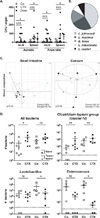
Fig. 2. Cyclophosphamide induces mucosa-associated microbial dysbiosis and bacterial translocation in secondary lymphoid organs
(A–B). At 48 hours post-CTX or Doxo, mesenteric lymph node (mLN) and spleen cells from naïve mice were cultivated in aerobic and anaerobic conditions and colonies were enumerated (A) from each mouse treated with NaCl (Co) (n=10–16), CTX (n=12–27) or Doxo (n=3–17) (3–4 experiments) and identified by mass spectrometry (B). In NaCl controls, attempts of bacterial identification mostly failed and yielded 67% L. murinus (not shown). Data were analyzed with the t-test. (C). The microbial composition (genus level) was analyzed by 454 pyrosequencing of the 16S rRNA gene from ilea and caeca of naïve mice and B16F10 tumor bearers. Principal Component Analyses (PCA) highlighted specific clustering of mice microbiota (each dot represents one mouse) depending on the treatment (NaCl: Co, grey dots; CTX-treated, black dots). A Monte Carlo rank test was applied to assess the significance of these clusterings. (D). Quantitative PCR (qPCR) analyses of various bacterial groups associated with small intestine mucosa were performed on CTX or NaCl (Co)-treated, naïve or MCA205 tumor-bearing mice. Absolute values were calculated for total bacteria, Lactobacilli, Enterococci and Clostridium group IV and normalized by the dilution and weight of the sample. Standard curves were generated from serial dilutions of a known concentration of genomic DNA from each bacterial group and by plotting threshold cycles (Ct) vs bacterial quantity (CFU). Points below the dotted lines were under the detection threshold. Data were analyzed with the linear model or generalized linear model. *, p<0.5, **, p<0.1, ***, p<0.001, ns, non significant.
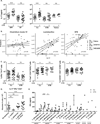
Fig. 3. CTX-induced pTh17 effectors and memory Th1 responses depend on gut microbiota
A). Splenocytes from CTX versus NaCl treated animals reared in germ-free (GF) or conventional specific pathogen-free (SPF) conditions (left panel) and treated or not with ATB or vancomycin (Vanco) (right panel) were cross-linked using anti-CD3+anti-CD28 Ab for 48h. IL-17 was measured by ELISA. Two to 3 experiments containing 2–9 mice/group are presented, each dot representing one mouse. (B). Correlations between the quantity of specific mucosal bacterial groups and the spleen Th17 signature. Each dot represents one mouse bearing no tumor (round dots), a B16F10 melanoma (diamond dots) or a MCA205 sarcoma (square dots), open dots featuring NaCl-treated mice and full dots indicating CTX-treated animals. (C). Intracellular analyses of splenocytes harvested from non-tumor-bearing mice after 7 days of either NaCl or CTX treatment, under ATB or water regimen as control. Means±SEM of percentages of IFNγ+ Th17 cells, T-bet+ cells among RORγt+ CD4+ T cells and CXCR3+ cells among CCR6+CD4+ T cells in 2 – 8 independent experiments, each dot representing one mouse. (D) Intracellular staining of total splenocytes harvested 7 days post-CTX treatment from naïve mice orally-reconstituted with the indicated bacterial species after ATB treatment. (E). 7 days post CTX or NaCl (Co) treatment, splenic CD4+ T cells were restimulated ex vivo with bone-marrow dendritic cells (BM-DCs) loaded with decreasing amounts of bacteria for 24 hours. IFNγ release, monitored by ELISA is shown. The numbers of responder mice (based on the NaCl baseline threshold) out of the total number of mice tested is indicated (n). Statistical comparisons were based on the paired t-test. Data were either analyzed with beta regression or linear model and correlation analyses from modified Kendall tau. *, p<0.05, ***, p<0.001, ns, non significant.
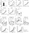
Fig. 4. Vancomycin blunts CTX-induced pTh17 differentiation which is mandatory for the tumoricidal activity of chemotherapy
(A). After a 3 week-long pretreatment with broad-spectrum ATB, DBA2 mice were inoculated with P815 mastocytomas (day 0), treated at day 6 with CTX (arrow) and tumor growth was monitored. Tumor growth kinetics are shown in Fig. S9A and percentages of tumor-free mice at sacrifice are depicted for two experiments of 11–14 mice/group. (B). MCA205 sarcoma were inoculated at day 0 in specific pathogen-free (SPF) or germ-free (GF) mice that were optionally mono-associated with segmented filamentous bacteria (SFB), treated with CTX (arrow) and monitored for growth kinetics (means±SEM). One representative experiment (n=5–8 mice/group) out of two to three is shown for GF mice and two pooled experiments (n=14 mice/group) for SPF mice (C). After a 3 week- conditioning with vancomycin or colistin, C57BL/6 mice were inoculated with MCA205 sarcomas (day 0), treated at day 12–15 with CTX (arrow) and tumor growth was monitored. Concatenated data (n=15–20 mice/group) from two independent experiments are shown for colistin treatment and one representative experiment (n=6 mice/group) for vancomycin treatment. (D). Eight week-old KP (_KrasLSL_-G12D/WT; _p53_Flox/Flox) mice received an adenovirus expressing the Cre recombinase (AdCre) by intranasal instillation to initiate lung adenocarcinoma (d0). Vancomycin was started for a subgroup of mice (“Chemo + Vanco”) on d77 post-AdCre. One week after the start of vancomycin, CTX-based chemotherapy was applied i.p. to mice that only received chemotherapy (“Chemo”) or those that received in parallel vancomycin (“Chemo + Vanco”). Mice received chemotherapy on d84, d91 and d98. A control group was left untreated ("Co"). Data show the evolution of total lung tumor volumes (mean±SEM) assessed by non invasive imaging between d73 and d100 in 6–12 mice/group. (E). As in Fig. 3C, we determined the number of pTh17 cells in spleens from untreated or vancomycin treated mice bearing established (15–17 days) MCA205 tumors, 7 days after CTX treatment. Each dot represents one mouse from 2 pooled experiments. (F). Flow cytometric analyses of CD3+ and CD4+IFNγ+ T cells were performed by gating on CD45+ live tumor-infiltrating lymphocytes (TILs) extracted from day 18 established MCA205 tumors (8 days post-CTX) in water or vancomycin-treated mice. Each dot representing one mouse from up to four pooled experiments. (G). MCA205 tumors established in WT mice pretreated for 3 weeks with water or vancomycin were injected with CTX (arrow), and tumor growth was monitored. At day 7 post-CTX, 3 million of ex vivo generated Th17 or pTh17 CD4+ T cells were injected intravenously. Up to three experiments comprising 2–10 mice/group were pooled. Data were either analyzed with the t-test, linear model or generalized linear model. *, p<0.5, **, p<0.1, ***, p<0.001, ns, non significant.
Comment in
- Biomedicine. Cancer therapies use a little help from microbial friends.
Pennisi E. Pennisi E. Science. 2013 Nov 22;342(6161):921. doi: 10.1126/science.342.6161.921. Science. 2013. PMID: 24264971 No abstract available. - Gut microbiota: Anti-cancer therapies affected by gut microbiota.
Greenhill C. Greenhill C. Nat Rev Gastroenterol Hepatol. 2014 Jan;11(1):1. doi: 10.1038/nrgastro.2013.238. Epub 2013 Dec 10. Nat Rev Gastroenterol Hepatol. 2014. PMID: 24322902 No abstract available. - Tumour immunology: Anticancer drugs need bugs.
Bordon Y. Bordon Y. Nat Rev Immunol. 2014 Jan;14(1):1. doi: 10.1038/nri3591. Epub 2013 Dec 13. Nat Rev Immunol. 2014. PMID: 24336106 No abstract available. - Chemotherapy, immunity and microbiota--a new triumvirate?
Karin M, Jobin C, Balkwill F. Karin M, et al. Nat Med. 2014 Feb;20(2):126-7. doi: 10.1038/nm.3473. Nat Med. 2014. PMID: 24504404 Free PMC article. - Tumour microenvironment: bacterial balance affects cancer treatment.
Lokody I. Lokody I. Nat Rev Cancer. 2014 Jan;14(1):10. doi: 10.1038/nrc3658. Nat Rev Cancer. 2014. PMID: 24505616 No abstract available.
Similar articles
- Gut microbiota: Anti-cancer therapies affected by gut microbiota.
Greenhill C. Greenhill C. Nat Rev Gastroenterol Hepatol. 2014 Jan;11(1):1. doi: 10.1038/nrgastro.2013.238. Epub 2013 Dec 10. Nat Rev Gastroenterol Hepatol. 2014. PMID: 24322902 No abstract available. - Tumour immunology: Anticancer drugs need bugs.
Bordon Y. Bordon Y. Nat Rev Immunol. 2014 Jan;14(1):1. doi: 10.1038/nri3591. Epub 2013 Dec 13. Nat Rev Immunol. 2014. PMID: 24336106 No abstract available. - Chemotherapy, immunity and microbiota--a new triumvirate?
Karin M, Jobin C, Balkwill F. Karin M, et al. Nat Med. 2014 Feb;20(2):126-7. doi: 10.1038/nm.3473. Nat Med. 2014. PMID: 24504404 Free PMC article. - Gut microbiome and anticancer immune response: really hot Sh*t!
Viaud S, Daillère R, Boneca IG, Lepage P, Langella P, Chamaillard M, Pittet MJ, Ghiringhelli F, Trinchieri G, Goldszmid R, Zitvogel L. Viaud S, et al. Cell Death Differ. 2015 Feb;22(2):199-214. doi: 10.1038/cdd.2014.56. Epub 2014 May 16. Cell Death Differ. 2015. PMID: 24832470 Free PMC article. Review. - Gut microbiota and cancer: How gut microbiota modulates activity, efficacy and toxicity of antitumoral therapy.
Gori S, Inno A, Belluomini L, Bocus P, Bisoffi Z, Russo A, Arcaro G. Gori S, et al. Crit Rev Oncol Hematol. 2019 Nov;143:139-147. doi: 10.1016/j.critrevonc.2019.09.003. Epub 2019 Sep 20. Crit Rev Oncol Hematol. 2019. PMID: 31634731 Review.
Cited by
- New Insights Into the Cancer-Microbiome-Immune Axis: Decrypting a Decade of Discoveries.
Jain T, Sharma P, Are AC, Vickers SM, Dudeja V. Jain T, et al. Front Immunol. 2021 Feb 23;12:622064. doi: 10.3389/fimmu.2021.622064. eCollection 2021. Front Immunol. 2021. PMID: 33708214 Free PMC article. Review. - The gut microbiota and gastrointestinal surgery.
Guyton K, Alverdy JC. Guyton K, et al. Nat Rev Gastroenterol Hepatol. 2017 Jan;14(1):43-54. doi: 10.1038/nrgastro.2016.139. Epub 2016 Oct 12. Nat Rev Gastroenterol Hepatol. 2017. PMID: 27729657 Review. - The interaction of anticancer therapies with tumor-associated macrophages.
Mantovani A, Allavena P. Mantovani A, et al. J Exp Med. 2015 Apr 6;212(4):435-45. doi: 10.1084/jem.20150295. Epub 2015 Mar 9. J Exp Med. 2015. PMID: 25753580 Free PMC article. Review. - Exploring the Potential Role of the Gut Microbiome in Chemotherapy-Induced Neurocognitive Disorders and Cardiovascular Toxicity.
Ciernikova S, Mego M, Chovanec M. Ciernikova S, et al. Cancers (Basel). 2021 Feb 13;13(4):782. doi: 10.3390/cancers13040782. Cancers (Basel). 2021. PMID: 33668518 Free PMC article. Review. - The Crosstalk between Microbiome and Immunotherapeutics: Myth or Reality.
Tojjari A, Abushukair H, Saeed A. Tojjari A, et al. Cancers (Basel). 2022 Sep 24;14(19):4641. doi: 10.3390/cancers14194641. Cancers (Basel). 2022. PMID: 36230563 Free PMC article. Review.
References
Publication types
MeSH terms
Substances
Grants and funding
- P01 DK071176/DK/NIDDK NIH HHS/United States
- P50 CA086355/CA/NCI NIH HHS/United States
- R01 AI084880/AI/NIAID NIH HHS/United States
- P01DK071176/DK/NIDDK NIH HHS/United States
LinkOut - more resources
Full Text Sources
Other Literature Sources


