Handheld simultaneous scanning laser ophthalmoscopy and optical coherence tomography system - PubMed (original) (raw)
Handheld simultaneous scanning laser ophthalmoscopy and optical coherence tomography system
Francesco Larocca et al. Biomed Opt Express. 2013.
Abstract
Scanning laser ophthalmoscopy (SLO) and optical coherence tomography (OCT) are widely used retinal imaging modalities that can assist in the diagnosis of retinal pathologies. The combination of SLO and OCT provides a more comprehensive imaging system and a method to register OCT images to produce motion corrected retinal volumes. While high quality, bench-top SLO-OCT systems have been discussed in the literature and are available commercially, there are currently no handheld designs. We describe the first design and fabrication of a handheld SLO/spectral domain OCT probe. SLO and OCT images were acquired simultaneously with a combined power under the ANSI limit. High signal-to-noise ratio SLO and OCT images were acquired simultaneously from a normal subject with visible motion artifacts. Fully automated motion estimation methods were performed in post-processing to correct for the inter- and intra-frame motion in SLO images and their concurrently acquired OCT volumes. The resulting set of reconstructed SLO images and the OCT volume were without visible motion artifacts. At a reduced field of view, the SLO resolved parafoveal cones without adaptive optics at a retinal eccentricity of 11° in subjects with good ocular optics. This system may be especially useful for imaging young children and subjects with less stable fixation.
Keywords: (080.3620) Lens system design; (110.4153) Motion estimation and optical flow; (110.4500) Optical coherence tomography; (170.0110) Imaging systems; (170.4460) Ophthalmic optics and devices; (170.4470) Ophthalmology; (170.5755) Retina scanning.
Figures
Fig. 1
A side view schematic of the handheld SLO-OCT design. All optical components are labeled and described in the legend. The SLO source is a 770 nm SLD with 15 nm bandwidth. The OCT source is an 840 nm SLD with 70 nm bandwidth. The optical paths shown are the superposition of both the illumination and collection paths. The illumination beam diameters for both SLO and OCT were ~2.5 mm. The collection beam diameter for the SLO was larger due to the larger NA and size of the collection fiber, and because backscattered light from the retina fills the pupil in the return path.
Fig. 2
Spot diagrams for the SLO (A) and the OCT (B) illumination on the retina spanning a 20° FOV. SLO and OCT are nearly diffraction limited at 7 and 7.5 µm (the Airy disk radii), respectively (Airy disk is shown by black circle on spot diagrams). Spot diagrams are color coded for 3 wavelengths spanning the bandwidth of the respective sources. SLO spot diagrams have increased astigmatism compared to OCT spot diagrams due to the transmission through the tilted dichroic mirror.
Fig. 3
Solidworks design of the handheld SLO-OCT probe. A) Side view showing the internal components of the probe. B) Isometric view of probe with case. Rotatable three-dimensional versions of these figures are included as
Media 1
(3D PDF) and
Media 2
(3D PDF) [alternate version available in U3D format as
Media 3
and
Media 4
].
Fig. 4
The handheld SLO-OCT probe. A) Tabletop mountable configuration on a patient positioning system from a Carl Zeiss slit-lamp. B) Handheld use of probe.
Fig. 5
A) SLO image (single frame) with red line representing location of B-scan. B) Single B-scan taken simultaneously with the SLO at 40 fps. C) Foveal SLO image (single frame) indicating the position where the SLO was optically zoomed to visualize parafoveal cones. D) Optically zoomed retinal image (5 frame average) via reduction of scan range to a 2.5° FOV at location shown by red box in C) at an 11° eccentricity. E) Digitally zoomed image at location shown by blue box in D) with a 1.5° FOV showing the cone photoreceptor mosaic.
Fig. 6
Single frame SLO image (A) and the corresponding LoG/Gabor filtered and thresholded SLO image (B).
Fig. 7
SLO patch-based registration. A) SLO reference frame to which all images are registered. B) Another SLO image from the same subject. C) Patch-based registration of SLO image in B) to the reference frame in A). The voids have been colored in green to better visualize the regions where no information was obtained. D) Spline interpolated motion determined from the patch-based registration in C) is applied to each line in the SLO image from B) and empty voids within the frame are linearly interpolated.
Fig. 8
SLO motion estimation with X-fast (A-C) and Y-fast (D-F) OCT. A slow horizontal drift is visualized in the X-fast motion estimation with a large saccade captured at around the 13 second point. Both vertical and horizontal drifts are apparent in the Y-fast motion estimation with two microsaccades captured at around the 0.5 and 11 second points.
Fig. 9
Raw and registered SVPs with and without interpolation based on motion estimation through SLO frame registration. A) Raw X-fast SVP. B) Registered X-fast SVP. C) Registered X-fast SVP after interpolation. D) Raw Y-fast SVP. E) Registered Y-fast SVP. F) Registered Y-fast SVP after interpolation. G) Combined X- and Y-fast SVPs after registration. H) Combined X- and Y-fast SVPs after registration and interpolation.
Fig. 10
Steps for axial registration with an X- and Y-fast OCT volume. A) The initial four B-scans taken near the corners of the composite volume. Red dots indicate points of lateral overlap on the surface of each initial B-scan. B) The initial four B-scans after axial translation and rotation. Note that the red dots at points of lateral overlap now overlap axially as well. C) The registration of an additional two X- and Y-fast B-scans. D) Final registered volume rendering of all X- and Y-fast B-scans. Orthogonal fly-throughs of this data are included as supplementary material (
Media 5
).
Fig. 11
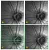
Comparison between registration with and without LoG/Gabor preprocessing. A) The reference frame. B) The target frame (to be registered with respect to reference frame). C) LoG/Gabor preprocessed reference frame. D) LoG/Gabor preprocessed target frame. E) Registration of A) and B) without LoG/Gabor preprocessing. F) Registration of A) and B) with the LoG/Gabor preprocessing shown in C) and D).
Similar articles
- True color scanning laser ophthalmoscopy and optical coherence tomography handheld probe.
LaRocca F, Nankivil D, Farsiu S, Izatt JA. LaRocca F, et al. Biomed Opt Express. 2014 Aug 27;5(9):3204-16. doi: 10.1364/BOE.5.003204. eCollection 2014 Sep 1. Biomed Opt Express. 2014. PMID: 25401032 Free PMC article. - Volumetric imaging of rod and cone photoreceptor structure with a combined adaptive optics-optical coherence tomography-scanning laser ophthalmoscope.
Wells-Gray EM, Choi SS, Zawadzki RJ, Finn SC, Greiner C, Werner JS, Doble N. Wells-Gray EM, et al. J Biomed Opt. 2018 Mar;23(3):1-15. doi: 10.1117/1.JBO.23.3.036003. J Biomed Opt. 2018. PMID: 29508564 Free PMC article. - Progress on Developing Adaptive Optics-Optical Coherence Tomography for In Vivo Retinal Imaging: Monitoring and Correction of Eye Motion Artifacts.
Zawadzki RJ, Capps AG, Kim DY, Panorgias A, Stevenson SB, Hamann B, Werner JS. Zawadzki RJ, et al. IEEE J Sel Top Quantum Electron. 2014 Mar;20(2):7100912. doi: 10.1109/JSTQE.2013.2288302. IEEE J Sel Top Quantum Electron. 2014. PMID: 25544826 Free PMC article. - [New examination methods for macular disorders--application of diagnosis and treatment].
Yoshida A. Yoshida A. Nippon Ganka Gakkai Zasshi. 2000 Dec;104(12):899-942. Nippon Ganka Gakkai Zasshi. 2000. PMID: 11193944 Review. Japanese. - Combinations of techniques in imaging the retina with high resolution.
Podoleanu AG, Rosen RB. Podoleanu AG, et al. Prog Retin Eye Res. 2008 Jul;27(4):464-99. doi: 10.1016/j.preteyeres.2008.03.002. Epub 2008 Mar 28. Prog Retin Eye Res. 2008. PMID: 18495519 Review.
Cited by
- The Development and Clinical Application of Innovative Optical Ophthalmic Imaging Techniques.
Alexopoulos P, Madu C, Wollstein G, Schuman JS. Alexopoulos P, et al. Front Med (Lausanne). 2022 Jun 30;9:891369. doi: 10.3389/fmed.2022.891369. eCollection 2022. Front Med (Lausanne). 2022. PMID: 35847772 Free PMC article. Review. - In vivo cellular-resolution retinal imaging in infants and children using an ultracompact handheld probe.
LaRocca F, Nankivil D, DuBose T, Toth CA, Farsiu S, Izatt JA. LaRocca F, et al. Nat Photonics. 2016;10:580-584. doi: 10.1038/nphoton.2016.141. Epub 2016 Aug 1. Nat Photonics. 2016. PMID: 29479373 Free PMC article. No abstract available. - Posterior Segment Optical Coherence Tomography in Uncooperative Paediatric Patients Using Exo-Illumination and Microscope-Integrated Optical Coherence Tomography.
Singh A, Dogra M, Moharana B, Singh R. Singh A, et al. Cureus. 2022 Dec 27;14(12):e32994. doi: 10.7759/cureus.32994. eCollection 2022 Dec. Cureus. 2022. PMID: 36712705 Free PMC article. - Ergonomic handheld OCT angiography probe optimized for pediatric and supine imaging.
Viehland C, Chen X, Tran-Viet D, Jackson-Atogi M, Ortiz P, Waterman G, Vajzovic L, Toth CA, Izatt JA. Viehland C, et al. Biomed Opt Express. 2019 Apr 29;10(5):2623-2638. doi: 10.1364/BOE.10.002623. eCollection 2019 May 1. Biomed Opt Express. 2019. PMID: 31143506 Free PMC article. - Fundus Photography in the 21st Century--A Review of Recent Technological Advances and Their Implications for Worldwide Healthcare.
Panwar N, Huang P, Lee J, Keane PA, Chuan TS, Richhariya A, Teoh S, Lim TH, Agrawal R. Panwar N, et al. Telemed J E Health. 2016 Mar;22(3):198-208. doi: 10.1089/tmj.2015.0068. Epub 2015 Aug 26. Telemed J E Health. 2016. PMID: 26308281 Free PMC article. Review.
References
- S. Ricco, M. Chen, H. Ishikawa, G. Wollstein, and J. Schuman, “Correcting motion artifacts in retinal spectral domain optical coherence tomography via image registration,” in Medical Image Computing and Computer-Assisted Intervention – MICCAI 2009, G.-Z. Yang, D. Hawkes, D. Rueckert, A. Noble, and C. Taylor, eds. (Springer Berlin / Heidelberg, 2009), pp. 100–107. - PMC - PubMed
- M. D. Robinson, S. J. Chiu, C. A. Toth, J. Izatt, J. Y. Lo, and S. Farsiu, “Novel applications of super-resolution in medical imaging,” in “Super-resolution imaging”, P. Milanfar, ed. (CRC Press 2010), pp. 383–412.
- Podoleanu A. G., Jackson D. A., “Combined optical coherence tomograph and scanning laser ophthalmoscope,” Electron. Lett. 34(11), 1088–1090 (1998).10.1049/el:19980793 - DOI
LinkOut - more resources
Full Text Sources
Other Literature Sources


