Embryonal tumor with abundant neuropil and true rosettes (ETANTR), ependymoblastoma, and medulloepithelioma share molecular similarity and comprise a single clinicopathological entity - PubMed (original) (raw)
. 2014 Aug;128(2):279-89.
doi: 10.1007/s00401-013-1228-0. Epub 2013 Dec 14.
Dominik Sturm, Marina Ryzhova, Volker Hovestadt, Marco Gessi, David T W Jones, Marc Remke, Paul Northcott, Arie Perry, Daniel Picard, Marc Rosenblum, Manila Antonelli, Eleonora Aronica, Ulrich Schüller, Martin Hasselblatt, Adelheid Woehrer, Olga Zheludkova, Ella Kumirova, Stephanie Puget, Michael D Taylor, Felice Giangaspero, V Peter Collins, Andreas von Deimling, Peter Lichter, Annie Huang, Torsten Pietsch, Stefan M Pfister, Marcel Kool
Affiliations
- PMID: 24337497
- PMCID: PMC4102829
- DOI: 10.1007/s00401-013-1228-0
Embryonal tumor with abundant neuropil and true rosettes (ETANTR), ependymoblastoma, and medulloepithelioma share molecular similarity and comprise a single clinicopathological entity
Andrey Korshunov et al. Acta Neuropathol. 2014 Aug.
Abstract
Three histological variants are known within the family of embryonal rosette-forming neuroepithelial brain tumors. These include embryonal tumor with abundant neuropil and true rosettes (ETANTR), ependymoblastoma (EBL), and medulloepithelioma (MEPL). In this study, we performed a comprehensive clinical, pathological, and molecular analysis of 97 cases of these rare brain neoplasms, including genome-wide DNA methylation and copy number profiling of 41 tumors. We identified uniform molecular signatures in all tumors irrespective of histological patterns, indicating that ETANTR, EBL, and MEPL comprise a single biological entity. As such, future WHO classification schemes should consider lumping these variants into a single diagnostic category, such as embryonal tumor with multilayered rosettes (ETMR). We recommend combined LIN28A immunohistochemistry and FISH analysis of the 19q13.42 locus for molecular diagnosis of this tumor category. Recognition of this distinct pediatric brain tumor entity based on the fact that the three histological variants are molecularly and clinically uniform will help to distinguish ETMR from other embryonal CNS tumors and to better understand the biology of these highly aggressive and therapy-resistant pediatric CNS malignancies, possibly leading to alternate treatment strategies.
Figures
Fig. 1
Microscopical appearance (a, d, g), FISH analysis of the 19q13.42 locus (b, e, h), LIN28A immunohistochemistry (c, f, i) of ETANTR (a–c), EBL (d–f) and MEPL (g–i). Amplification of 19q13.42 (b, e, h) and LIN28A immunoexpression (c, f, i) was detected in all three histological ETMR subtypes. For the FISH analysis the C19MC 19q13.42 probe (green signals) and a reference 19p13 probe were used (red signals)
Fig. 2
Two examples of primary ETANTR (a, c) with further tumor transformation in either EBL (b) or MEPL (d) histology as it has been identified during analysis of the recurrence samples
Fig. 3
Overall survival curves generated for ETANTR (32 cases, blue), EBL (17 cases, red), and MEPL (6 cases, green). No differences in survival time were found (log-rank, p = 0.63)
Fig. 4
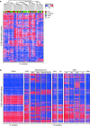
Cluster analyses of DNA methylation profiles of ETMR alone and compared to various other pediatric brain tumors and normal cerebellum. a Unsupervised cluster analysis of ETMR samples only shows that DNA methylation profiles of the histological variants ETANTR, EBL and MEPL are not distinct. Also, clusters outlined do not differ in terms of clinical findings, including age, gender, tumor location and outcome. b DNA methylation profiles of ETMRs are distinct from other pediatric brain tumors and normal cerebellum
Fig. 5
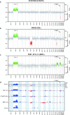
Copy number plots generated from 450 k methylation data. Amplifications and gains are indicated in green, losses in red. a Example of an ETANTR showing amplification 19q13.42, gain of 2 and loss of 19q13.3. b Example of an EBL showing amplification 19q13.42, gain of 2, and losses of 6q and 17p. c Example of a MEPL showing amplification 19q13.42, trisomy of 2, 17 and 19. d Summarizing profiles for all 41 cases analyzed
Comment in
- Embryonal tumor with multilayered rosettes (ETMR): signed, sealed, delivered ….
Wesseling P. Wesseling P. Acta Neuropathol. 2014 Aug;128(2):305-8. doi: 10.1007/s00401-014-1320-0. Acta Neuropathol. 2014. PMID: 25012402 No abstract available.
Similar articles
- Embryonal tumor with abundant neuropil and true rosettes with only one structure suggestive of an ependymoblastic rosette.
Nobusawa S, Orimo K, Horiguchi K, Ikota H, Yokoo H, Hirato J, Nakazato Y. Nobusawa S, et al. Pathol Int. 2014 Sep;64(9):472-7. doi: 10.1111/pin.12196. Epub 2014 Sep 3. Pathol Int. 2014. PMID: 25186165 - Analysis of chromosome 19q13.42 amplification in embryonal brain tumors with ependymoblastic multilayered rosettes.
Nobusawa S, Yokoo H, Hirato J, Kakita A, Takahashi H, Sugino T, Tasaki K, Itoh H, Hatori T, Shimoyama Y, Nakazawa A, Nishizawa S, Kishimoto H, Matsuoka K, Nakayama M, Okura N, Nakazato Y. Nobusawa S, et al. Brain Pathol. 2012 Sep;22(5):689-97. doi: 10.1111/j.1750-3639.2012.00574.x. Epub 2012 Mar 6. Brain Pathol. 2012. PMID: 22324795 Free PMC article. - Clinicopathological characteristics and outcomes in embryonal tumor with multilayered rosettes: A decade long experience from a tertiary care centre in North India.
Gupta K, Sood R, Salunke P, Chatterjee D, Madan R, Ahuja CK, Jain R, Trehan A, Radotra BD. Gupta K, et al. Ann Diagn Pathol. 2021 Aug;53:151745. doi: 10.1016/j.anndiagpath.2021.151745. Epub 2021 Apr 19. Ann Diagn Pathol. 2021. PMID: 33964610 - Embryonal tumor with multilayered rosettes: diagnostic tools update and review of the literature.
Ceccom J, Bourdeaut F, Loukh N, Rigau V, Milin S, Takin R, Richer W, Uro-Coste E, Couturier J, Bertozzi AI, Delattre O, Delisle MB. Ceccom J, et al. Clin Neuropathol. 2014 Jan-Feb;33(1):15-22. doi: 10.5414/NP300636. Clin Neuropathol. 2014. PMID: 23863344 Review. - [A new entity in WHO classification of tumors of the central nervous system--embryonic tumor with abundant neuropil and true rosettes: case report and review of literature].
Ryzhova MV, Zheludkova OG, Ozerov SS, Shishkina LV, Panina TN, Gorelyshev SK, Novikov AI, Melikian AG, Kushel' IuV, Korshunov AE. Ryzhova MV, et al. Zh Vopr Neirokhir Im N N Burdenko. 2011;75(4):25-33; discussion 33. Zh Vopr Neirokhir Im N N Burdenko. 2011. PMID: 22379850 Review. Russian.
Cited by
- Transcriptomic Analysis Revealed an Emerging Role of Alternative Splicing in Embryonal Tumor with Multilayered Rosettes.
Hesham D, El-Naggar S. Hesham D, et al. Genes (Basel). 2020 Sep 22;11(9):1108. doi: 10.3390/genes11091108. Genes (Basel). 2020. PMID: 32971786 Free PMC article. - Genomics of adult and pediatric solid tumors.
Rahal Z, Abdulhai F, Kadara H, Saab R. Rahal Z, et al. Am J Cancer Res. 2018 Aug 1;8(8):1356-1386. eCollection 2018. Am J Cancer Res. 2018. PMID: 30210910 Free PMC article. Review. - The 2016 World Health Organization Classification of tumours of the Central Nervous System: what the paediatric neuroradiologist needs to know.
Chhabda S, Carney O, D'Arco F, Jacques TS, Mankad K. Chhabda S, et al. Quant Imaging Med Surg. 2016 Oct;6(5):486-489. doi: 10.21037/qims.2016.10.01. Quant Imaging Med Surg. 2016. PMID: 27942466 Free PMC article. - Embryonal tumors with multilayered rosettes in children: the SFCE experience.
Horwitz M, Dufour C, Leblond P, Bourdeaut F, Faure-Conter C, Bertozzi AI, Delisle MB, Palenzuela G, Jouvet A, Scavarda D, Vinchon M, Padovani L, Gaudart J, Branger DF, Andre N. Horwitz M, et al. Childs Nerv Syst. 2016 Feb;32(2):299-305. doi: 10.1007/s00381-015-2920-2. Epub 2015 Oct 5. Childs Nerv Syst. 2016. PMID: 26438544 - Preclinical drug screen reveals topotecan, actinomycin D, and volasertib as potential new therapeutic candidates for ETMR brain tumor patients.
Schmidt C, Schubert NA, Brabetz S, Mack N, Schwalm B, Chan JA, Selt F, Herold-Mende C, Witt O, Milde T, Pfister SM, Korshunov A, Kool M. Schmidt C, et al. Neuro Oncol. 2017 Nov 29;19(12):1607-1617. doi: 10.1093/neuonc/nox093. Neuro Oncol. 2017. PMID: 28482026 Free PMC article.
References
- Al-Hussaini M, Abuirmeileh N, Swaidan M, Al-Jumaily U, Rajjal H, Musharbash A, Hashem S, Sultan I. Embryonal tumor with abundant neuropil and true rosettes: a report of three cases of a rare tumor, with an unusual case showing rhabdomyoblastic and melanocytic differentiation. Neuropathology. 2011;31(6):620–625. doi: 10.1111/j.1440-1789.2011.01213.x. - DOI - PubMed
- Buccoliero AM, Castiglione F, Rossi Degl’Innocenti D, Franchi A, Paglierani M, Sanzo M, Cetica V, Giunti L, Sardi I, Genitori L, Taddei GL. Embryonal tumor with abundant neuropil and true rosettes: morphological, immunohistochemical, ultrastructural and molecular study of a case showing features of medulloepithelioma and areas of mesenchymal and epithelial differentiation. Neuropathology. 2010;30(1):84–91. doi: 10.1111/j.1440-1789.2009.01040.x. - DOI - PubMed
- Ceccom J, Bourdeaut F, Loukh N, Rigau V, Milin S, Takin R, Richer W, Uro-Coste E, Couturier J, Bertozzi AI, Delattre O, Delisle MB (2013) Embryonal tumor with multilayered rosettes: diagnostic tools update and review of the literature. Clin Neuropathol (Epub ahead of print) - PubMed
MeSH terms
LinkOut - more resources
Full Text Sources
Other Literature Sources
Medical
Research Materials


