Microglial proliferation in the brain of chronic alcoholics with hepatic encephalopathy - PubMed (original) (raw)
Microglial proliferation in the brain of chronic alcoholics with hepatic encephalopathy
Claude V Dennis et al. Metab Brain Dis. 2014 Dec.
Abstract
Hepatic encephalopathy (HE) is a common complication of chronic alcoholism and patients show neurological symptoms ranging from mild cognitive dysfunction to coma and death. The HE brain is characterized by glial changes, including microglial activation, but the exact pathogenesis of HE is poorly understood. During a study investigating cell proliferation in the subventricular zone of chronic alcoholics, a single case with widespread proliferation throughout their adjacent grey and white matter was noted. This case also had concomitant HE raising the possibility that glial proliferation might be a pathological feature of the disease. In order to explore this possibility fixed postmortem human brain tissue from chronic alcoholics with cirrhosis and HE (n = 9), alcoholics without HE (n = 4) and controls (n = 4) were examined using immunohistochemistry and cytokine assays. In total, 4/9 HE cases had PCNA- and a second proliferative marker, Ki-67-positive cells throughout their brain and these cells co-stained with the microglial marker, Iba1. These cases were termed 'proliferative HE' (pHE). The microglia in pHEs displayed an activated morphology with hypertrophied cell bodies and short, thickened processes. In contrast, the microglia in white matter regions of the non-proliferative HE cases were less activated and appeared dystrophic. pHEs were also characterized by higher interleukin-6 levels and a slightly higher neuronal density . These findings suggest that microglial proliferation may form part of an early neuroprotective response in HE that ultimately fails to halt the course of the disease because underlying etiological factors such as high cerebral ammonia and systemic inflammation remain.
Figures
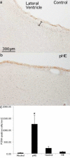
Fig. 1. Proliferation in HE alcoholics with cirrhosis
PCNA immunostaining of 48μm sections of the rostral caudate nucleus shows (a) a typical staining pattern in a neurologically normal individual with immunopositive cells in the ependymal layer and subventricular zone (SVZ) but not the underlying parenchyma. The approximate width of the SVZ is indicated (black spacer). (b) a pHE case with PCNA+ cells throughout the caudate nucleus underlying the SVZ. (c) a significantly higher number of PCNA+ cells in pHEs compared to all other groups. 200x magnification.
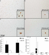
Fig. 2. PCNA labels a greater number of cells in pHEs compared to Ki-67
7μm serial sections of the SFG (a and b) and PCG (c and d) of a pHE case show PCNA+ cells (a and c) and Ki-67+ cells (b and d). 200× magnification and 600× for insets. (e) Quantification of Ki-67+ and PCNA+ cells in pHE cases shows a greater number of PCNA+ cells compared to Ki-67 in both the SFG and PCG. (f) pHEs have substantially more Ki-67+ cells in both regions compared to the other three groups.
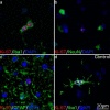
Fig. 3. Phenotyping proliferative cells
Immunofluorescence double labeling shows (a) Ki-67+ cells in a pHE case co-localized with the microglial marker Iba1 but not (b) the neuronal marker NeuN or (c) the astrocytic marker GFAP. (d) A rare Ki-67+ cell in a neurologically normal individual co-localizes with Iba1. 400× magnification.
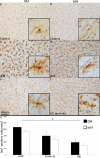
Fig. 4. Morphological differences in Iba1+ cells in HE
Iba1+ cells in 48μm sections of a neurologically normal individual have a small cell body with long, fine processes (a) extending radially in the GM and (b) tangentially along the WM tracts. A pHE case displays microglial hypertrophy and hyperplasia in both their (c) GM and (d) WM. A non-proliferative HE case with (e) a subtle increase in the size of the cell body in the GM but (f) extensive, widespread fragmentation of the microglial cells in the WM. (g) A comparison of Iba1+ cells shows a significant increase in both the GM (p = 0.04) and WM (p < 0.001) in pHEs compared to HE cases. Data represent the mean ± SEM of Iba1+ cells/mm2. 200× magnification with 600× insets.
Fig. 5. Reduction in neuronal density in the SFG of HE cases
In comparison to controls there was a reduction of neuronal density in (a) the SFG of HE cases but not alcoholics (b) There was no difference in the neuronal density of HE cases compared to controls in the PCG. Data are presented as mean neuronal density/mm2 ± SEM.
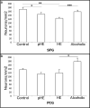
Fig. 6. IL-6 levels are elevated in the cortex of pHEs
IL-6 levels were higher in pHEs in both (a) the SFG and (b) the PCG.
Similar articles
- The effects of chronic alcoholism on cell proliferation in the human brain.
Sutherland GT, Sheahan PJ, Matthews J, Dennis CV, Sheedy DS, McCrossin T, Curtis MA, Kril JJ. Sutherland GT, et al. Exp Neurol. 2013 Sep;247:9-18. doi: 10.1016/j.expneurol.2013.03.020. Epub 2013 Mar 28. Exp Neurol. 2013. PMID: 23541433 Free PMC article. - Microglia activation in hepatic encephalopathy in rats and humans.
Zemtsova I, Görg B, Keitel V, Bidmon HJ, Schrör K, Häussinger D. Zemtsova I, et al. Hepatology. 2011 Jul;54(1):204-15. doi: 10.1002/hep.24326. Hepatology. 2011. PMID: 21452284 - The effects of chronic smoking on the pathology of alcohol-related brain damage.
McCorkindale AN, Sheedy D, Kril JJ, Sutherland GT. McCorkindale AN, et al. Alcohol. 2016 Jun;53:35-44. doi: 10.1016/j.alcohol.2016.04.002. Epub 2016 Apr 30. Alcohol. 2016. PMID: 27286935 Free PMC article. - Cerebral dysfunction in chronic alcoholism: role of alcoholic liver disease.
Butterworth RF. Butterworth RF. Alcohol Alcohol Suppl. 1994;2:259-65. Alcohol Alcohol Suppl. 1994. PMID: 8974345 Review. - Hepatic encephalopathy in alcoholic cirrhosis.
Butterworth RF. Butterworth RF. Handb Clin Neurol. 2014;125:589-602. doi: 10.1016/B978-0-444-62619-6.00034-3. Handb Clin Neurol. 2014. PMID: 25307598 Review.
Cited by
- Inflammatory Biomarkers in Addictive Disorders.
Morcuende A, Navarrete F, Nieto E, Manzanares J, Femenía T. Morcuende A, et al. Biomolecules. 2021 Dec 3;11(12):1824. doi: 10.3390/biom11121824. Biomolecules. 2021. PMID: 34944470 Free PMC article. Review. - Increased expression of M1 and M2 phenotypic markers in isolated microglia after four-day binge alcohol exposure in male rats.
Peng H, Geil Nickell CR, Chen KY, McClain JA, Nixon K. Peng H, et al. Alcohol. 2017 Aug;62:29-40. doi: 10.1016/j.alcohol.2017.02.175. Epub 2017 Jun 20. Alcohol. 2017. PMID: 28755749 Free PMC article. - The Direct Contribution of Astrocytes and Microglia to the Pathogenesis of Hepatic Encephalopathy.
Jaeger V, DeMorrow S, McMillin M. Jaeger V, et al. J Clin Transl Hepatol. 2019 Dec 28;7(4):352-361. doi: 10.14218/JCTH.2019.00025. Epub 2019 Nov 13. J Clin Transl Hepatol. 2019. PMID: 31915605 Free PMC article. Review. - Chronic alcohol induced mechanical allodynia by promoting neuroinflammation: A mouse model of alcohol-evoked neuropathic pain.
Borgonetti V, Roberts AJ, Bajo M, Galeotti N, Roberto M. Borgonetti V, et al. Br J Pharmacol. 2023 Sep;180(18):2377-2392. doi: 10.1111/bph.16091. Epub 2023 May 11. Br J Pharmacol. 2023. PMID: 37050867 Free PMC article. - Microglial-specific transcriptome changes following chronic alcohol consumption.
McCarthy GM, Farris SP, Blednov YA, Harris RA, Mayfield RD. McCarthy GM, et al. Neuropharmacology. 2018 Jan;128:416-424. doi: 10.1016/j.neuropharm.2017.10.035. Epub 2017 Oct 31. Neuropharmacology. 2018. PMID: 29101021 Free PMC article.
References
- Albrecht J, Norenberg MD. Glutamine: a Trojan horse in ammonia neurotoxicity. Hepatology. 2006;44(4):788–794. doi:10.1002/hep.21357. - PubMed
- Australian Bureau of Statistics Alcohol Consumption in Australia: A Snapshot, 2004-05. [14th April 2012];Australian Bureau of Statistics. 2006 http://www.abs.gov.au/AUSSTATS/abs@.nsf/Previousproducts/4832.0.55.001Ma....
- Bhardwaj RD, Curtis MA, Spalding KL, Buchholz BA, Fink D, Bjork-Eriksson T, Nordborg C, Gage FH, Druid H, Eriksson PS, Frisen J. Neocortical neurogenesis in humans is restricted to development. Proc Natl Acad Sci U S A. 2006;103(33):12564–12568. doi:0605177103 [pii] 10.1073/pnas.0605177103. - PMC - PubMed
- Brumback RA, Lapham LW. DNA synthesis in Alzheimer type II astrocytosis. The question of astrocytic proliferation and mitosis in experimentally induced hepatic encephalopathy. Archives of neurology. 1989;46(8):845–848. - PubMed
Publication types
MeSH terms
Substances
LinkOut - more resources
Full Text Sources
Other Literature Sources
Medical
Miscellaneous
