Hydroxymethylation at gene regulatory regions directs stem/early progenitor cell commitment during erythropoiesis - PubMed (original) (raw)
. 2014 Jan 16;6(1):231-244.
doi: 10.1016/j.celrep.2013.11.044. Epub 2013 Dec 27.
Hui Liu # 1, Alexis Rodriguez # 2, Aparna Vasanthakumar 1, Sriram Sundaravel 1, Donne Bennett D Caces 1, Timothy J Looney 3 4, Li Zhang 3 4, Janet B Lepore 1, Trisha Macrae 1, Robert Duszynski 1, Alan H Shih 5, Chun-Xiao Song 6 7, Miao Yu 6 7, Yiting Yu 8, Robert Grossman 2, Brigitte Raumann 2, Amit Verma 8, Chuan He 6 7, Ross L Levine 5, Don Lavelle 9 10, Bruce T Lahn 3 4, Amittha Wickrema 1, Lucy A Godley 1
Affiliations
- PMID: 24373966
- PMCID: PMC3976649
- DOI: 10.1016/j.celrep.2013.11.044
Hydroxymethylation at gene regulatory regions directs stem/early progenitor cell commitment during erythropoiesis
Jozef Madzo et al. Cell Rep. 2014.
Abstract
Hematopoietic stem cell differentiation involves the silencing of self-renewal genes and induction of a specific transcriptional program. Identification of multiple covalent cytosine modifications raises the question of how these derivatized bases influence stem cell commitment. Using a replicative primary human hematopoietic stem/progenitor cell differentiation system, we demonstrate dynamic changes of 5-hydroxymethylcytosine (5-hmC) during stem cell commitment and differentiation to the erythroid lineage. Genomic loci that maintain or gain 5-hmC density throughout erythroid differentiation contain binding sites for erythroid transcription factors and several factors not previously recognized as erythroid-specific factors. The functional importance of 5-hmC was demonstrated by impaired erythroid differentiation, with augmentation of myeloid potential, and disrupted 5-hmC patterning in leukemia patient-derived CD34+ stem/early progenitor cells with TET methylcytosine dioxygenase 2 (TET2) mutations. Thus, chemical conjugation and affinity purification of 5-hmC-enriched sequences followed by sequencing serve as resources for deciphering functional implications for gene expression during stem cell commitment and differentiation along a particular lineage.
Copyright © 2014 The Authors. Published by Elsevier Inc. All rights reserved.
Figures
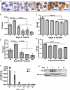
Figure 1. Dynamic Changes in 5-hmC and 5-mC Levels during Erythroid Differentiation
(A) Photomicrographs of hematoxylin and benzidine-stained cells cytospun onto slides on the days indicated. Changes in cellular morphology as well as acquisition of HB (brown) during the differentiation program are seen in the micrographs. (B and C) Total 5-hmC (B) and 5-mC (C) content in cells at days 0, 3, 7, 10, 13, and 17 during in vitro erythroid differentiation, measured by LC-MS. The average (± SD) of two independent biological replicates obtained from two independent donor CD34+ cells is shown. Repeated measures ANOVA was used to determine statistical significance. (D) Total 5-hmC content in the erythroid fractions isolated from baboon bone marrow. “R6” is the CD117+ CD36 bRBC fraction of cells, which form granulocytic, mixed, and large BFUe colonies in methylcellulose. This population contains both early progenitors and later nonerythroid progenitors. “R7” is the CD117+CD36+ fraction, which forms CFUe and late BFUe in methylcellulose (10%–20% erythroid colony-forming cells). “R8” is the CD117– CD36+ bRBC– fraction, which does not form colonies in methylcellulose. “RBC+” is the fraction of erythroid precursors that do not form colonies and that were purified using Miltenyi Biotec columns with the baboon-specific red blood cell (RBC) antibody (BD Biosciences). All numbers were normalized to RBC+ values. White blood cell (WBC) was used as a control. Data are represented as the mean of two independent biological replicates ± SD. (E) Total 5-mC content in the erythroid fractions isolated from baboon bone marrow, as described in (D). Data are represented as the mean of two independent biological replicates ± SD. (F) qRT-PCR for TET1 (white bars), TET2 (gray bars), and TET3 (black bars) expression normalized to 18S, at days 0, 3, 7, 10, 13, and 17 during in vitro erythroid differentiation. The average (± SD) of two independent biological replicates is shown. (G) Representative western blot showing expression of TET2 protein in nuclear extracts from cells at days 0, 3, 7, and 10. The blot was stripped and reprobed for histone H3, the loading control. See also Figure S1 and Table S1.
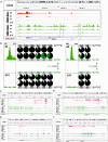
Figure 2. Dynamic Changes of 5-hmC Peaks and RNA-Seq in Regions Surrounding Stem and Progenitor Cell Genes
(A) RNA-seq (red) and hMe-Seal (green) tracks in the CD34 gene. (B) Pie charts depict results of bisulfite sequencing (outer rows) and TAB-seq (inner rows) showing quantitation of modified cytosines over regions of the CD34 gene on day 0 (top) and day 10 (bottom). For bisulfite sequencing, the percentage of unprotected cytosines is shown in white and of protected cytosines in black; for TAB-seq, the percentage of unmodified cytosines is shown in white, of 5-mC in black, and of 5-hmC in green. p values were calculated by using the χ2 test to compare 5-hmC percentages at days 0 and 10. (C–F) RNA-seq (red) and hMe-Seal (green) tracks in regions surrounding CD38 (C), CD45 (D), CD90 (E), and CD133 (F). See also Figures S2 and S3.
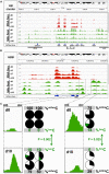
Figure 3. Dynamic Changes of 5-hmC Peaks and RNA-Seq in Regions Surrounding the HB Gene Cluster
(A) RNA-seq (red) and hMe-Seal (green) tracks in the region surrounding the HB cluster. Black bars indicate known regulatory regions: HPFH, BCL11A-BS (BCL11A binding site), and LCR. (B) Results in (A) magnified to show the HBB gene. (C) Pie charts depict results of bisulfite sequencing (outer rows) and TAB-seq (inner rows) showing quantitation of modified cytosines over regions of HBB on day 0 (top) and day 10 (bottom). For bisulfite sequencing, the percentage of unprotected cytosines is shown in white and of protected cytosines in black; for TAB-seq, the percentage of unmodified cytosines is shown in white, of 5-mC in black, and of 5-hmC in green. p values were calculated by using the χ2 test to compare 5-hmC percentages at days 0 and 10. See also Figures S2 and S3.
Figure 4. Functional Significance of 5-hmC Gains
(A) Total number of gained 5-hmC peaks between days 0 and 3 (days 0–3, white), 3 and 7 (days 3–7, hatched), and 7 and 10 (days 7–10, striped) with respect to relative position to annotated gene elements. (B) Relative number of 5-hmC peaks from (A) normalized to the base pair lengths of each element. (C) Comparison of the normalized 5-hmC reads surrounding the TSS, indicated by a vertical dotted line, ±1,000 bp for all known annotated genes at day 0 (left) versus day 10 (right), plotted by the Metaseq suite (Dale, 2013). (D) Percentage of the overlap among publicly available GATA1, GATA2, and KLF1 ChIP-seq data (Fujiwara et al., 2009; Kang et al., 2012; Tallack et al., 2010) and 5-hmC gained peaks over days 0–3 (white), 3–7 (hatched), and 7–10 (striped) time points with significance (p ≤ 10–5). See also Figure S4.
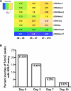
Figure 5. Overlap of Histone Marks as well as DNA Methylation Dynamics with 5-hmC Peaks that Were Gained between Days 0 and 3, 3 and 7, and 7 and 10
(A) Overlap of the distribution of particular histone modifications (deposited by Xu et al., 2012) and gain of 5-hmC density throughout erythroid differentiation. Scores were calculated as the ratio of actual overlap relative to a normal distribution generated by random permutations (yellow, high normalized enrichment; green, medium normalized enrichment; and blue, low normalized enrichment). All normalized enrichment scores except H3K9me3 reached significance of at least 10–5. ‡, levels of significance for H3K9me3 versus 5-hmC enrichment were days 0–3 (p = 0.066), 3–7 (p < 10–5), and 7–10 (p = 0.075). (B) Percentage of overlap between 5-hmC-specific peaks identified during erythroid differentiation and modified cytosines identified using the HELP assay (Yu et al., 2013).
Figure 6. TET2 Mutations and 5-hmC Deficiency Impede Erythroid Differentiation and Enhance Myeloid Development
(A) Bone marrow-derived CD34+ cells from healthy nonmalignant TET2WT donors (top panel) or from patients with CMML with TET2WT (middle panel) or TET2mut (bottom panel) were differentiated down the erythroid (left) or myeloid lineage (right). Differentiation was determined by FACS at day 14. Erythroid differentiation was determined by expression of Glycophorin A and CD71, and myeloid differentiation by expression of CD15 and CD33. Photomicrographs of cytospins prepared at each time point are shown to the right of each FACS plot. (B) Differential differentiation potential was quantified by calculating a myeloid:erythroid (M:E) ratio for each sample. Myeloid expression was defined by the CD15+ and CD15+CD33+ populations, and erythroid differentiation was defined using the Glycophorin A+ CD71+ population. Samples from healthy nonmalignant TET2wt cells are shown in orange, those from patients with TET2wt CMML are shown in white, and those from patients with TET2mut CMML are shown in blue. (C) Total 5-hmC levels measured at day 10 of in vitro erythroid differentiation, as determined by LC/MS. Data are represented as mean ± SEM. (D) Total 5-mC levels measured in these same samples, as determined by LC/MS. Data are represented as mean ± SEM. (E) Overlap in 5-hmC reads in nonmalignant CD34+ cells (orange) versus CD34+ cells derived from a patient with _TET2_-mutated CMML (blue) at day 10 after in vitro erythroid differentiation. Inset is a Venn diagram showing the number of base pairs covered by 5-hmC peaks in the respective samples. (F) RNA-seq (top track, gray) and hMe-Seal tracks (for nonmalignant CD34+ cells, middle track, orange; versus patient with _TET2_-mutated CMML, bottom track, blue) in the region surrounding GATA1. See also Figure S5.
Similar articles
- Hydroxymethylcytosine and demethylation of the γ-globin gene promoter during erythroid differentiation.
Ruiz MA, Rivers A, Ibanez V, Vaitkus K, Mahmud N, DeSimone J, Lavelle D. Ruiz MA, et al. Epigenetics. 2015;10(5):397-407. doi: 10.1080/15592294.2015.1039220. Epigenetics. 2015. PMID: 25932923 Free PMC article. - Inhibition of TET2-mediated conversion of 5-methylcytosine to 5-hydroxymethylcytosine disturbs erythroid and granulomonocytic differentiation of human hematopoietic progenitors.
Pronier E, Almire C, Mokrani H, Vasanthakumar A, Simon A, da Costa Reis Monte Mor B, Massé A, Le Couédic JP, Pendino F, Carbonne B, Larghero J, Ravanat JL, Casadevall N, Bernard OA, Droin N, Solary E, Godley LA, Vainchenker W, Plo I, Delhommeau F. Pronier E, et al. Blood. 2011 Sep 1;118(9):2551-5. doi: 10.1182/blood-2010-12-324707. Epub 2011 Jul 6. Blood. 2011. PMID: 21734233 Free PMC article. - MEIS1 regulates early erythroid and megakaryocytic cell fate.
Zeddies S, Jansen SB, di Summa F, Geerts D, Zwaginga JJ, van der Schoot CE, von Lindern M, Thijssen-Timmer DC. Zeddies S, et al. Haematologica. 2014 Oct;99(10):1555-64. doi: 10.3324/haematol.2014.106567. Epub 2014 Aug 8. Haematologica. 2014. PMID: 25107888 Free PMC article. - A regulatory network governing Gata1 and Gata2 gene transcription orchestrates erythroid lineage differentiation.
Moriguchi T, Yamamoto M. Moriguchi T, et al. Int J Hematol. 2014 Nov;100(5):417-24. doi: 10.1007/s12185-014-1568-0. Epub 2014 Mar 18. Int J Hematol. 2014. PMID: 24638828 Review. - Metabolic regulation of hematopoietic stem cell commitment and erythroid differentiation.
Oburoglu L, Romano M, Taylor N, Kinet S. Oburoglu L, et al. Curr Opin Hematol. 2016 May;23(3):198-205. doi: 10.1097/MOH.0000000000000234. Curr Opin Hematol. 2016. PMID: 26871253 Review.
Cited by
- Increased iron uptake by splenic hematopoietic stem cells promotes TET2-dependent erythroid regeneration.
Tseng YJ, Kageyama Y, Murdaugh RL, Kitano A, Kim JH, Hoegenauer KA, Tiessen J, Smith MH, Uryu H, Takahashi K, Martin JF, Samee MAH, Nakada D. Tseng YJ, et al. Nat Commun. 2024 Jan 15;15(1):538. doi: 10.1038/s41467-024-44718-0. Nat Commun. 2024. PMID: 38225226 Free PMC article. - DNMT3A and TET2 compete and cooperate to repress lineage-specific transcription factors in hematopoietic stem cells.
Zhang X, Su J, Jeong M, Ko M, Huang Y, Park HJ, Guzman A, Lei Y, Huang YH, Rao A, Li W, Goodell MA. Zhang X, et al. Nat Genet. 2016 Sep;48(9):1014-23. doi: 10.1038/ng.3610. Epub 2016 Jul 18. Nat Genet. 2016. PMID: 27428748 Free PMC article. - TET2 deficiency leads to stem cell factor-dependent clonal expansion of dysfunctional erythroid progenitors.
Qu X, Zhang S, Wang S, Wang Y, Li W, Huang Y, Zhao H, Wu X, An C, Guo X, Hale J, Li J, Hillyer CD, Mohandas N, Liu J, Yazdanbakhsh K, Vinchi F, Chen L, Kang Q, An X. Qu X, et al. Blood. 2018 Nov 29;132(22):2406-2417. doi: 10.1182/blood-2018-05-853291. Epub 2018 Sep 25. Blood. 2018. PMID: 30254129 Free PMC article. - Tet-mediated DNA hydroxymethylation regulates retinal neurogenesis by modulating cell-extrinsic signaling pathways.
Seritrakul P, Gross JM. Seritrakul P, et al. PLoS Genet. 2017 Sep 19;13(9):e1006987. doi: 10.1371/journal.pgen.1006987. eCollection 2017 Sep. PLoS Genet. 2017. PMID: 28926578 Free PMC article. - Erythroid Krüppel-Like Factor (KLF1): A Surprisingly Versatile Regulator of Erythroid Differentiation.
Bieker JJ, Philipsen S. Bieker JJ, et al. Adv Exp Med Biol. 2024;1459:217-242. doi: 10.1007/978-3-031-62731-6_10. Adv Exp Med Biol. 2024. PMID: 39017846 Review.
References
- Dale R. Metaseq 0.5. 2013 http://pythonhosted.org/metaseq.
Publication types
MeSH terms
Substances
Grants and funding
- HL116336/HL/NHLBI NIH HHS/United States
- R01 CA129831/CA/NCI NIH HHS/United States
- R01 HL116336/HL/NHLBI NIH HHS/United States
- HHMI/Howard Hughes Medical Institute/United States
- CA129831-03S1/CA/NCI NIH HHS/United States
- F32-DK092030/DK/NIDDK NIH HHS/United States
- R01 HL114561/HL/NHLBI NIH HHS/United States
- P30 CA008748/CA/NCI NIH HHS/United States
- CA129831/CA/NCI NIH HHS/United States
- F32 DK092030/DK/NIDDK NIH HHS/United States
LinkOut - more resources
Full Text Sources
Other Literature Sources
Molecular Biology Databases


