Co-occurrence of anaerobic bacteria in colorectal carcinomas - PubMed (original) (raw)
Co-occurrence of anaerobic bacteria in colorectal carcinomas
René L Warren et al. Microbiome. 2013.
Abstract
Background: Numerous cancers have been linked to microorganisms. Given that colorectal cancer is a leading cause of cancer deaths and the colon is continuously exposed to a high diversity of microbes, the relationship between gut mucosal microbiome and colorectal cancer needs to be explored. Metagenomic studies have shown an association between Fusobacterium species and colorectal carcinoma. Here, we have extended these studies with deeper sequencing of a much larger number (n = 130) of colorectal carcinoma and matched normal control tissues. We analyzed these data using co-occurrence networks in order to identify microbe-microbe and host-microbe associations specific to tumors.
Results: We confirmed tumor over-representation of Fusobacterium species and observed significant co-occurrence within individual tumors of Fusobacterium, Leptotrichia and Campylobacter species. This polymicrobial signature was associated with over-expression of numerous host genes, including the gene encoding the pro-inflammatory chemokine Interleukin-8. The tumor-associated bacteria we have identified are all Gram-negative anaerobes, recognized previously as constituents of the oral microbiome, which are capable of causing infection. We isolated a novel strain of Campylobacter showae from a colorectal tumor specimen. This strain is substantially diverged from a previously sequenced oral Campylobacter showae isolate, carries potential virulence genes, and aggregates with a previously isolated tumor strain of Fusobacterium nucleatum.
Conclusions: A polymicrobial signature of Gram-negative anaerobic bacteria is associated with colorectal carcinoma tissue.
Figures
Figure 1
Microbial abundance in CRC and unaffected control gut mucosa tissue measured by RNA-Seq. (A) Phylogenetic abundance inferred from unique metatranscriptomics read pair mapping. Genera (n = 57) comprising, collectively, 99% of the microbial sequence data are shown. Values presented are the mean percent abundance ± SE, on a log10 scale. Genera are ordered top to bottom by decreasing read pair abundance in control tissue. The names of genera nominally over-represented in tumor (P <0.05) are red, and the names of genera nominally under-represented in tumor (P <0.05) are green. Genera indicated with an asterisk are significantly over-represented in tumor samples after multiple hypothesis testing correction (q <0.05); (B) Species distribution of uniquely mapped Fusobacterium, Leptotrichia, Campylobacter and Selenomonas normalized sequence pairs.
Figure 2
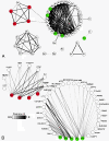
Bacterial co-occurrence and correlation with host gene expression. (A) The co-occurrence of microbes was inferred by pairwise correlation of sequence counts for all genera in Figure 1. Significance was determined by 1,000 iterations of random re-assignment of sequence read pairs to subjects, with re-calculation of Pearson correlation coefficients. Interactions of significantly differentially abundant genera are illustrated here in a network diagram, constructed using Cytoscape [25]. Pearson R values ranged from a low of 0.51 (Holdemania-Haemophilus) to a high of 0.97 (_Fusobacterium_-Selenomonas). Each prefix within a node of the network indicates a bacterial genus: Sp: Sphingobium; Sh: Sphingopyxis; Si: Sphingomonas; La: Lactobacillus; Bi: Bifidobacterium; No: Novosphingobium; Se: Selenomonas; Fu: Fusobacterium; Ro: Roseburia; Eg: Eggerthella; Bl: Blautia; Cl: Clostridium; Ve: Veillonella; Ha: Haemophilus; Co: Coprococcus; Ps: Pseudoflavonifractor; Do: Dorea; Ru: Ruminococcus; Bu: Butyrivibrio; Cs: Clostridiales; Ca: Campylobacter; Le: Leptotrichia; Pa: Parabacteroides; Ba: Bacteroides; Rm: Ruminococcaceae; Er: Erysipelotrichaceae; En: Enterococcus; Br: Burkholderia; Ph: Phascolarctobacterium; Lw: Lawsonia; Al: Alistipes; Su: Subdoligranulum; Od: Odoribacter; Po: Porphyromonas; Bk: Burkholderiales; Go: Gordonibacter; Gr: Granulicatella; Di: Dialister; Fa: Faecalibacterium; Eu: Eubacterium; De: Desulfovibrio; Pr: Parvimonas; Pe: Peptostreptococcus; An: Anaerotruncus; Ci: Collinsella; Or: Oribacterium; Ho: Holdemania; Es: Escherichia; Pv: Prevotella; Me: Methylobacterium; Bd: Bradyrhizobium; Ra: Ralstonia. (B) The host factor and microbe interaction was inferred by comparing the relative read pair abundance in the tumor vs. the control samples for each patient. The red and green nodes correspond to microbes with at least nominally significant differential abundance between tumor and control tissues.
Figure 3
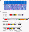
Genome sequence analyses of C. showae tumor strain CC57C. (A) Sequence alignments between the assemblies of the CRC tumor-associated C. showae CC57C genome and that of its closest known relative reference, C. showae RM3277. Velvet contigs from the CC57C strain were aligned to the HMP reference strain (NCBI Reference Sequence: NZ_ACVQ00000000.1) using cross_match (
), ordered and oriented based on the sequence alignments. The black and grey rectangles represent each genome sequences and indicate the absence and presence of alignments, in that order. Co-linear and inverted sequence alignment blocks are shown in blue and pink, respectively. In average, the CC57C genome assembly is 92.5% identical to that of the RM3277 strain, with 85.4% sequence coverage (represented by the dark blue and lack of bars in the histogram above the alignment, respectively). (B) Gene organization of the type IV secretion system (T4SS) operon in A. tumefaciens (plasmid pTiAB2/73 vir region AF329849), H. pylori strain 26695 (NC_000915.1), C. rectus (NZ_ACFU0100008) and C. showae CC57C (AOTD01000166). Components of the T4SS found in the CC57C strain are color-coded in other organisms and include VirB4 (red), VirB8 (blue), VirB9 (green), VirB10 (orange), VirB11 (yellow) and VirD4 (purple). Other annotated genes within the CC57C operon and with homologs in C. rectus are shown using a dark grey scale. Annotated genes in C. rectus, H. pylori and A. tumefaciens are shown in light grey. Hypothetical protein coding genes are depicted in white. CC57C contig AOTD01000166 with the T4SS shown in (B) does not have any similarity to the C. showae RM3277 strain and thus is not represented in the alignment shown in (A).
Figure 4
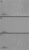
F. nucleatum and C. showae co-aggregation. Phase-contrast microscopy images of (A) F. nucleatum CC53 alone, (B) C. showae CC57C alone and (C) a mixture of CC53 and CC57C, following incubation in co-aggregation buffer (see methods). The long, thin cells of CC53 readily self-aggregate when incubated in aggregation buffer alone (panel A), but also show aggregative ability with the much smaller CC57C coccobacilli (panel C). Images were taken using a Leica DM750 microscope fitted with a 100× oil objective, using the Leica Application Suite LAS EZ Version 1.7.0 software.
Similar articles
- Fusobacterium nucleatum Increases Proliferation of Colorectal Cancer Cells and Tumor Development in Mice by Activating Toll-Like Receptor 4 Signaling to Nuclear Factor-κB, and Up-regulating Expression of MicroRNA-21.
Yang Y, Weng W, Peng J, Hong L, Yang L, Toiyama Y, Gao R, Liu M, Yin M, Pan C, Li H, Guo B, Zhu Q, Wei Q, Moyer MP, Wang P, Cai S, Goel A, Qin H, Ma Y. Yang Y, et al. Gastroenterology. 2017 Mar;152(4):851-866.e24. doi: 10.1053/j.gastro.2016.11.018. Epub 2016 Nov 19. Gastroenterology. 2017. PMID: 27876571 Free PMC article. - Diets That Promote Colon Inflammation Associate With Risk of Colorectal Carcinomas That Contain Fusobacterium nucleatum.
Liu L, Tabung FK, Zhang X, Nowak JA, Qian ZR, Hamada T, Nevo D, Bullman S, Mima K, Kosumi K, da Silva A, Song M, Cao Y, Twombly TS, Shi Y, Liu H, Gu M, Koh H, Li W, Du C, Chen Y, Li C, Li W, Mehta RS, Wu K, Wang M, Kostic AD, Giannakis M, Garrett WS, Hutthenhower C, Chan AT, Fuchs CS, Nishihara R, Ogino S, Giovannucci EL. Liu L, et al. Clin Gastroenterol Hepatol. 2018 Oct;16(10):1622-1631.e3. doi: 10.1016/j.cgh.2018.04.030. Epub 2018 Apr 24. Clin Gastroenterol Hepatol. 2018. PMID: 29702299 Free PMC article. - Association of Fusobacterium nucleatum with immunity and molecular alterations in colorectal cancer.
Nosho K, Sukawa Y, Adachi Y, Ito M, Mitsuhashi K, Kurihara H, Kanno S, Yamamoto I, Ishigami K, Igarashi H, Maruyama R, Imai K, Yamamoto H, Shinomura Y. Nosho K, et al. World J Gastroenterol. 2016 Jan 14;22(2):557-66. doi: 10.3748/wjg.v22.i2.557. World J Gastroenterol. 2016. PMID: 26811607 Free PMC article. Review. - Opportunistic detection of Fusobacterium nucleatum as a marker for the early gut microbial dysbiosis.
Huh JW, Roh TY. Huh JW, et al. BMC Microbiol. 2020 Jul 13;20(1):208. doi: 10.1186/s12866-020-01887-4. BMC Microbiol. 2020. PMID: 32660414 Free PMC article. - Fusobacterium nucleatum: an emerging gut pathogen?
Allen-Vercoe E, Strauss J, Chadee K. Allen-Vercoe E, et al. Gut Microbes. 2011 Sep 1;2(5):294-8. doi: 10.4161/gmic.2.5.18603. Epub 2011 Sep 1. Gut Microbes. 2011. PMID: 22067936 Review.
Cited by
- Bacterial community structure alterations within the colorectal cancer gut microbiome.
Loftus M, Hassouneh SA, Yooseph S. Loftus M, et al. BMC Microbiol. 2021 Mar 31;21(1):98. doi: 10.1186/s12866-021-02153-x. BMC Microbiol. 2021. PMID: 33789570 Free PMC article. - Extremely small and incredibly close: Gut microbes as modulators of inflammation and targets for therapeutic intervention.
Piazzesi A, Putignani L. Piazzesi A, et al. Front Microbiol. 2022 Aug 22;13:958346. doi: 10.3389/fmicb.2022.958346. eCollection 2022. Front Microbiol. 2022. PMID: 36071979 Free PMC article. Review. - Co-enrichment of cancer-associated bacterial taxa is correlated with immune cell infiltrates in esophageal tumor tissue.
Greathouse KL, Stone JK, Vargas AJ, Choudhury A, Padgett RN, White JR, Jung A, Harris CC. Greathouse KL, et al. Sci Rep. 2024 Jan 31;14(1):2574. doi: 10.1038/s41598-023-48862-3. Sci Rep. 2024. PMID: 38296990 Free PMC article. - Urogenital Microbiota:Potentially Important Determinant of PD-L1 Expression in Male Patients with Non-muscle Invasive Bladder Cancer.
Chen C, Huang Z, Huang P, Li K, Zeng J, Wen Y, Li B, Zhao J, Wu P. Chen C, et al. BMC Microbiol. 2022 Jan 4;22(1):7. doi: 10.1186/s12866-021-02407-8. BMC Microbiol. 2022. PMID: 34983384 Free PMC article. - Characterization of Mucosa-Associated Microbiota in Matched Cancer and Non-neoplastic Mucosa From Patients With Colorectal Cancer.
Leung PHM, Subramanya R, Mou Q, Lee KT, Islam F, Gopalan V, Lu CT, Lam AK. Leung PHM, et al. Front Microbiol. 2019 Jun 12;10:1317. doi: 10.3389/fmicb.2019.01317. eCollection 2019. Front Microbiol. 2019. PMID: 31244818 Free PMC article.
References
- Moore RA, Warren RL, Freeman JD, Gustavsen JA, Chénard C, Friedman JM, Suttle CA, Zhao Y, Holt RA. The sensitivity of massively parallel sequencing for detecting candidate infectious agents associated with human tissue. PLoS One. 2011;1:e19838. doi: 10.1371/journal.pone.0019838. - DOI - PMC - PubMed
- Kostic AD, Gevers D, Pedamallu CS, Michaud M, Duke F, Earl AM, Ojesina AI, Jung J, Bass AJ, Tabernero J, Baselga J, Liu C, Shivdasani RA, Ogino S, Birren BW, Huttenhower C, Garrett WS, Meyerson M. Genomic analysis identifies association of Fusobacterium with colorectal carcinoma. Genome Res. 2012;1:292–298. doi: 10.1101/gr.126573.111. - DOI - PMC - PubMed
LinkOut - more resources
Full Text Sources
Other Literature Sources


