Phylogenic studies on the olfactory system in vertebrates - PubMed (original) (raw)
Phylogenic studies on the olfactory system in vertebrates
Kazuyuki Taniguchi et al. J Vet Med Sci. 2014 Jun.
Abstract
The olfactory receptor organs and their primary centers are classified into several types. The receptor organs are divided into fish-type olfactory epithelium (OE), mammal-type OE, middle chamber epithelium (MCE), lower chamber epithelium (LCE), recess epithelium, septal olfactory organ of Masera (SO), mammal-type vomeronasal organ (VNO) and snake-type VNO. The fish-type OE is observed in flatfish and lungfish, while the mammal-type OE is observed in amphibians, reptiles, birds and mammals. The MCE and LCE are unique to Xenopus and turtles, respectively. The recess epithelium is unique to lungfish. The SO is observed only in mammals. The mammal-type VNO is widely observed in amphibians, lizards and mammals, while the snake-type VNO is unique to snakes. The VNO itself is absent in turtles and birds. The mammal-type OE, MCE, LCE and recess epithelium seem to be descendants of the fish-type OE that is derived from the putative primitive OE. The VNO may be derived from the recess epithelium or fish-type OE and differentiate into the mammal-type VNO and snake-type VNO. The primary olfactory centers are divided into mammal-type main olfactory bulbs (MOB), fish-type MOB and mammal-type accessory olfactory bulbs (AOB). The mammal-type MOB first appears in amphibians and succeeds to reptiles, birds and mammals. The fish-type MOB, which is unique to fish, may be the ancestor of the mammal-type MOB. The mammal-type AOB is observed in amphibians, lizards, snakes and mammals and may be the remnant of the fish-type MOB.
Figures
Fig. 1.
Schematic drawings of the phylogenic tree of the vertebrates showing the presence (O) or absence (X) of the vomeronasal organ. Modified after Sarnat and Netsky (1974) [Ref. 32]. Asterisks (*) indicate extinct taxa.
Fig. 2.
Schematic drawings of the parasagittal section of the nasal cavity in the rat. aob: accessory olfactory bulb, et: endoturbinates, in: incisor, mob: main olfactory bulb, ns: nasal septum, so: septal olfactory organ of Masera, vno: vomeronasal organ. An arrow indicates the opening of the vomeronasal organ. The shadow represents the distribution of the olfactory epithelium.
Fig. 3.
Lamellar structure of the main (MOB) and accessory olfactory bulb (AOB) in the rat. ①~⑥: MOB, 1’~6’: AOB, ①: olfactory nerve layer, ②: glomerular layer, ③: external plexiform layer, ④: mitral cell layer, ⑤: internal plexiform layer, ⑥: granule cell layer, 1′: vomeronasal nerve layer, 2′: glomerular layer, 3′: mitral/tufted cell layer, 4′: plexiform layer, 5′: lateral olfactory tract, 6′: granule cell layer.
Fig. 4.
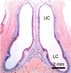
Transverse section of the nasal cavity in the turtle. LC: lower chamber, UC: upper chamber.
Fig. 5.
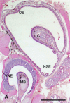
Frontal section of the nasal region in the snake. C: nasal concha, MB: mushroom body, NSE: non-sensory epithelium, OE: olfactory epithelium, VNE: vomeronasal sensory epithelium. Bar=500_µ_m.
Fig. 6.
Three nasal chambers in Xenopus laevis. Blue: upper chamber lined with the olfactory epithelium. Green: middle chamber lined with the middle chamber epithelium. Red: lower chamber lined with the vomeronasal sensory epithelium.
Fig. 7.
The olfactory organ of lungfish stained with HE. (A) Sagittal section of the nasal sac of the lungfish showing many lamellae hanging from the roof. Recesses (arrows) are observed at the base of the lamellae and lined with the recess epithelium. A: anterior, D: dorsal, P: posterior, V: ventral. (B) The lamellar olfactory epithelium covering the lamellae. Neighboring lamellar olfactory epithelia are separated by non-sensory epithelium (asterisk). BC: basal cell, ORC: olfactory receptor cell, SP: supporting cell.
Fig. 8.
Phylogeny of the olfactory receptor organs in vertebrates. CE: chamber epithelium, OE: olfactory epithelium, SO: septal olfactory organ of Masera, VNO: vomeronasal organ.
Similar articles
- Type 2 vomeronasal receptor expression in the olfactory organ of African lungfish, Protopterus annectens.
Nakamuta S, Zhang Z, Nikaido M, Yokoyama T, Yamamoto Y, Nakamuta N. Nakamuta S, et al. Cell Tissue Res. 2024 Nov;398(2):79-91. doi: 10.1007/s00441-024-03918-2. Epub 2024 Sep 30. Cell Tissue Res. 2024. PMID: 39347998 - Distribution of cells expressing vomeronasal receptors in the olfactory organ of turtles.
Abdali SS, Nakamuta S, Yamamoto Y, Nakamuta N. Abdali SS, et al. J Vet Med Sci. 2020 Aug 19;82(8):1068-1079. doi: 10.1292/jvms.20-0207. Epub 2020 Jul 28. J Vet Med Sci. 2020. PMID: 32727968 Free PMC article. - Type 1 vomeronasal receptors expressed in the olfactory organs of two African lungfish, Protopterus annectens and Protopterus amphibius.
Nakamuta S, Yamamoto Y, Miyazaki M, Sakuma A, Nikaido M, Nakamuta N. Nakamuta S, et al. J Comp Neurol. 2023 Jan;531(1):116-131. doi: 10.1002/cne.25416. Epub 2022 Sep 26. J Comp Neurol. 2023. PMID: 36161277 - Phylogenic outline of the olfactory system in vertebrates.
Taniguchi K, Saito S, Taniguchi K. Taniguchi K, et al. J Vet Med Sci. 2011 Feb;73(2):139-47. doi: 10.1292/jvms.10-0316. Epub 2010 Sep 22. J Vet Med Sci. 2011. PMID: 20877153 Review. - Vomeronasal Receptors in Vertebrates and the Evolution of Pheromone Detection.
Silva L, Antunes A. Silva L, et al. Annu Rev Anim Biosci. 2017 Feb 8;5:353-370. doi: 10.1146/annurev-animal-022516-022801. Epub 2016 Nov 28. Annu Rev Anim Biosci. 2017. PMID: 27912243 Review.
Cited by
- Type 2 vomeronasal receptor expression in the olfactory organ of African lungfish, Protopterus annectens.
Nakamuta S, Zhang Z, Nikaido M, Yokoyama T, Yamamoto Y, Nakamuta N. Nakamuta S, et al. Cell Tissue Res. 2024 Nov;398(2):79-91. doi: 10.1007/s00441-024-03918-2. Epub 2024 Sep 30. Cell Tissue Res. 2024. PMID: 39347998 - Synchrotron microtomography of a Nothosaurus marchicus skull informs on nothosaurian physiology and neurosensory adaptations in early Sauropterygia.
Voeten DFAE, Reich T, Araújo R, Scheyer TM. Voeten DFAE, et al. PLoS One. 2018 Jan 3;13(1):e0188509. doi: 10.1371/journal.pone.0188509. eCollection 2018. PLoS One. 2018. PMID: 29298295 Free PMC article. - Number of olfactory receptor neurons in the Chinese soft-shelled turtle.
Abdali SS, Kurasawa K, Nakamuta S, Yamamoto Y, Nakamuta N. Abdali SS, et al. J Vet Med Sci. 2017 Sep 29;79(9):1569-1572. doi: 10.1292/jvms.17-0326. Epub 2017 Aug 5. J Vet Med Sci. 2017. PMID: 28781329 Free PMC article. - Coding of pheromones by vomeronasal receptors.
Tirindelli R. Tirindelli R. Cell Tissue Res. 2021 Jan;383(1):367-386. doi: 10.1007/s00441-020-03376-6. Epub 2021 Jan 12. Cell Tissue Res. 2021. PMID: 33433690 Review. - Distribution of cells expressing vomeronasal receptors in the olfactory organ of turtles.
Abdali SS, Nakamuta S, Yamamoto Y, Nakamuta N. Abdali SS, et al. J Vet Med Sci. 2020 Aug 19;82(8):1068-1079. doi: 10.1292/jvms.20-0207. Epub 2020 Jul 28. J Vet Med Sci. 2020. PMID: 32727968 Free PMC article.
References
- Estes R. D.1972. The role of the vomeronasal organ in mammalian reproduction. Mammalia 36: 315–341. doi: 10.1515/mamm.1972.36.3.315 - DOI
- Franceschini F., Sbarbati A., Zancanaro C.1991. The vomeronasal organ in the frog, Rana esculenta. An electron microscopy study. J. Submicrosc. Cytol. Pathol. 23: 221–231 - PubMed
MeSH terms
Substances
LinkOut - more resources
Full Text Sources
Other Literature Sources


