Alternative radical pairs for cryptochrome-based magnetoreception - PubMed (original) (raw)
Alternative radical pairs for cryptochrome-based magnetoreception
Alpha A Lee et al. J R Soc Interface. 2014.
Abstract
There is growing evidence that the remarkable ability of animals, in particular birds, to sense the direction of the Earth's magnetic field relies on magnetically sensitive photochemical reactions of the protein cryptochrome. It is generally assumed that the magnetic field acts on the radical pair [FAD•- TrpH•+] formed by the transfer of an electron from a group of three tryptophan residues to the photo-excited flavin adenine dinucleotide cofactor within the protein. Here, we examine the suitability of an [FAD•- Z•] radical pair as a compass magnetoreceptor, where Z• is a radical in which the electron spin has no hyperfine interactions with magnetic nuclei, such as hydrogen and nitrogen. Quantum spin dynamics simulations of the reactivity of [FAD•- Z•] show that it is two orders of magnitude more sensitive to the direction of the geomagnetic field than is [FAD•- TrpH•+] under the same conditions (50 µT magnetic field, 1 µs radical lifetime). The favourable magnetic properties of [FAD•- Z•] arise from the asymmetric distribution of hyperfine interactions among the two radicals and the near-optimal magnetic properties of the flavin radical. We close by discussing the identity of Z• and possible routes for its formation as part of a spin-correlated radical pair with an FAD radical in cryptochrome.
Keywords: animal navigation; flavin; magnetic compass; radical pair mechanism; spin dynamics.
Figures
Figure 1.
Representations of the seven largest hyperfine tensors in (a) FAD_•−_ and (b) TrpH_•_+ superimposed on the structures of the parent molecules. The orientation of TrpH relative to FAD is that of Trp-342 relative to the FAD cofactor in the cryptochrome from D. melanogaster [28,29]. The hyperfine tensors, which are listed in the electronic supplementary material, tables S1 and S2, were calculated in Gaussian-03 [30] at the UB3LYP/EPR-III level of theory. The adenine group of FAD is omitted and the ribityl side chain is truncated after the first carbon. Only the side chain and the β-CH2 group of TrpH are shown.
Figure 2.
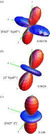
Singlet yield anisotropy plots for the radical pairs (a) [FAD_•−_ TrpH_•_+], (b) [Z_•_ TrpH_•_+] and (c) [FAD_•−_ Z_•], where Z_• is a radical with no hyperfine interactions. The spherical average of the reaction yield,  i.e. the part of Φ_S that is independent of the magnetic field direction, has been subtracted to reveal the anisotropic component, which contains the directional information. The distance in any direction from the centre of the pattern to the surface is proportional to the value of
  when the magnetic field has that direction. Red/blue regions correspond to reaction yields larger/smaller than
  FAD_•− and TrpH_•_+ each have the seven hyperfine interactions shown in figure 1. The values of Δ_Φ_S for the three radical pairs are as shown. The three anisotropy patterns are not drawn to scale. The coordinate system is that of FAD_•−_ (figure 1_a_). The orientation of TrpH_•_+ relative to FAD_•−_ is as shown in figure 1. Details of the simulations are given in the electronic supplementary material.
Figure 3.
Singlet yield anisotropy plots for radical pairs containing a single hyperfine interaction selected from those in FAD_•−_ and TrpH_•_+ (electronic supplementary material, tables S1 and S2). The values of Δ_Φ_S for the 10 spin systems are as shown. The anisotropy patterns are not drawn to scale. The coordinate system is that of FAD_•−_ (figure 1_a_). The orientation of TrpH_•_+ relative to FAD_•−_ is as shown in figure 1. Details of the simulations are given in the electronic supplementary material.
Figure 4.
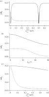
Calculations of the reaction yield anisotropy Δ_Φ_S of simple model systems. (a) Two nitrogens in the same radical (solid line) and in different radicals (dashed line). Axx = Ayy = 0 for both nuclei, parallel hyperfine _z_-axes,  The graphs show Δ_Φ_S as a function of
  the isotropic hyperfine coupling of the second nitrogen. (b) Two nitrogens in the same radical (solid line) and in different radicals (dashed line). Axx = Ayy = 0 for both nuclei,
 The graphs show Δ_Φ_S as a function of the angle _ξ_12 between the _z_-axes of the two hyperfine tensors. (c) Two nitrogens and a hydrogen with an isotropic hyperfine interaction,
  The hydrogen is either in the same radical as the nitrogens (solid line) or in the other radical (dashed line). Axx = Ayy = 0 for both nitrogens,
 parallel hyperfine axes. The graphs show Δ_Φ_S as a function of
Figure 5.
Ascorbyl radical. The isotropic 1H hyperfine interactions are as indicated [52]. (Online version in colour.)
Similar articles
- Viability of superoxide-containing radical pairs as magnetoreceptors.
Player TC, Hore PJ. Player TC, et al. J Chem Phys. 2019 Dec 14;151(22):225101. doi: 10.1063/1.5129608. J Chem Phys. 2019. PMID: 31837685 - Electron spin relaxation in cryptochrome-based magnetoreception.
Kattnig DR, Solov'yov IA, Hore PJ. Kattnig DR, et al. Phys Chem Chem Phys. 2016 May 14;18(18):12443-56. doi: 10.1039/c5cp06731f. Epub 2016 Mar 29. Phys Chem Chem Phys. 2016. PMID: 27020113 - Separation of photo-induced radical pair in cryptochrome to a functionally critical distance.
Solov'yov IA, Domratcheva T, Schulten K. Solov'yov IA, et al. Sci Rep. 2014 Jan 24;4:3845. doi: 10.1038/srep03845. Sci Rep. 2014. PMID: 24457842 Free PMC article. - The Magnetic Compass of Birds: The Role of Cryptochrome.
Wiltschko R, Nießner C, Wiltschko W. Wiltschko R, et al. Front Physiol. 2021 May 19;12:667000. doi: 10.3389/fphys.2021.667000. eCollection 2021. Front Physiol. 2021. PMID: 34093230 Free PMC article. Review. - The Radical-Pair Mechanism of Magnetoreception.
Hore PJ, Mouritsen H. Hore PJ, et al. Annu Rev Biophys. 2016 Jul 5;45:299-344. doi: 10.1146/annurev-biophys-032116-094545. Epub 2016 May 16. Annu Rev Biophys. 2016. PMID: 27216936 Review.
Cited by
- Radical pairs may play a role in xenon-induced general anesthesia.
Smith J, Zadeh Haghighi H, Salahub D, Simon C. Smith J, et al. Sci Rep. 2021 Mar 18;11(1):6287. doi: 10.1038/s41598-021-85673-w. Sci Rep. 2021. PMID: 33737599 Free PMC article. - Isotope Substitution Effects on the Magnetic Compass Properties of Cryptochrome-Based Radical Pairs: A Computational Study.
Pažėra GJ, Benjamin P, Mouritsen H, Hore PJ. Pažėra GJ, et al. J Phys Chem B. 2023 Feb 2;127(4):838-845. doi: 10.1021/acs.jpcb.2c05335. Epub 2023 Jan 20. J Phys Chem B. 2023. PMID: 36669149 Free PMC article. - Cryptochrome magnetoreception: four tryptophans could be better than three.
Wong SY, Wei Y, Mouritsen H, Solov'yov IA, Hore PJ. Wong SY, et al. J R Soc Interface. 2021 Nov;18(184):20210601. doi: 10.1098/rsif.2021.0601. Epub 2021 Nov 10. J R Soc Interface. 2021. PMID: 34753309 Free PMC article. - Ascorbic acid may not be involved in cryptochrome-based magnetoreception.
Nielsen C, Kattnig DR, Sjulstok E, Hore PJ, Solov'yov IA. Nielsen C, et al. J R Soc Interface. 2017 Dec;14(137):20170657. doi: 10.1098/rsif.2017.0657. J R Soc Interface. 2017. PMID: 29263128 Free PMC article. - Magnetosensitivity of Model Flavin-Tryptophan Radical Pairs in a Dynamic Protein Environment.
Benjamin PL, Gerhards L, Solov'yov IA, Hore PJ. Benjamin PL, et al. J Phys Chem B. 2025 Jun 19;129(24):5937-5947. doi: 10.1021/acs.jpcb.5c01187. Epub 2025 Jun 4. J Phys Chem B. 2025. PMID: 40464336 Free PMC article.
References
Publication types
MeSH terms
Substances
LinkOut - more resources
Full Text Sources
Other Literature Sources


