Physical activity when young provides lifelong benefits to cortical bone size and strength in men - PubMed (original) (raw)
Physical activity when young provides lifelong benefits to cortical bone size and strength in men
Stuart J Warden et al. Proc Natl Acad Sci U S A. 2014.
Abstract
The skeleton shows greatest plasticity to physical activity-related mechanical loads during youth but is more at risk for failure during aging. Do the skeletal benefits of physical activity during youth persist with aging? To address this question, we used a uniquely controlled cross-sectional study design in which we compared the throwing-to-nonthrowing arm differences in humeral diaphysis bone properties in professional baseball players at different stages of their careers (n = 103) with dominant-to-nondominant arm differences in controls (n = 94). Throwing-related physical activity introduced extreme loading to the humeral diaphysis and nearly doubled its strength. Once throwing activities ceased, the cortical bone mass, area, and thickness benefits of physical activity during youth were gradually lost because of greater medullary expansion and cortical trabecularization. However, half of the bone size (total cross-sectional area) and one-third of the bone strength (polar moment of inertia) benefits of throwing-related physical activity during youth were maintained lifelong. In players who continued throwing during aging, some cortical bone mass and more strength benefits of the physical activity during youth were maintained as a result of less medullary expansion and cortical trabecularization. These data indicate that the old adage of "use it or lose it" is not entirely applicable to the skeleton and that physical activity during youth should be encouraged for lifelong bone health, with the focus being optimization of bone size and strength rather than the current paradigm of increasing mass. The data also indicate that physical activity should be encouraged during aging to reduce skeletal structural decay.
Keywords: exercise; intracortical remodeling; osteoporosis; peak bone mass.
Conflict of interest statement
The authors declare no conflict of interest.
Figures
Fig. 1.
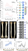
Overhand throwing loads the humeral diaphysis, inducing skeletal adaptation. (A and B) Median (and median absolute deviation) cross-sectional tensile (A) and shear (B) strains in the humerus of the throwing arm of an MLB player toward the end of the cocking stage of a fastball pitch showed strains throughout the diaphysis. Both tensile and shear strains were increased when the same forces were applied to the collateral, nonthrowing humerus. (C) Cross-sectional distribution of peak tensile strain in the bilateral humerus demonstrated reduced strains in the throwing arm. (D) Reconstructed CT images of the bilateral humerii in a representative MLB/MiLB player demonstrated a more robust diaphysis with visibly broader diameter on the throwing side. (E) Cross-sectional images of the humerii in D revealed substantially greater total and cortical bone areas and cortical thickness and smaller medullary area in the throwing arm. (F) Throwing substantially increased torsional bone strength (indicated by density-weighted polar moment of inertia) along the entire diaphysis, with strength nearly doubled toward the distal diaphysis. Data show the mean percent difference and 95% CI (shaded area) between the throwing arm and the nonthrowing arm in throwers normalized to the differences between the dominant arm and the nondominant arm in controls (‡P < 0.001, unpaired t test). (G) The distal diaphysis in throwers had a more circular cross-section than seen in controls, as indicated by a maximum:minimum (IMAX:IMIN) second moment of area ratio closer to 1 (*P < 0.05, ANCOVA with the contralateral arm as the covariate).
Fig. 2.
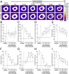
Physical activity-related mechanical loading during youth conferred lifelong benefits in cortical bone size and estimated strength but not in cortical bone mass. (A) Peripheral QCT images of the midshaft humerus in representative former throwers show increased medullary expansion and cortex trabecularization (arrows) in the throwing arm with increasing years of detraining but maintenance of loading effects on overall bone cross-sectional size. (B_–_I) Graphs show the maintenance of the effects of physical activity during youth on cortical volumetric bone mineral density (B); cortical bone mineral content (C); trabecular/subcortical bone mineral content (D); total cross-sectional area (E); cortical cross-sectional area (F); medullary cross-sectional area (G); cortical thickness (H); and density-weighted polar moment of inertia (I). Data show the mean difference and 95% CI between the throwing and nonthrowing arms in former throwers normalized to the differences between the dominant and nondominant arms in controls. CIs greater or less than 0% indicate differences between the throwing and nonthrowing arms in throwers that are greater or less, respectively, than the differences between the dominant and nondominant arms in controls (*P < 0.05, †P < 0.01 and ‡P < 0.001, unpaired t test). Source data are provided in
Table S3
.
Fig. 3.
Continued physical activity during aging maintained a proportion of the benefits in bone mass and more of the benefits in bone strength induced during youth. (A) Peripheral QCT images of the midshaft humerii in representative individuals showed less medullary expansion and cortical trabecularization (arrows) in the throwing arm of the continuing thrower than in the throwing arm of the former thrower. (B) Cortical bone mineral content and density-weighted polar moment of inertia were greater in continuing throwers than in former throwers and controls; cortical thickness was greater in continuing throwers than in former throwers; and trabecular/subcortical bone mineral content and medullary area were smaller in continuing throwers than in former throwers. Data show the mean percent difference and 95% CI between the throwing arm and the nonthrowing arm in throwers normalized to the differences between the dominant arm and the nondominant arm in controls. CIs greater than 0% indicate greater differences between the throwing arm and the nonthrowing arm in throwers than between the dominant arm and the nondominant arm in controls (†P < 0.01; ‡P < 0.001); § indicates significant differences between continuing and former throwers (P < 0.05; one-way ANOVA with Tukey post hoc comparisons).
Similar articles
- Physical activity completed when young has residual bone benefits at 94 years of age: a within-subject controlled case study.
Warden SJ, Mantila Roosa SM. Warden SJ, et al. J Musculoskelet Neuronal Interact. 2014 Jun;14(2):239-43. J Musculoskelet Neuronal Interact. 2014. PMID: 24879028 Free PMC article. - Progressive skeletal benefits of physical activity when young as assessed at the midshaft humerus in male baseball players.
Warden SJ, Weatherholt AM, Gudeman AS, Mitchell DC, Thompson WR, Fuchs RK. Warden SJ, et al. Osteoporos Int. 2017 Jul;28(7):2155-2165. doi: 10.1007/s00198-017-4029-9. Epub 2017 Apr 10. Osteoporos Int. 2017. PMID: 28396902 Free PMC article. - Throwing enhances humeral shaft cortical bone properties in pre-pubertal baseball players: a 12-month longitudinal pilot study.
Weatherholt AM, Warden SJ. Weatherholt AM, et al. J Musculoskelet Neuronal Interact. 2018 Jun 1;18(2):191-199. J Musculoskelet Neuronal Interact. 2018. PMID: 29855441 Free PMC article. - Why do bone strength and "mass" in aging adults become unresponsive to vigorous exercise? Insights of the Utah paradigm.
Frost HM. Frost HM. J Bone Miner Metab. 1999;17(2):90-7. doi: 10.1007/s007740050070. J Bone Miner Metab. 1999. PMID: 10340635 Review. - Physical activity and bone mass: exercises in futility?
Forwood MR, Burr DB. Forwood MR, et al. Bone Miner. 1993 May;21(2):89-112. doi: 10.1016/s0169-6009(08)80012-8. Bone Miner. 1993. PMID: 8358253 Review.
Cited by
- Focal enhancement of the skeleton to exercise correlates with responsivity of bone marrow mesenchymal stem cells rather than peak external forces.
Wallace IJ, Pagnotti GM, Rubin-Sigler J, Naeher M, Copes LE, Judex S, Rubin CT, Demes B. Wallace IJ, et al. J Exp Biol. 2015 Oct;218(Pt 19):3002-9. doi: 10.1242/jeb.118729. Epub 2015 Jul 31. J Exp Biol. 2015. PMID: 26232415 Free PMC article. - Intranuclear Actin Regulates Osteogenesis.
Sen B, Xie Z, Uzer G, Thompson WR, Styner M, Wu X, Rubin J. Sen B, et al. Stem Cells. 2015 Oct;33(10):3065-76. doi: 10.1002/stem.2090. Stem Cells. 2015. PMID: 26140478 Free PMC article. - Mechanism and Treatment Strategy of Osteoporosis after Transplantation.
Song L, Xie XB, Peng LK, Yu SJ, Peng YT. Song L, et al. Int J Endocrinol. 2015;2015:280164. doi: 10.1155/2015/280164. Epub 2015 Jul 27. Int J Endocrinol. 2015. PMID: 26273295 Free PMC article. Review. - Adaptation of the proximal humerus to physical activity: A within-subject controlled study in baseball players.
Warden SJ, Carballido-Gamio J, Avin KG, Kersh ME, Fuchs RK, Krug R, Bice RJ. Warden SJ, et al. Bone. 2019 Apr;121:107-115. doi: 10.1016/j.bone.2019.01.008. Epub 2019 Jan 8. Bone. 2019. PMID: 30634064 Free PMC article. - Peak power and body mass as predictors of tibial bone strength in healthy male and female adults.
Denys AT, Bugayong JC, Juhala CC, Ma EJ, Carvalho KE, Kwong SM, Yingling VR. Denys AT, et al. J Musculoskelet Neuronal Interact. 2022 Jun 1;22(2):154-160. J Musculoskelet Neuronal Interact. 2022. PMID: 35642695 Free PMC article.
References
- Kannus P, et al. Effect of starting age of physical activity on bone mass in the dominant arm of tennis and squash players. Ann Intern Med. 1995;123(1):27–31. - PubMed
- Rizzoli R, Bianchi ML, Garabédian M, McKay HA, Moreno LA. Maximizing bone mineral mass gain during growth for the prevention of fractures in the adolescents and the elderly. Bone. 2010;46(2):294–305. - PubMed
- Erlandson MC, et al. Higher premenarcheal bone mass in elite gymnasts is maintained into young adulthood after long-term retirement from sport: A 14-year follow-up. J Bone Miner Res. 2012;27(1):104–110. - PubMed
- Kontulainen S, et al. Good maintenance of exercise-induced bone gain with decreased training of female tennis and squash players: A prospective 5-year follow-up study of young and old starters and controls. J Bone Miner Res. 2001;16(2):195–201. - PubMed
Publication types
MeSH terms
LinkOut - more resources
Full Text Sources
Other Literature Sources
Medical


