Microscale assembly directed by liquid-based template - PubMed (original) (raw)
Microscale assembly directed by liquid-based template
Pu Chen et al. Adv Mater. 2014.
Abstract
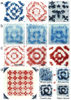
A liquid surface established by standing waves is used as a dynamically reconfigurable template to assemble microscale materials into ordered, symmetric structures in a scalable and parallel manner. The broad applicability of this technology is illustrated by assembling diverse materials from soft matter, rigid bodies, individual cells, cell spheroids and cell-seeded microcarrier beads.
Keywords: bottom-up; directed assembly; liquid-based template; microscale materials; tissue engineering.
© 2014 WILEY-VCH Verlag GmbH & Co. KGaA, Weinheim.
Figures
Figure 1. Principle demonstration of liquid-based templated assembly
a, Schematics of assembly based on liquid-based template. b, Top down of the standing waves simulated according to equation (S2) in the supplementary information. Color bar indicates wave amplitude. c, Numerical simulation of drift energy for 200 μm polystyrene divinylbenzene beads on the standing waves based on equation (1). d–e, Assembly of polystyrene divinylbenzene beads on the nodal regions of standing waves with different coverage rate of beads on the air–liquid surface (d, 53% e, 2.5%). f, Numerical simulation of drift energy for 200 μm copper-zinc powder on the standing waves based on Equation (1). g, Assembly of copper-zinc powder on the antinodes of the standing waves. h, Assembly of complementary pattern by using copper-zinc powder (yellow regions) and polystyrene divinylbenzene beads (red regions). Chamber dimensions are 20 mm × 20 mm × 1.5 mm for all the experiments and simulations. Scale bars, 2 mm.
Figure 2
Diversity of the structures created by liquid-based templated assembly. a, Chamber shape effect on the assembly. b, Numerical simulation of the waveforms generated in the square chamber. Square waveform (SQ), stripe waveform (ST) and crystalline waveforms (CR1 and CR2) were obtained using equation S2, S3 and S4 respectively. c–d, Typical assembled structure and corresponding drift energy (numerical simulation) under each waveforms. Each panel in c and d represents the dashed square in the corresponding panel of b. e, Principle demonstration of the symmetric modes within the square waveform. The color bar depicts the amplitude of the standing waves. Each code represents the symmetric modes. λ is the wavelength, and (λ/4, λ/4) indicates translation of the standing waves by λ/4 in both x-axis and y-axis directions. The symmetric axes are indicated with white dash-dotted lines. f, Harmonic order within each symmetric modes. All of the scale bars indicate 2 mm. Codes under the assembled structures are applied in the phase diagrams.
Figure 3. Dynamical reconfigurability of liquid-based templated assembly
a, Schematic demonstration of dynamic reconfiguration of the assembled structures: (_f_A, _a_A) and (_f_B, _a_B) are vibrational frequencies and accelerations for the formation of structures A and B, respectively. By tuning the initial chamber with (_f_A, _a_A) and (_f_B, _a_B), assembly of the structures A and B from dispersed floaters can be performed, as well as the reversible transitions between the structures. b, Dynamic process of the assembly. I–VI show different stages of the assembly: I. before assembly; II. during assembly; III. formation of the ring-shaped structure; IV. intermediate state; V. formation of “H”-shaped structure; VI. restoration of the ring-shaped structure. All of the experiments were performed in10 mm × 10 mm × 1.5 mm chamber using beads with 200 μm in diameter. Scale bars, 2 mm. Note: The snapshots in top-down view and side view were recorded separately and approximately represent the events in the corresponding column. c, Time evolution of the assembly fraction during the assembly process. Vibration was applied at time zero. * Resetting vibrational parameters: the vibrational frequency was first increased from 46 Hz to 60 Hz, and the acceleration was then increased from 1.46 g to 1.9 g. ** Resetting vibrational parameters: the acceleration was first decreased from 1.9 g to 1.46 g, and the vibrational frequency was then decreased from 60 Hz to 46 Hz.
Figure 4. Broad applicability of liquid-based templated assembly
a, Assembly of various materials. From left to right: GelMA hydrogel units (blue) and PDMS blocks (red) on nodal regions; silicon chiplets on antinodes. b, Assembly of size-varied materials. PEG hydrogel units with sizes of 0.5, 1, and 2 mm. c, Scalable assembly. Chamber sizes are 10, 20 and 30 mm respectively. Floater size is 200 μm for all. d, Parallel assembly in a 5-by-5 chamber array. Dimensions of each chamber are 10 mm × 10 mm × 1.5 mm. e, Photo crosslinking of the assembled structure. Once the hydrogels assembled, the crosslinking was performed to immobilize the hydrogels and the assembled pattern. Scale bars: 4 mm.
Figure 5. Liquid-based templated assembly for tissue engineering
a–d, Assembly of cell-seeded microcarrier beads. a, Microcarrier beads with CFSE (Green) stained NIH 3T3 fibroblast cells after assembly and crosslinking. b, Live/dead assays on the cells seeded on the microcarrier beads, 3-day culture after chemical crosslinking. Green color indicates live cells (calcein-AM), red color indicates dead cells (ethidium homodimer-1). c–d, Formation of 3D neural structures on the assembled microcarrier beads after 14-day cell culture. e–h, Scaffold-free assembly of cells spheroids (mean size: 200 μm). f is magnified region in e, marked with red dashed lines. g–h, assembled structures from cell spheroids, bright field recorded by digital SLR camera. i–l, Scaffold-free assembly of fibroblast cells and cytocompatibility tests. i is magnified region in g, marked with red dashed lines. The cells were stained by cell tracker CFSE (Green). k, Cell viability test under assembly onset acceleration at various vibrational frequencies (n = 6); l, Cell proliferation test with Alamar blue. Cells experienced by 15-second agitations at 50, 100 and 200 Hz. The treated cells were seeded in a 64-well plate with a seeding density of 200 cells/well for 11-day cell culture. Data was presented as mean ± S.D. (n = 8).
Similar articles
- 3D-to-3D Microscale Shape-Morphing from Configurable Helices with Controlled Chirality.
Zhao Z, He Y, Meng X, Ye C. Zhao Z, et al. ACS Appl Mater Interfaces. 2021 Dec 29;13(51):61723-61732. doi: 10.1021/acsami.1c15711. Epub 2021 Dec 16. ACS Appl Mater Interfaces. 2021. PMID: 34913686 - Surface Tension-Assisted Additive Manufacturing of Tubular, Multicomponent Biomaterials.
Guzzi EA, Ragelle H, Tibbitt MW. Guzzi EA, et al. Methods Mol Biol. 2021;2147:149-160. doi: 10.1007/978-1-0716-0611-7_13. Methods Mol Biol. 2021. PMID: 32840818 - Assessing cellular response to functionalized α-helical peptide hydrogels.
Mehrban N, Abelardo E, Wasmuth A, Hudson KL, Mullen LM, Thomson AR, Birchall MA, Woolfson DN. Mehrban N, et al. Adv Healthc Mater. 2014 Sep;3(9):1387-91. doi: 10.1002/adhm.201400065. Epub 2014 Mar 24. Adv Healthc Mater. 2014. PMID: 24659615 Free PMC article. - Directed assembly of cell-laden hydrogels for engineering functional tissues.
Kachouie NN, Du Y, Bae H, Khabiry M, Ahari AF, Zamanian B, Fukuda J, Khademhosseini A. Kachouie NN, et al. Organogenesis. 2010 Oct-Dec;6(4):234-44. doi: 10.4161/org.6.4.12650. Organogenesis. 2010. PMID: 21220962 Free PMC article. Review. - Double network hydrogel for tissue engineering.
Gu Z, Huang K, Luo Y, Zhang L, Kuang T, Chen Z, Liao G. Gu Z, et al. Wiley Interdiscip Rev Nanomed Nanobiotechnol. 2018 Nov;10(6):e1520. doi: 10.1002/wnan.1520. Epub 2018 Apr 17. Wiley Interdiscip Rev Nanomed Nanobiotechnol. 2018. PMID: 29664220 Review.
Cited by
- Dynamic interface printing.
Vidler C, Halwes M, Kolesnik K, Segeritz P, Mail M, Barlow AJ, Koehl EM, Ramakrishnan A, Caballero Aguilar LM, Nisbet DR, Scott DJ, Heath DE, Crozier KB, Collins DJ. Vidler C, et al. Nature. 2024 Oct;634(8036):1096-1102. doi: 10.1038/s41586-024-08077-6. Epub 2024 Oct 30. Nature. 2024. PMID: 39478212 Free PMC article. - Capillary-based, multifunctional manipulation of particles and fluids via focused surface acoustic waves.
Pei Z, Tian Z, Yang S, Shen L, Hao N, Naquin TD, Li T, Sun L, Rong W, Huang TJ. Pei Z, et al. J Phys D Appl Phys. 2024 Aug;57(30):305401. doi: 10.1088/1361-6463/ad415a. Epub 2024 May 7. J Phys D Appl Phys. 2024. PMID: 38800708 - Differential proteomics profile of microcapillary networks in response to sound pattern-driven local cell density enhancement.
Di Marzio N, Tognato R, Bella ED, De Giorgis V, Manfredi M, Cochis A, Alini M, Serra T. Di Marzio N, et al. Biomater Biosyst. 2024 Mar 29;14:100094. doi: 10.1016/j.bbiosy.2024.100094. eCollection 2024 Jun. Biomater Biosyst. 2024. PMID: 38596510 Free PMC article. - FastSkin® Concept: A Novel Treatment for Complex Acute and Chronic Wound Management.
di Summa PG, Di Marzio N, Jafari P, Jaconi ME, Nesic D. di Summa PG, et al. J Clin Med. 2023 Oct 16;12(20):6564. doi: 10.3390/jcm12206564. J Clin Med. 2023. PMID: 37892702 Free PMC article. - Sound-based assembly of three-dimensional cellularized and acellularized constructs.
Tognato R, Parolini R, Jahangir S, Ma J, Florczak S, Richards RG, Levato R, Alini M, Serra T. Tognato R, et al. Mater Today Bio. 2023 Aug 19;22:100775. doi: 10.1016/j.mtbio.2023.100775. eCollection 2023 Oct. Mater Today Bio. 2023. PMID: 37674778 Free PMC article.
References
- Whitesides GM, Grzybowski B. Science. 2002;295:2418. - PubMed
- Grzybowski BA, Wilmer CE, Kim J, Browne KP, Bishop KJM. Soft Matter. 2009;5:1110.
- Lu Y, Yin Y, Xia Y. Adv Mater. 2001;13:34.
Publication types
MeSH terms
Substances
Grants and funding
- U54 EB015408/EB/NIBIB NIH HHS/United States
- R21HL112114/HL/NHLBI NIH HHS/United States
- R21 HL112114/HL/NHLBI NIH HHS/United States
- U54EB15408/EB/NIBIB NIH HHS/United States
- R01 EB015776/EB/NIBIB NIH HHS/United States
- R01EB015776-01A1/EB/NIBIB NIH HHS/United States
- R15 HL115556/HL/NHLBI NIH HHS/United States
- R15HL115556/HL/NHLBI NIH HHS/United States
LinkOut - more resources
Full Text Sources
Other Literature Sources


