TPX2 levels modulate meiotic spindle size and architecture in Xenopus egg extracts - PubMed (original) (raw)
TPX2 levels modulate meiotic spindle size and architecture in Xenopus egg extracts
Kara J Helmke et al. J Cell Biol. 2014.
Abstract
The spindle segregates chromosomes in dividing eukaryotic cells, and its assembly pathway and morphology vary across organisms and cell types. We investigated mechanisms underlying differences between meiotic spindles formed in egg extracts of two frog species. Small Xenopus tropicalis spindles resisted inhibition of two factors essential for assembly of the larger Xenopus laevis spindles: RanGTP, which functions in chromatin-driven spindle assembly, and the kinesin-5 motor Eg5, which drives antiparallel microtubule (MT) sliding. This suggested a role for the MT-associated protein TPX2 (targeting factor for Xenopus kinesin-like protein 2), which is regulated by Ran and binds Eg5. Indeed, TPX2 was threefold more abundant in X. tropicalis extracts, and elevated TPX2 levels in X. laevis extracts reduced spindle length and sensitivity to Ran and Eg5 inhibition. Higher TPX2 levels recruited Eg5 to the poles, where MT density increased. We propose that TPX2 levels modulate spindle architecture through Eg5, partitioning MTs between a tiled, antiparallel array that promotes spindle expansion and a cross-linked, parallel architecture that concentrates MTs at spindle poles.
© 2014 Helmke and Heald.
Figures
Figure 1.
X. tropicalis and X. laevis spindles differ in morphology and sensitivity to inhibition of Ran and Eg5. (A) Spinning disk confocal images of X. laevis and X. tropicalis spindle midzones in live spindle reactions on polyethylene glycol–coated glass. Bar, 5 µm. Mean line scan intensity of MTs across the length of the spindle (as described in Materials and methods), showing reduced MT density in the center of the spindle of X. tropicalis; n = 15 spindles for X. laevis and n = 19 for X. tropicalis from three extracts. (B) Inhibition of the RanGTP pathway with 1 µM of the dominant-negative mutant RanT24N disrupted spindle assembly in X. laevis but not X. tropicalis egg extracts. Bar, 10 µm. (C) Line scan quantification of TPX2 immunofluorescence relative to tubulin intensity from >50 spindles in each condition from one representative experiment. TPX2 intensity was higher in X. tropicalis spindles (dark blue) and remained unchanged upon addition of 1 µM RanT24N (light blue), whereas X. laevis spindles recruited less TPX2 (red), which was lost upon RanT24N treatment (pink). Although spindle assembly was strongly inhibited in X. laevis upon RanT24N treatment, the remaining bipolar MT structures formed in these reactions were used for line scan analysis. (D) Representative images of spindle assembly reactions in X. laevis and X. tropicalis egg extracts in the presence of increasing amounts of monastrol to inhibit Eg5. X. laevis spindles began to shorten and collapse with increasing monastrol, whereas X. tropicalis spindles showed little collapse and were resistant to monastrol except at the highest concentrations. Bar, 10 µm.
Figure 2.
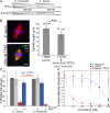
TPX2 levels correlate with RanGTP and Eg5 dependence and spindle size. (A) Representative Western blot of dilution series of X. tropicalis and X. laevis egg extracts showing higher levels of TPX2 present in X. tropicalis. β-Tubulin is shown as a loading control. TPX2 is ∼82 kD, but migrates on a gel at ∼100 kD. Band intensities from Western blots of three extracts each were quantified (Odyssey software), and on average TPX2 levels were approximately threefold higher in X. tropicalis compared with X. laevis, giving an estimated concentration of 300 nM (X. laevis TPX2 concentration is ∼100 nM; Gruss et al., 2001). (B, left) Addition of 200 nM MBP-TPX2 to X. laevis extracts decreased spindle size. Polar localization of recombinant protein is detected by immunofluorescence against the MBP tag. Bar, 10 µm. (right) Quantification of spindle length with addition of 200 nM MBP-TPX2 compared with control addition of 200 nM MBP. Mean ± SD; n ≥ 671 spindles in each condition from three separate extracts; ***, P < 0.0001 from unpaired t test. (C) Quantification of spindle bipolarity as an indicator of proper spindle formation upon inhibition of the RanGTP pathway in X. laevis and X. tropicalis egg extracts. For each condition, all MT structures near condensed DNA were scored for morphology with n ≥ 100 for each extract in three separate experiments. Percentage of bipolarity was calculated from total structures scored. Mean ± SD. Dark gray bars indicate control extracts with no treatment. With 5 µM RanT24N addition, spindle formation in X. laevis egg extracts was impaired and the fraction of bipolar structures decreased to 7.3% (left red bar) with the remaining structures monopolar (39%) or lacking MTs (45.8%). When X. laevis extracts were supplemented with 200 nM MBP-TPX2, spindle bipolarity with RanT24N treatment was strongly rescued (left blue bar). TPX2 addition alone did not affect bipolarity (left light gray bar). X. tropicalis only showed a modest decrease with RanT24N treatment, from 54 to 46% (right blue bar). However, when X. tropicalis was immunodepleted of TPX2, bipolarity was almost completely lost upon RanT24N treatment, which reduced bipolar spindle assembly to 2.7% (right red bar). TPX2 immunodepletion from X. tropicalis extracts did not affect spindle bipolarity (right light gray bar). (D) Quantification of spindle bipolarity of X. laevis, X. laevis + 200 nM MBP-TPX2, and X. tropicalis MT structures upon monastrol treatment at the indicated concentration. MT structures were scored as in C but normalized to the 0-µM condition for each extract and plotted as the mean ± standard error. Four-parameter logistic fit is shown for both X. tropicalis (blue dashed line; IC50 of 211 µM) and X. laevis (red dashed line; IC50 of 21 µM), but no fit could be calculated for the X. laevis + TPX2 condition as it was resistant to monastrol treatment at the highest concentration.
Figure 3.
TPX2 MT nucleation activity is regulated by a 7–amino acid sequence that contributes to spindle pole morphology but not length. (A) Domain schematic of the X. laevis TPX2 protein. The first 39 amino acids are unstructured and interact with Aurora A (orange; Bayliss et al., 2003) and the last 35 amino acids interact with Eg5 (green; Bayliss et al., 2003; Eckerdt et al., 2008). A mapped nuclear localization signal is at amino acid 284. Three highly conserved domains are denoted by dark purple shading (Goshima, 2011). Zoom-in shows conservation between X. laevis and human of a 7–amino acid sequence missing from the X. tropicalis orthologue. (B) Addition of 200 nM of recombinant TPX2 mutants to X. laevis extract. (top) 20× field of view showing increased MT aster structures nucleated upon addition of X. tropicalis TPX2 or X. laevis Δ7 TPX2 compared with control or X. laevis TPX2. Arrowheads indicate MT asters. (bottom) Spindle morphology after TPX2 mutant addition. X. tropicalis TPX2 and X. laevis Δ7 TPX2 induced formation of radial astral MTs emanating from the poles not seen in control spindles or with X. laevis TPX2 addition. Bars, 10 µm. (C) Quantification of nucleation activity of recombinant TPX2 proteins. For each condition, spindle reactions were sedimented onto coverslips as described in Materials and methods and the number of MT aster structures was counted in 10 microscope fields, repeated in three separate extracts. Boxplot of number of asters per field with median marked by gray line, first and third quartiles marked by box edges, and data maxima and minima noted by whiskers. (D) Quantification of pole-to-pole spindle length, not including astral MTs, with addition of 200 nM MBP-TPX2 proteins compared with control of addition of 200 nM MBP. Mean ± SD; n ≥ 671 spindles in each condition from three separate extracts. For each TPX2 protein compared with MBP control, P < 0.0001 from unpaired t test.
Figure 4.
TPX2 regulates spindle size and MT distribution through interaction with Eg5. (A) Addition of TPX2 domain truncation mutants to X. laevis extract. The TPX2ΔAurA mutant localized like full-length TPX2 and had similar effects on spindle size, whereas TPX2ΔEg5 did not localize to the spindle or cause a decrease in spindle length. Bar, 10 µm. Mean ± SD; n ≥ 166 spindles in each condition from three separate extracts preserved by squashing under coverslips with spindle fixative. ΔEg5 TPX2 mean spindle length was not significantly different from the MBP control, but was significantly different from full-length TPX2 by unpaired t test; ***, P < 0.0001. (B) Representative images of Eg5 localization and MT density changes in X. laevis spindles upon addition of 200 nM MBP-TPX2 and in comparison to X. tropicalis. Left panels are merged, middle panels show rhodamine-tubulin, and right panels show Eg5 immunofluorescence. Bar, 10 µm. (C) Comparison of phenotypes in X. laevis extracts with the addition of either 200 nM MBP-TPX2 or 50 µM monastrol to inhibit Eg5. Although both treatments reduced spindle length, the insets demonstrate a significant decrease in MT density in the center of the spindle with TPX2 addition but not monastrol treatment. Bars, 10 µm. (D) Line scan quantification of Eg5 immunofluorescence intensity in X. tropicalis and X. laevis spindles with and without addition of TPX2 mutants (see Materials and methods). Spindle lengths were normalized for statistical analysis. Mean ± standard error; n ≥ 169 spindles for each condition from three extracts. (E) Line scan quantification of MT density measured by rhodamine-tubulin intensity in X. tropicalis and X. laevis spindles with and without addition of TPX2 (see Materials and methods). Spindle lengths were normalized for statistical analysis. Mean ± standard error; n ≥ 156 spindles for each condition from three extracts.
Figure 5.
Model of TPX2 regulation of spindle size and MT distribution via Eg5. MTs with plus ends to the right are red, whereas MTs with the opposite orientation are green. An increase in TPX2 (purple) concentration recruits more Eg5 (orange) to the spindle poles, reducing the extent of MT overlap and increasing MT cross-linking at spindle poles, thereby causing spindle length to decrease.
Similar articles
- Characterization of the TPX2 domains involved in microtubule nucleation and spindle assembly in Xenopus egg extracts.
Brunet S, Sardon T, Zimmerman T, Wittmann T, Pepperkok R, Karsenti E, Vernos I. Brunet S, et al. Mol Biol Cell. 2004 Dec;15(12):5318-28. doi: 10.1091/mbc.e04-05-0385. Epub 2004 Sep 22. Mol Biol Cell. 2004. PMID: 15385625 Free PMC article. - Cdk11 is a RanGTP-dependent microtubule stabilization factor that regulates spindle assembly rate.
Yokoyama H, Gruss OJ, Rybina S, Caudron M, Schelder M, Wilm M, Mattaj IW, Karsenti E. Yokoyama H, et al. J Cell Biol. 2008 Mar 10;180(5):867-75. doi: 10.1083/jcb.200706189. Epub 2008 Mar 3. J Cell Biol. 2008. PMID: 18316407 Free PMC article. - Hepatoma up-regulated protein is required for chromatin-induced microtubule assembly independently of TPX2.
Casanova CM, Rybina S, Yokoyama H, Karsenti E, Mattaj IW. Casanova CM, et al. Mol Biol Cell. 2008 Nov;19(11):4900-8. doi: 10.1091/mbc.e08-06-0624. Epub 2008 Sep 17. Mol Biol Cell. 2008. PMID: 18799614 Free PMC article. - The mechanism of spindle assembly: functions of Ran and its target TPX2.
Gruss OJ, Vernos I. Gruss OJ, et al. J Cell Biol. 2004 Sep 27;166(7):949-55. doi: 10.1083/jcb.200312112. J Cell Biol. 2004. PMID: 15452138 Free PMC article. Review. - Regulation of Aurora-A kinase on the mitotic spindle.
Kufer TA, Nigg EA, Silljé HH. Kufer TA, et al. Chromosoma. 2003 Dec;112(4):159-63. doi: 10.1007/s00412-003-0265-1. Epub 2003 Nov 21. Chromosoma. 2003. PMID: 14634755 Review.
Cited by
- Use of Xenopus cell-free extracts to study size regulation of subcellular structures.
Jevtić P, Milunović-Jevtić A, Dilsaver MR, Gatlin JC, Levy DL. Jevtić P, et al. Int J Dev Biol. 2016;60(7-8-9):277-288. doi: 10.1387/ijdb.160158dl. Int J Dev Biol. 2016. PMID: 27759156 Free PMC article. Review. - TPX2 phosphorylation maintains metaphase spindle length by regulating microtubule flux.
Fu J, Bian M, Xin G, Deng Z, Luo J, Guo X, Chen H, Wang Y, Jiang Q, Zhang C. Fu J, et al. J Cell Biol. 2015 Aug 3;210(3):373-83. doi: 10.1083/jcb.201412109. J Cell Biol. 2015. PMID: 26240182 Free PMC article. - Changes in seam number and location induce holes within microtubules assembled from porcine brain tubulin and in Xenopus egg cytoplasmic extracts.
Guyomar C, Bousquet C, Ku S, Heumann JM, Guilloux G, Gaillard N, Heichette C, Duchesne L, Steinmetz MO, Gibeaux R, Chrétien D. Guyomar C, et al. Elife. 2022 Dec 12;11:e83021. doi: 10.7554/eLife.83021. Elife. 2022. PMID: 36503602 Free PMC article. - Understanding eukaryotic chromosome segregation from a comparative biology perspective.
Oliferenko S. Oliferenko S. J Cell Sci. 2018 Jul 20;131(14):jcs203653. doi: 10.1242/jcs.203653. J Cell Sci. 2018. PMID: 30030298 Free PMC article. Review. - A versatile multivariate image analysis pipeline reveals features of Xenopus extract spindles.
Grenfell AW, Strzelecka M, Crowder ME, Helmke KJ, Schlaitz AL, Heald R. Grenfell AW, et al. J Cell Biol. 2016 Apr 11;213(1):127-36. doi: 10.1083/jcb.201509079. Epub 2016 Apr 4. J Cell Biol. 2016. PMID: 27044897 Free PMC article.
References
Publication types
MeSH terms
Substances
LinkOut - more resources
Full Text Sources
Other Literature Sources
Miscellaneous


