Anatomic changes in Schlemm's canal and collector channels in normal and primary open-angle glaucoma eyes using low and high perfusion pressures - PubMed (original) (raw)
Anatomic changes in Schlemm's canal and collector channels in normal and primary open-angle glaucoma eyes using low and high perfusion pressures
Cheryl R Hann et al. Invest Ophthalmol Vis Sci. 2014.
Abstract
Purpose: To examine the anatomy of Schlemm's canal (SC) and collector channels (CCs) in normal human and primary open-angle glaucoma (POAG) eyes under low and high perfusion pressure.
Methods: In normal (n = 3) and POAG (n = 3) eye pairs, one eye was perfused at 10 mm Hg while the fellow eye was perfused at 20 mm Hg for 2 hours. Eyes were perfusion fixed at like pressures, dissected into quadrants, embedded in Epon Araldite, and scanned by three-dimensional micro-computed tomography (3D micro-CT). Schlemm's canal volume, CC orifice area, diameter, and number were measured using ANALYZE software.
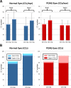
Results: Normal eyes showed a larger SC volume (3.3-fold) and CC orifice area (9962.8 vs. 8825.2 μm(2)) and a similar CC diameter (34.3 ± 17.8 vs. 32.7 ± 13.0 μm) at 10 mm Hg compared to 20 mm Hg. In POAG eyes, SC volume (2.0-fold), CC orifice area (8049.2 μm(2)-6468.4 μm(2)), and CC diameter (36.2 ± 19.1 vs. 29.0 ± 13.8 μm) were increased in 10 mm Hg compared to 20 mm Hg perfusion pressures. Partial and total CC occlusions were present in normal and POAG eyes, with a 3.7-fold increase in total occlusions in POAG eyes compared to normal eyes at 20 mm Hg. Visualization of CCs increased by 24% in normal and by 21% in POAG eyes at 20 mm Hg compared to 10 mm Hg. Schlemm's canal volume, CC area, and CC diameter were decreased in POAG eyes compared to normal eyes at like pressures.
Conclusions: Compensatory mechanisms for transient and short periods of increased pressure appear to be diminished in POAG eyes. Variable response to pressure change in SC and CCs may be a contributing factor to outflow facility change in POAG eyes.
Keywords: POAG; Schlemm's canal; anterior segment; collector channel; glaucoma.
Copyright 2014 The Association for Research in Vision and Ophthalmology, Inc.
Figures
Figure 1
Analysis of Schlemm's canal (SC) and collector channels (CCs) within the distal outflow pathway. (A) Three-dimensional micro-CT reconstruction of a nasal quadrant from one of the primary open-angle glaucoma (POAG) eyes in the study. The quadrant reconstruction is oriented with the corneal endothelial surface up. Ciliary body processes of the pars plicata are visible posterior to Schlemm's canal (SC), which is shown in red. Collector channel orifices are shown in bright aqua and are indicated with black asterisks. A vertical black line indicates where a radial section was removed from the volume for (B). Scale bar: 500 μm. (B) A 6-μm radial section from 3D micro-CT volume shown in (A) at black vertical line. Schlemm's canal lumen is shown in red. Scale bar: 500 μm. TM, trabecular meshwork.
Figure 2
Three-dimensional micro-CT reconstruction of SC and CCs. (A) Three-dimensional reconstruction of 6-μm 3D micro-CT images of a nasal quadrant wedge isolated from a normal eye perfused at 10 mm Hg. Four CCs (aqua and asterisks) were identified in the serpentine-appearing SC (red) anterior to ciliary body (CB). Inset shows CC orifice opening in SC. (B) Three-dimensional reconstruction of 6-μm 3D micro-CT images of a nasal quadrant wedge from the fellow eye in (A) perfused at 20 mm Hg. Schlemm's canal (red) becomes more discontinuous and displays less anastomotic areas at elevated pressure. Eight CCs (aqua and asterisks) are identified in this wedge. (C) Three-dimensional reconstruction of 6-μm 3D micro-CT images of a superior quadrant wedge isolated from a POAG eye perfused at 10 mm Hg. Schlemm's canal appears more discontinuous than in normal eyes perfused at 10 mm Hg, and anastomosing channels are less frequent. Four CC orifices (aqua and asterisks) were identified anterior to ciliary body (CB). (D) Three-dimensional reconstruction of 6-μm 3D micro-CT images of a superior quadrant wedge from the fellow eye in (C) perfused at 20 mm Hg. Schlemm's canal (red) becomes more discontinuous, and adherent areas between inner and outer wall are more prevalent. Five CCs (aqua and asterisks) are identified in this wedge. (A–D) Inset shows magnified representative CC. Scale bars: 1000 μm.
Figure 3
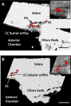
Types of CC orifices. (A) Three-dimensional micro-CT volume of SC with a funnel-shaped CC orifice that extends from middle of SC and travels posteriorly. A portion of the intrascleral parallel vessel (PV) leading away from the orifice is visible. This type of orifice was always observed to be wider initially, then becoming narrower as it extended away from SC. Inset contains a single radial section (6 μm) from the surface of the 3D volume. (B) Three-dimensional micro-CT volume of SC with a tubular-shaped CC orifice that extends a short distance and joins an intrascleral vessel traveling adjacent to SC. Collector channel orifices at SC and at connecting vessel are the same size. Inset contains a single radial 6-μm section from the surface of the volume showing the tubular orifice. Scale bars: 500 μm (A, B).
Figure 4
Collector channels in normal and POAG eyes. (A) Average number of CCs increases in normal (n = 3) and POAG (n = 3) eyes when pressure increases from 10 to 20 mm Hg. Number of open and partially open CCs increases in normal eyes from 10 to 20 mm Hg but does not change in POAG eyes. (B) Number of totally occluded CCs increases nearly 4-fold in POAG eyes at 20 mm Hg compared to normal eyes at 20 mm Hg.
Figure 5
Juxtacanalicular expansion into CC orifice. (A) Three-dimensional micro-CT image (2 μm) showing JCT expansion (asterisk) into CC (imaged from eye perfused at 20 mm Hg). Long arrows indicate boundaries of CC orifice. Inner wall adhesion to outer wall of SC was observed in region just past the orifice (short arrow). (B). Correlative 1-μm plastic section of same region as in (A) with expanded JCT (asterisk), CC orifice (long arrows), and area of adhesion of JCT (short arrow) just past the orifice. Regions similar to this that were adherent in several sections but cleared were evaluated as open CCs. (C) Three-dimensional micro-CT images (2 μm) of an occluded CC (asterisk; imaged from eye perfused at 20 mm Hg), triangular in shape, and its orifice filled with light gray material. Area of adhesion can be seen posterior to occluded CC. (D) Correlative 1-μm toluidine blue–stained plastic section of (C) showing anterior CC (asterisk) filled with light blue occlusive material extending into the sclera. Area of adhesion can be seen posterior to the CC orifice. Ocl CC, occluded collector channel. Scale bars: 50 μm (A–C); 20 μm (D). TM, trabecular meshwork.
Similar articles
- Schlemm's Canal and Trabecular Meshwork in Eyes with Primary Open Angle Glaucoma: A Comparative Study Using High-Frequency Ultrasound Biomicroscopy.
Yan X, Li M, Chen Z, Zhu Y, Song Y, Zhang H. Yan X, et al. PLoS One. 2016 Jan 4;11(1):e0145824. doi: 10.1371/journal.pone.0145824. eCollection 2016. PLoS One. 2016. PMID: 26726880 Free PMC article. - Schlemm's canal and primary open angle glaucoma: correlation between Schlemm's canal dimensions and outflow facility.
Allingham RR, de Kater AW, Ethier CR. Allingham RR, et al. Exp Eye Res. 1996 Jan;62(1):101-9. doi: 10.1006/exer.1996.0012. Exp Eye Res. 1996. PMID: 8674505 - Schlemm's canal and trabecular meshwork morphology in high myopia.
Chen Z, Song Y, Li M, Chen W, Liu S, Cai Z, Chen L, Xiang Y, Zhang H, Wang J. Chen Z, et al. Ophthalmic Physiol Opt. 2018 May;38(3):266-272. doi: 10.1111/opo.12451. Ophthalmic Physiol Opt. 2018. PMID: 29691920 - Aqueous outflow regulation: Optical coherence tomography implicates pressure-dependent tissue motion.
Xin C, Wang RK, Song S, Shen T, Wen J, Martin E, Jiang Y, Padilla S, Johnstone M. Xin C, et al. Exp Eye Res. 2017 May;158:171-186. doi: 10.1016/j.exer.2016.06.007. Epub 2016 Jun 11. Exp Eye Res. 2017. PMID: 27302601 Free PMC article. Review. - Ab interno Schlemm's Canal Surgery.
Francis BA, Akil H, Bert BB. Francis BA, et al. Dev Ophthalmol. 2017;59:127-146. doi: 10.1159/000458492. Epub 2017 Apr 25. Dev Ophthalmol. 2017. PMID: 28442693 Review.
Cited by
- Simultaneous influence of sympathetic autonomic stress on Schlemm's canal, intraocular pressure and ocular circulation.
Chen W, Chen Z, Xiang Y, Deng C, Zhang H, Wang J. Chen W, et al. Sci Rep. 2019 Dec 27;9(1):20060. doi: 10.1038/s41598-019-56562-0. Sci Rep. 2019. PMID: 31882796 Free PMC article. - Expression Profiling of Human Schlemm's Canal Endothelial Cells From Eyes With and Without Glaucoma.
Cai J, Perkumas KM, Qin X, Hauser MA, Stamer WD, Liu Y. Cai J, et al. Invest Ophthalmol Vis Sci. 2015 Oct;56(11):6747-53. doi: 10.1167/iovs.15-17720. Invest Ophthalmol Vis Sci. 2015. PMID: 26567786 Free PMC article. - Canaloplasty in the Treatment of Primary Open-Angle Glaucoma: Patient Selection and Perspectives.
Byszewska A, Konopińska J, Kicińska AK, Mariak Z, Rękas M. Byszewska A, et al. Clin Ophthalmol. 2019 Dec 31;13:2617-2629. doi: 10.2147/OPTH.S155057. eCollection 2019. Clin Ophthalmol. 2019. PMID: 32021062 Free PMC article. Review. - Iridocorneal angle imaging of a human donor eye by spectral-domain optical coherence tomography.
Luo S, Holland G, Khazaeinezhad R, Bradford S, Joshi R, Juhasz T. Luo S, et al. Sci Rep. 2023 Aug 24;13(1):13861. doi: 10.1038/s41598-023-37248-0. Sci Rep. 2023. PMID: 37620338 Free PMC article. - Quantification of Focal Outflow Enhancement Using Differential Canalograms.
Loewen RT, Brown EN, Scott G, Parikh H, Schuman JS, Loewen NA. Loewen RT, et al. Invest Ophthalmol Vis Sci. 2016 May 1;57(6):2831-8. doi: 10.1167/iovs.16-19541. Invest Ophthalmol Vis Sci. 2016. PMID: 27227352 Free PMC article.
References
- Chader GJ. Key needs and opportunities for treating glaucoma. Invest Ophthalmol Vis Sci. 2012; 53: 2456–2460 - PubMed
- Grant WM. Further studies on facility of flow through the trabecular meshwork. AMA Arch Ophthalmol. 1958; 60: 523–533 - PubMed
- Grierson I, Lee WR. Changes in the monkey outflow apparatus at graded levels of intraocular pressure: a qualitative analysis by light microscopy and scanning electron microscopy. Exp Eye Res. 1974; 19: 21–33 - PubMed
Publication types
MeSH terms
Grants and funding
- EY 21727/EY/NEI NIH HHS/United States
- F32 EY007065/EY/NEI NIH HHS/United States
- EY 07065/EY/NEI NIH HHS/United States
- R01 EY021727/EY/NEI NIH HHS/United States
- R01 EY007065/EY/NEI NIH HHS/United States
LinkOut - more resources
Full Text Sources
Other Literature Sources


