Cell microenvironment engineering and monitoring for tissue engineering and regenerative medicine: the recent advances - PubMed (original) (raw)
Review
Cell microenvironment engineering and monitoring for tissue engineering and regenerative medicine: the recent advances
Julien Barthes et al. Biomed Res Int. 2014.
Abstract
In tissue engineering and regenerative medicine, the conditions in the immediate vicinity of the cells have a direct effect on cells' behaviour and subsequently on clinical outcomes. Physical, chemical, and biological control of cell microenvironment are of crucial importance for the ability to direct and control cell behaviour in 3-dimensional tissue engineering scaffolds spatially and temporally. In this review, we will focus on the different aspects of cell microenvironment such as surface micro-, nanotopography, extracellular matrix composition and distribution, controlled release of soluble factors, and mechanical stress/strain conditions and how these aspects and their interactions can be used to achieve a higher degree of control over cellular activities. The effect of these parameters on the cellular behaviour within tissue engineering context is discussed and how these parameters are used to develop engineered tissues is elaborated. Also, recent techniques developed for the monitoring of the cell microenvironment in vitro and in vivo are reviewed, together with recent tissue engineering applications where the control of cell microenvironment has been exploited. Cell microenvironment engineering and monitoring are crucial parts of tissue engineering efforts and systems which utilize different components of the cell microenvironment simultaneously can provide more functional engineered tissues in the near future.
Figures
Figure 1
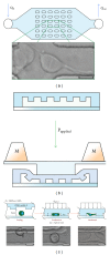
The effect of microlevel mechanical confinement on the division of HeLa cells. (a) and (b) show the macroscopic structure of the microfluidic system and the cross-section of PDMS posts. By the application of pressure on the posts, cells can be confined within the area between the posts (the distance between the posts is 40 _μ_m) (c). The confinement caused significant changes in the behavior of the cells during mitosis, such as delays in mitosis, and led to daughter cells of different sizes and multidaughter cells following mitosis. Reproduced from [4].
Figure 2
Bone formation via endochondral pathway. An in vitro formed artificial cartilage successfully forms a bone containing bone marrow within 12 weeks. The in vitro grown tissue is a cartilaginous one as evidenced by the extensive safranin O staining; over time, the cartilaginous tissue has been gradually replaced by bone tissue, as can be seen by the extensive Masson's Trichrome staining. Micro-CT images also showed the development of a bone like structure within 12 weeks. Reproduced from [8].
Figure 3
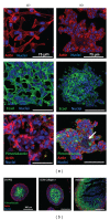
Manipulating the cell microenvironment in 3D via encapsulation within hydrogels. Encapsulation of prostate cancer cells within PEG hydrogels resulted in more pronounced cell-cell contacts as evidenced by E-cadherin staining (a) and also formation of a necrotic core within the cell aggregates as shown by pimonidazole staining (b). All scale bars are 75 _μ_m for (a) and 100 _μ_m for (b). Reproduced from [9].
Figure 4
The main types of soluble factors that have distinct effects on the cellular behaviour at both single cell and tissue level. Controlled delivery of such factors and their regulated presence in cell microenvironment is an indispensable tool in tissue engineering research. Reproduced from [10].
Similar articles
- Nanotopography-guided tissue engineering and regenerative medicine.
Kim HN, Jiao A, Hwang NS, Kim MS, Kang DH, Kim DH, Suh KY. Kim HN, et al. Adv Drug Deliv Rev. 2013 Apr;65(4):536-58. doi: 10.1016/j.addr.2012.07.014. Epub 2012 Aug 18. Adv Drug Deliv Rev. 2013. PMID: 22921841 Free PMC article. Review. - Advances in tissue engineering through stem cell-based co-culture.
Paschos NK, Brown WE, Eswaramoorthy R, Hu JC, Athanasiou KA. Paschos NK, et al. J Tissue Eng Regen Med. 2015 May;9(5):488-503. doi: 10.1002/term.1870. Epub 2014 Feb 3. J Tissue Eng Regen Med. 2015. PMID: 24493315 Review. - Biomolecule delivery to engineer the cellular microenvironment for regenerative medicine.
Bishop CJ, Kim J, Green JJ. Bishop CJ, et al. Ann Biomed Eng. 2014 Jul;42(7):1557-72. doi: 10.1007/s10439-013-0932-1. Epub 2013 Oct 30. Ann Biomed Eng. 2014. PMID: 24170072 Free PMC article. Review. - Nanoscale surfacing for regenerative medicine.
Yang Y, Leong KW. Yang Y, et al. Wiley Interdiscip Rev Nanomed Nanobiotechnol. 2010 Sep-Oct;2(5):478-95. doi: 10.1002/wnan.74. Wiley Interdiscip Rev Nanomed Nanobiotechnol. 2010. PMID: 20803682 Review. - Cell/tissue microenvironment engineering and monitoring in tissue engineering, regenerative medicine, and in vitro tissue models.
Vrana NE, Hasirci V, McGuinness GB, Ndreu-Halili A. Vrana NE, et al. Biomed Res Int. 2014;2014:951626. doi: 10.1155/2014/951626. Epub 2014 Aug 26. Biomed Res Int. 2014. PMID: 25247195 Free PMC article. Review. No abstract available.
Cited by
- A sequential 3D bioprinting and orthogonal bioconjugation approach for precision tissue engineering.
Yu C, Miller KL, Schimelman J, Wang P, Zhu W, Ma X, Tang M, You S, Lakshmipathy D, He F, Chen S. Yu C, et al. Biomaterials. 2020 Nov;258:120294. doi: 10.1016/j.biomaterials.2020.120294. Epub 2020 Aug 9. Biomaterials. 2020. PMID: 32805500 Free PMC article. - Exploiting Matrix Stiffness to Overcome Drug Resistance.
Aydin HB, Ozcelikkale A, Acar A. Aydin HB, et al. ACS Biomater Sci Eng. 2024 Aug 12;10(8):4682-4700. doi: 10.1021/acsbiomaterials.4c00445. Epub 2024 Jul 5. ACS Biomater Sci Eng. 2024. PMID: 38967485 Free PMC article. Review. - Lung cancer stem cells-origin, characteristics and therapy.
Prabavathy D, Swarnalatha Y, Ramadoss N. Prabavathy D, et al. Stem Cell Investig. 2018 Mar 14;5:6. doi: 10.21037/sci.2018.02.01. eCollection 2018. Stem Cell Investig. 2018. PMID: 29682513 Free PMC article. Review. - Accelerated Chondrogenic Differentiation of Human Perivascular Stem Cells with NELL-1.
Li CS, Zhang X, Péault B, Jiang J, Ting K, Soo C, Zhou YH. Li CS, et al. Tissue Eng Part A. 2016 Feb;22(3-4):272-85. doi: 10.1089/ten.TEA.2015.0250. Epub 2016 Jan 27. Tissue Eng Part A. 2016. PMID: 26700847 Free PMC article. - Nanoparticles in tissue engineering: applications, challenges and prospects.
Hasan A, Morshed M, Memic A, Hassan S, Webster TJ, Marei HE. Hasan A, et al. Int J Nanomedicine. 2018 Sep 24;13:5637-5655. doi: 10.2147/IJN.S153758. eCollection 2018. Int J Nanomedicine. 2018. PMID: 30288038 Free PMC article. Review.
References
- Satyam A, Kumar P, Fan X, et al. Macromolecular crowding meets tissue engineering by self-assembly: a paradigm shift in regenerative medicine. Advanced Materials. 2014;26(19):3024–3034. - PubMed
- Fujie T, Mori Y, Ito S, et al. Micropatterned polymeric nanosheets for local delivery of an engineered epithelial monolayer. Advanced Materials. 2014;26:1699–1705. - PubMed
- Metallo CM, Mohr JC, Detzel CJ, de Pablo JJ, van Wie BJ, Palecek SP. Engineering the stem cell microenvironment. Biotechnology Progress. 2007;23(1):18–23. - PubMed
Publication types
MeSH terms
LinkOut - more resources
Full Text Sources
Other Literature Sources


