Computed tomography angiography or magnetic resonance angiography for detection of intracranial vascular malformations in patients with intracerebral haemorrhage - PubMed (original) (raw)
Review
Computed tomography angiography or magnetic resonance angiography for detection of intracranial vascular malformations in patients with intracerebral haemorrhage
Colin B Josephson et al. Cochrane Database Syst Rev. 2014.
Abstract
Background: Intracranial vascular malformations (brain or pial/dural arteriovenous malformations/fistulae, and aneurysms) are the leading cause of intracerebral haemorrhage (ICH) in young adults. Early identification of the intracranial vascular malformation may improve outcome if treatment can prevent ICH recurrence. Catheter intra-arterial digital subtraction angiography (IADSA) is considered the reference standard for the detection an intracranial vascular malformation as the cause of ICH. Computed tomography angiography (CTA) and magnetic resonance angiography (MRA) are less invasive than IADSA and may be as accurate for identifying some causes of ICH.
Objectives: To evaluate the diagnostic test accuracy of CTA and MRA versus IADSA for the detection of intracranial vascular malformations as a cause of ICH.
Search methods: We searched MEDLINE (1948 to August 2013), EMBASE (1980 to August 2013), MEDION (August 2013), the Database of Abstracts of Reviews of Effects (DARE; August 2013), the Health Technology Assessment Database (HTA; August 2013), ClinicalTrials.gov (August 2013), and WHO ICTRP (International Clinical Trials Register Portfolio; August 2013). We also performed a cited reference search for forward tracking of relevant articles on Google Scholar (http://scholar.google.com/), screened bibliographies, and contacted authors to identify additional studies.
Selection criteria: We selected studies reporting data that could be used to construct contingency tables that compared CTA or MRA, or both, with IADSA in the same patients for the detection of intracranial vascular malformations following ICH.
Data collection and analysis: Two authors (CBJ and RA-SS) independently extracted data on study characteristics and measures of test accuracy. Two authors (CBJ and PMW) independently extracted data on test characteristics. We obtained data restricted to the subgroup undergoing IADSA in studies using multiple reference standards. We combined data using the bivariate model. We generated forest plots of the sensitivity and specificity of CTA and MRA and created a summary receiver operating characteristic plot.
Main results: Eleven studies (n = 927 participants) met our inclusion criteria. Eight studies compared CTA with IADSA (n = 526) and three studies compared MRA with IADSA (n = 401). Methodological quality varied considerably among studies, with partial verification bias in 7/11 (64%) and retrospective designs in 5/10 (50%). In studies of CTA, the pooled estimate of sensitivity was 0.95 (95% confidence interval (CI) 0.90 to 0.97) and specificity was 0.99 (95% CI 0.95 to 1.00). The results remained robust in a sensitivity analysis in which only studies evaluating adult patients (≥ 16 years of age) were included. In studies of MRA, the pooled estimate of sensitivity was 0.98 (95% CI 0.80 to 1.00) and specificity was 0.99 (95% CI 0.97 to 1.00). An indirect comparison of CTA and MRA using a bivariate model incorporating test type as one of the parameters failed to reveal a statistically significant difference in sensitivity or specificity between the two imaging modalities (P value = 0.6).
Authors' conclusions: CTA and MRA appear to have good sensitivity and specificity following ICH for the detection of intracranial vascular malformations, although several of the included studies had methodological shortcomings (retrospective designs and partial verification bias in particular) that may have increased apparent test accuracy.
Conflict of interest statement
CBJ, AK, and RA‐SS have no known disclosures to report. Siemens Medical, who make all modalities of imaging equipment, are a minor sponsor of an educational neurointerventional meeting co‐organised by PMW (all of the grant subsidises the course and no fee goes to the organisers).
Figures
1
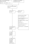
Study flow diagram.
2
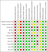
Methodological quality summary: review authors' judgements about each methodological quality item for each included study.
3
Forest plot of the paired sensitivity and specificity values for the detection of an intracranial vascular malformation following intracerebral haemorrhage using computed tomography angiography (CTA) or magnetic resonance angiography (MRA) compared to a reference standard of catheter intra‐arterial digital subtraction angiography.
4
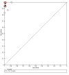
Pooled estimates of sensitivity and specificity for computed tomography angiography (black) and magnetic resonance angiography (red) plotted in receiver operator characteristic space of studies compared with catheter intra‐arterial digital subtraction angiography for the detection of intracranial vascular malformations following intracerebral haemorrhage.
1. Test
CTA.
2. Test
MRA.
Update of
- doi: 10.1002/14651858.CD009372
Similar articles
- Duplex ultrasound for diagnosing symptomatic carotid stenosis in the extracranial segments.
Cassola N, Baptista-Silva JC, Nakano LC, Flumignan CD, Sesso R, Vasconcelos V, Carvas Junior N, Flumignan RL. Cassola N, et al. Cochrane Database Syst Rev. 2022 Jul 11;7(7):CD013172. doi: 10.1002/14651858.CD013172.pub2. Cochrane Database Syst Rev. 2022. PMID: 35815652 Free PMC article. Review. - Diagnostic yield and accuracy of CT angiography, MR angiography, and digital subtraction angiography for detection of macrovascular causes of intracerebral haemorrhage: prospective, multicentre cohort study.
van Asch CJ, Velthuis BK, Rinkel GJ, Algra A, de Kort GA, Witkamp TD, de Ridder JC, van Nieuwenhuizen KM, de Leeuw FE, Schonewille WJ, de Kort PL, Dippel DW, Raaymakers TW, Hofmeijer J, Wermer MJ, Kerkhoff H, Jellema K, Bronner IM, Remmers MJ, Bienfait HP, Witjes RJ, Greving JP, Klijn CJ; DIAGRAM Investigators. van Asch CJ, et al. BMJ. 2015 Nov 9;351:h5762. doi: 10.1136/bmj.h5762. BMJ. 2015. PMID: 26553142 Free PMC article. - Is four-dimensional CT angiography as effective as digital subtraction angiography in the detection of the underlying causes of intracerebral haemorrhage: a systematic review.
Denby CE, Chatterjee K, Pullicino R, Lane S, Radon MR, Das KV. Denby CE, et al. Neuroradiology. 2020 Mar;62(3):273-281. doi: 10.1007/s00234-019-02349-z. Epub 2020 Jan 4. Neuroradiology. 2020. PMID: 31901972 Free PMC article. - The non-invasive detection of intracranial aneurysms: are neuroradiologists any better than other observers?
White PM, Wardlaw JM, Lindsay KW, Sloss S, Patel DK, Teasdale EM. White PM, et al. Eur Radiol. 2003 Feb;13(2):389-96. doi: 10.1007/s00330-002-1520-1. Epub 2002 Jun 28. Eur Radiol. 2003. PMID: 12599005 Clinical Trial. - Intracranial vascular malformations.
Anzalone N, Scomazzoni F, Strada L, Patay Z, Scotti G. Anzalone N, et al. Eur Radiol. 1998;8(5):685-90. doi: 10.1007/s003300050460. Eur Radiol. 1998. PMID: 9601953 Review.
Cited by
- An Unusual Case of Cerebral Arteriovenous Malformation in Pregnancy.
Kow CS, Yang L, Tan WC, Leong RW, Ng YP, Arunachalam S, Kanagalingam D. Kow CS, et al. J Med Cases. 2022 Mar;13(3):104-108. doi: 10.14740/jmc3862. Epub 2022 Mar 5. J Med Cases. 2022. PMID: 35356390 Free PMC article. - Yield of angiographic examinations in isolated intraventricular hemorrhage: A case series and systematic review of the literature.
Hilkens NA, van Asch CJ, Rinkel GJ, Klijn CJ. Hilkens NA, et al. Eur Stroke J. 2016 Dec;1(4):288-293. doi: 10.1177/2396987316666589. Epub 2016 Aug 26. Eur Stroke J. 2016. PMID: 31008290 Free PMC article. - Medical vs. invasive therapy in AVM-related epilepsy: Systematic review and meta-analysis.
Josephson CB, Sauro K, Wiebe S, Clement F, Jette N. Josephson CB, et al. Neurology. 2016 Jan 5;86(1):64-71. doi: 10.1212/WNL.0000000000002240. Epub 2015 Dec 7. Neurology. 2016. PMID: 26643547 Free PMC article. Review. - Thrombolytic removal of intraventricular haemorrhage in treatment of severe stroke: results of the randomised, multicentre, multiregion, placebo-controlled CLEAR III trial.
Hanley DF, Lane K, McBee N, Ziai W, Tuhrim S, Lees KR, Dawson J, Gandhi D, Ullman N, Mould WA, Mayo SW, Mendelow AD, Gregson B, Butcher K, Vespa P, Wright DW, Kase CS, Carhuapoma JR, Keyl PM, Diener-West M, Muschelli J, Betz JF, Thompson CB, Sugar EA, Yenokyan G, Janis S, John S, Harnof S, Lopez GA, Aldrich EF, Harrigan MR, Ansari S, Jallo J, Caron JL, LeDoux D, Adeoye O, Zuccarello M, Adams HP Jr, Rosenblum M, Thompson RE, Awad IA; CLEAR III Investigators. Hanley DF, et al. Lancet. 2017 Feb 11;389(10069):603-611. doi: 10.1016/S0140-6736(16)32410-2. Epub 2017 Jan 10. Lancet. 2017. PMID: 28081952 Free PMC article. Clinical Trial. - Stroke Controversies and Debates: Imaging in Intracerebral Hemorrhage.
Bower MM, Giles JA, Sansing LH, Carhuapoma JR, Woo D. Bower MM, et al. Stroke. 2024 Nov;55(11):2765-2771. doi: 10.1161/STROKEAHA.123.043480. Epub 2024 Oct 2. Stroke. 2024. PMID: 39355925 No abstract available.
References
References to studies included in this review
Delgado Almandoz 2009 {published and unpublished data}
Eshwar Chandra 1998 {published and unpublished data}
- Eshwar Chandra N, Khandelwal N, Bapuraj JR, Mathuriya SN, Vasista RK, Kak VK, et al. Spontaneous intracranial hematomas: role of dynamic CT and angiography. Acta Neurologica Scandinavica 1998;98(3):176‐81. - PubMed
Lummel 2012 {published data only}
- Lummel N, Lutz J, Bruckmann H, Linn J. The value of magnetic resonance imaging for the detection of the bleeding source in non‐traumatic intracerebral haemorrhages: a comparison with conventional digital subtraction angiography. Neuroradiology 2012;54(7):673‐80. [PUBMED: 21918851] - PubMed
Ma 2012 {published data only}
- Ma J, Hao L, You C, Huang S, Ma L, Leong C. Accuracy of computed tomography angiography in detecting the underlying vascular abnormalities for spontaneous intracerebral hemorrhage: a comparative study and meta‐analysis. Neurology India 2012;60(3):299‐303. - PubMed
Murai 1999 {published and unpublished data}
- Murai Y, Takagi R, Ikeda Y, Yamamoto Y, Teramoto A. Three‐dimensional computerized tomography angiography in patients with hyperacute intracerebral hemorrhage. Journal of Neurosurgery 1999;91(3):424‐31. - PubMed
Romero 2009 {published and unpublished data}
- Romero JM, Artunduaga M, Forero NP, Delgado J, Sarfaraz K, Goldstein JN, et al. Accuracy of CT angiography for the diagnosis of vascular abnormalities causing intraparenchymal hemorrhage in young patients. Emergency Radiology 2009;16(3):195‐201. - PubMed
Wong 2010 {published and unpublished data}
- Wong GK, Siu DY, Ahuja AT, King AD, Yu SC, Zhu XL, et al. Comparisons of DSA and MR angiography with digital subtraction angiography in 151 patients with subacute spontaneous intracerebral hemorrhage. Journal of Clinical Neuroscience 2010;17(5):601‐5. - PubMed
Wong 2011 {published data only (unpublished sought but not used)}
- Wong GK, Siu DY, Abrigo JM, Poon WS, Tsang FC, Zhu XL, et al. Computed tomographic angiography and venography for young or nonhypertensive patients with acute spontaneous intracerebral hemorrhage. Stroke 2011;42(1):211‐3. - PubMed
Yeung 2009 {published and unpublished data}
- Yeung R, Ahmad T, Aviv RI, Tilly LN, Fox AJ, Symons SP. Comparison of CTA to DSA in determining the etiology of spontaneous ICH. Canadian Journal of Neurological Sciences 2009;36(2):176‐80. - PubMed
Yoon 2009 {published and unpublished data}
Zhou 2012 {published data only}
- Zhou M‐L, Feng J, Qu T‐R. Comparison of application value of DSA and MRA in diagnosis of subacute spontaneous intracerebral hemorrhage. Journal of Jilin University Medicine Edition 2012;38(6):1209‐13.
References to studies excluded from this review
Awad 1992 {published data only}
- Awad IA, Mckenzie R, Magdinec M, Masaryk T. Application of magnetic resonance angiography to neurosurgical practice: a critical review of 150 cases. Neurological Research 1992;14(5):360‐8. [PUBMED: 1362250] - PubMed
Bekelis 2012 {published data only}
- Bekelis K, Desai A, Zhao W, Gibson D, Gologorsky D, Eskey C, et al. Computed tomography angiography: improving diagnostic yield and cost effectiveness in the initial evaluation of spontaneous nonsubarachnoid intracerebral hemorrhage. Journal of Neurosurgery 2012;117(4):761‐6. [PUBMED: 22880718] - PubMed
Bowen 2007 {published data only}
- Bowen BC. MR angiography versus CT angiography in the evaluation of neurovascular disease. Radiology 2007;245(2):357‐60; discussion 60‐1. [PUBMED: 17940298] - PubMed
Campeau 2012 {published data only}
- Campeau NG, Huston J 3rd. Vascular disorders‐‐magnetic resonance angiography: brain vessels. Neuroimaging Clinics of North America 2012;22(2):207‐33. [PUBMED: 22548929] - PubMed
Chandra 2012 {published data only}
- Chandra T, Pukenas B, Mohan S, Melhem E. Contrast‐enhanced magnetic resonance angiography. Magnetic Resonance Imaging Clinics of North America 2012;20(4):687‐98. [PUBMED: 23088945] - PubMed
Dammert 2004 {published data only}
- Dammert S, Krings T, Moller‐Hartmann W, Ueffing E, Hans FJ, Willmes K, et al. Detection of intracranial aneurysms with multislice CT: comparison with conventional angiography. Neuroradiology 2004;46(6):427‐34. [PUBMED: 15105978] - PubMed
Elhammady 2008 {published data only}
- Elhammady MS, Baskaya MK, Heros RC. Early elective surgical exploration of spontaneous intracerebral hematomas of unknown origin. Journal of Neurosurgery 2008;109(6):1005‐11. [PUBMED: 19035712] - PubMed
Evans 2005 {published data only}
- Evans AL, Coley SC, Wilkinson ID, Griffiths PD. First‐line investigation of acute intracerebral hemorrhage using dynamic magnetic resonance angiography. Acta Radiologica 2005;46(6):625‐30. [PUBMED: 16334846] - PubMed
Fasulakis 2003 {published data only}
- Fasulakis S, Andronikou S. Comparison of MR angiography and conventional angiography in the investigation of intracranial arteriovenous malformations and aneurysms in children. Pediatric Radiology 2003;33(6):378‐84. [PUBMED: 12768254] - PubMed
Griffiths 2006 {published data only}
- Griffiths PD, Wilkinson ID. MR imaging of recent non‐traumatic intracranial hemorrhage: early experience at 3 T. Neuroradiology 2006;48(4):247‐54. [PUBMED: 16489468] - PubMed
Gross 2012 {published data only}
- Gross BA, Frerichs KU, Du R. Sensitivity of CT angiography, T2‐weighted MRI, and magnetic resonance angiography in detecting cerebral arteriovenous malformations and associated aneurysms. Journal of Clinical Neuroscience 2012;19(8):1093‐5. [PUBMED: 22705129] - PubMed
Hünerbein 2003 {published data only}
- Hünerbein R, Klötzer JP, Ladanyi T, Skutta B, Mennel HD, Kailing A, et al. Spontaneous intracerebral bleeding requiring surgery ‐ role of CT angiography in the emergency preoperative diagnosis. Klinische Neuroradiologie 2003;13:194‐202.
Kadkhodayan 2012 {published data only}
- Kadkhodayan Y, Delgado Almandoz JE, Kelly JE, Kale SP, Jagadeesan BD, Moran CJ, et al. Yield of catheter angiography in patients with intracerebral hemorrhage with and without intraventricular extension. Journal of Neurointerventional Surgery 2012;4(5):358‐63. [PUBMED: 21990524] - PubMed
Kamel 2013 {published data only}
- Kamel H, Navi BB, Hemphill JC 3rd. A rule to identify patients who require magnetic resonance imaging after intracerebral hemorrhage. Neurocritical Care 2013;18(1):59‐63. [PUBMED: 21761271] - PubMed
Khosravani 2013 {published data only}
Kidwell 2010 {published data only}
- Kidwell CS, Wintermark M. The role of CT and MRI in the emergency evaluation of persons with suspected stroke. Current Neurology and Neuroscience Reports 2010;10(1):21‐8. [PUBMED: 20425222] - PubMed
Lee 2007 {published data only}
- Lee HJ, Kong MH, Hong HJ, Kang DS, Song KY. The usefulness of 3D‐CT angiography as a screening tool for vascular abnormalities in spontaneous ICH patients. Journal of Korean Neurosurgical Society 2007;41(4):230‐5.
Leung 2012 {published data only}
- Leung KW, Fung KH. Computed tomographic angiography findings in spontaneous intracranial haemorrhage: correlation with digital subtraction angiography. Hong Kong Journal of Radiology 2012;15(2):125‐30.
Li 2013 {published data only}
- Li X‐M, Cao D‐R, She D‐J, Jiang F. 320‐detector row CT angiography in diagnosis of cerebral arteriovenous malformation with hemorrhage. Chinese Journal of Medical Imaging Technology 2013;29(5):697‐700.
Pott 1992 {published data only}
- Pott M, Huber M, Assheuer J, Bewermeyer H. Comparison of MRI, CT and angiography in cerebral arteriovenous malformations. Bildgebung = Imaging 1992;59(2):98‐102. [PUBMED: 1511219] - PubMed
Sasiadek 2000 {published data only}
- Sasiadek M, Hendrich B, Turek T, Kowalewski K, Maksymowicz H. Our own experience with CT angiography in early diagnosis of cerebral vascular malformations. Neurologia i Neurochirurgia Polska 2000;34(6 Suppl):48‐55. [PUBMED: 11452855] - PubMed
Sasiadek 2002 {published data only}
- Sasiadek M, Kowalewski K, Turek T, Hendrich B, Podkowa J, Maksymowicz H. Efficiency of CT‐angiography in the diagnosis of intracranial aneurysms. Medical Science Monitor 2002;8(6):MT99‐MT104. [PUBMED: 12070447] - PubMed
Sha 2008 {published data only}
- Sha L, Bian J, Cheng S‐L, Huang D, Dong J‐D. Compared study of diagnostic value of CE‐MRA and TOF‐MRA in cerebral and cervical aneurysm. Journal of Dalian Medical University 2008;30(1):56‐60.
Truwit 2007 {published data only}
- Truwit CL. CT angiography versus MR angiography in the evaluation of acute neurovascular disease. Radiology 2007;245(2):362‐6. [PUBMED: 17940299] - PubMed
Wijman 2012 {published data only}
- Wijman CA, Snider RW, Venkatasubramanian C, Finley‐Caulfield A, Buckwalter M, Eyngorn I, et al. Diagnostic accuracy of MRI in spontaneous intra‐cerebral hemorrhage (DASH) ‐ final results. Stroke 2012;43:A105.
Zheng 2012 {published data only}
- Zheng T, Wang S, Barras C, Davis S, Yan B. Vascular imaging adds value in investigation of basal ganglia hemorrhage. Journal of Clinical Neuroscience 2012;19(2):277‐80. [PUBMED: 22118795] - PubMed
Additional references
Al‐Shahi 2001
- Al‐Shahi R, Warlow C. A systematic review of the frequency and prognosis of arteriovenous malformations of the brain in adults. Brain 2001;124(Pt 10):1900‐26. [PUBMED: 11571210] - PubMed
Al‐Shahi Salman 2009
- Al‐Shahi Salman R, Labovitz DL, Stapf C. Spontaneous intracerebral haemorrhage. BMJ 2009;339:b2586. [PUBMED: 19633038] - PubMed
Bossuyt 2003
Brazzelli 2009
- Brazzelli M, Sandercock PA, Chappell FM, Celani MG, Righetti E, Arestis N, et al. Magnetic resonance imaging versus computed tomography for detection of acute vascular lesions in patients presenting with stroke symptoms. Cochrane Database of Systematic Reviews 2009, Issue 4. [DOI: 10.1002/14651858.CD007424.pub2] - DOI - PubMed
Chappell 2003
- Chappell ET, Moure FC, Good MC. Comparison of computed tomographic angiography with digital subtraction angiography in the diagnosis of cerebral aneurysms: a meta‐analysis. Neurosurgery 2003;52(3):624‐31; discussion 630‐1. [PUBMED: 12590688] - PubMed
Cordonnier 2010
- Cordonnier C, Klijn CJ, Beijnum J, Al‐Shahi Salman R. Radiological investigation of spontaneous intracerebral hemorrhage: systematic review and trinational survey. Stroke 2010;41(4):685‐90. [PUBMED: 20167915] - PubMed
Deeks 2005
- Deeks JJ, Macaskill P, Irwig L. The performance of tests of publication bias and other sample size effects in systematic reviews of diagnostic test accuracy was assessed. Journal of Clinical Epidemiology 2005;58(9):882‐93. [PUBMED: 16085191] - PubMed
Gluud 2005
Hadizadeh 2008
- Hadizadeh DR, Falkenhausen M, Gieseke J, Meyer B, Urbach H, Hoogeveen R, et al. Cerebral arteriovenous malformation: Spetzler‐Martin classification at subsecond‐temporal‐resolution four‐dimensional MR angiography compared with that at DSA. Radiology 2008;246(1):205‐13. - PubMed
Hino 1998
- Hino A, Fujimoto M, Yamaki T, Iwamoto Y, Katsumori T. Value of repeat angiography in patients with spontaneous subcortical hemorrhage. Stroke 1998;29(12):2517‐21. [PUBMED: 9836762] - PubMed
Lijmer 1999
- Lijmer JG, Mol BW, Heisterkamp S, Bonsel GJ, Prins MH, Meulen JH, et al. Empirical evidence of design‐related bias in studies of diagnostic tests. JAMA 1999;282(11):1061‐6. [PUBMED: 10493205] - PubMed
Lovelock 2007
- Lovelock CE, Molyneux AJ, Rothwell PM. Change in incidence and aetiology of intracerebral haemorrhage in Oxfordshire, UK, between 1981 and 2006: a population‐based study. Lancet Neurology 2007;6(6):487‐93. [PUBMED: 17509483] - PubMed
Macaskill 2010
- Macaskill P, Gatsonis C, Deeks JJ, Harbord RM, Takwoingi Y. Chapter 10: Analysing and presenting results. In: Deeks JJ, Bossuyt PM, Gatsonis C editor(s). Cochrane Handbook for Systematic Reviews of Diagnostic Test Accuracy Version 1.0. The Cochrane Collaboration, 2010.
Mallett 2012
- Mallett S, Halligan S, Thompson M, Collins GS, Altman DG. Interpreting diagnostic accuracy studies for patient care. BMJ 2012;345:e3999. [PUBMED: 22750423] - PubMed
Manninen 2012
RevMan 2012 [Computer program]
- The Nordic Cochrane Centre, The Cochrane Collaboration. Review Manager (RevMan). Version 5.2. Copenhagen: The Nordic Cochrane Centre, The Cochrane Collaboration, 2012.
Rutjes 2006
Scott 2009
- Scott RM, Smith ER. Moyamoya disease and moyamoya syndrome. New England Journal of Medicine 2009;360(12):1226‐37. [PUBMED: 19297575] - PubMed
StataCorp 2011 [Computer program]
- StataCorp LP. Stata Statistical Software: Release 12. College Station, TX, USA: StataCorp LP, 2011.
Taschner 2008
- Taschner CA, Gieseke J, Thuc V, Rachdi H, Reyns N, Gauvrit JY, et al. Intracranial arteriovenous malformation: time‐resolved, contrast‐enhanced MR angiography with combination of parallel imaging, keyhole acquisition, and k‐space sampling techniques at 1.5 T. Radiology 2008;246(3):871‐9. - PubMed
Van Asch 2010
- Asch CJ, Luitse MJ, Rinkel GJ, Tweel I, Algra A, Klijn CJ. Incidence, case fatality, and functional outcome of intracerebral haemorrhage over time, according to age, sex, and ethnic origin: a systematic review and meta‐analysis. Lancet Neurology 2010;9(2):167‐76. [PUBMED: 20056489] - PubMed
Van Beijnum 2009
- Beijnum J, Lovelock CE, Cordonnier C, Rothwell PM, Klijn CJ, Al‐Shahi Salman R. Outcome after spontaneous and arteriovenous malformation‐related intracerebral haemorrhage: population‐based studies. Brain 2009;132(Pt 2):537‐43. [PUBMED: 19042932] - PubMed
Van Gelder 2003
- Gelder JM. Computed tomographic angiography for detecting cerebral aneurysms: implications of aneurysm size distribution for the sensitivity, specificity, and likelihood ratios. Neurosurgery 2003;53(3):597‐605; discussion 605‐6. [PUBMED: 12943576] - PubMed
Westerlaan 2011
- Westerlaan HE, Dijk MJ, Jansen‐van der Weide MC, Groot JC, Groen RJ, Mooij JJ, et al. Intracranial aneurysms in patients with subarachnoid hemorrhage: CT angiography as a primary examination tool for diagnosis‐‐systematic review and meta‐analysis. Radiology 2011;258(1):134‐45. [PUBMED: 20935079] - PubMed
White 2000
- White PM, Wardlaw JM, Easton V. Can noninvasive imaging accurately depict intracranial aneurysms? A systematic review. Radiology 2000;217(2):361‐70. [PUBMED: 11058629] - PubMed
Whiting 2003
Willems 2011
Willems 2012
Wong 2012
- Wong GK, Siu DY, Abrigo JM, Ahuja AT, Poon WS. Computed tomographic angiography for patients with acute spontaneous intracerebral hemorrhage. Journal of Clinical Neuroscience 2012;19(4):498‐500. [PUBMED: 22321368] - PubMed
Publication types
MeSH terms
LinkOut - more resources
Full Text Sources
Other Literature Sources
Medical
Miscellaneous


