Human COL7A1-corrected induced pluripotent stem cells for the treatment of recessive dystrophic epidermolysis bullosa - PubMed (original) (raw)
. 2014 Nov 26;6(264):264ra163.
doi: 10.1126/scitranslmed.3009540.
Hanson Hui Zhen 2, Bahareh Haddad, Elizaveta Bashkirova 3, Sandra P Melo 2, Pei Wang 4, Thomas L Leung 4, Zurab Siprashvili 2, Andrea Tichy 2, Jiang Li 2, Mohammed Ameen 2, John Hawkins 3, Susie Lee 2, Lingjie Li 2, Aaron Schwertschkow 5, Gerhard Bauer 5, Leszek Lisowski 6, Mark A Kay 6, Seung K Kim 4, Alfred T Lane 2, Marius Wernig 7, Anthony E Oro 8
Affiliations
- PMID: 25429056
- PMCID: PMC4428910
- DOI: 10.1126/scitranslmed.3009540
Human COL7A1-corrected induced pluripotent stem cells for the treatment of recessive dystrophic epidermolysis bullosa
Vittorio Sebastiano et al. Sci Transl Med. 2014.
Erratum in
- Sci Transl Med. 2014 Dec 17;6(267):267er8. Derafshi, Bahareh Haddad [corrected to Haddad, Bahareh]
Abstract
Patients with recessive dystrophic epidermolysis bullosa (RDEB) lack functional type VII collagen owing to mutations in the gene COL7A1 and suffer severe blistering and chronic wounds that ultimately lead to infection and development of lethal squamous cell carcinoma. The discovery of induced pluripotent stem cells (iPSCs) and the ability to edit the genome bring the possibility to provide definitive genetic therapy through corrected autologous tissues. We generated patient-derived COL7A1-corrected epithelial keratinocyte sheets for autologous grafting. We demonstrate the utility of sequential reprogramming and adenovirus-associated viral genome editing to generate corrected iPSC banks. iPSC-derived keratinocytes were produced with minimal heterogeneity, and these cells secreted wild-type type VII collagen, resulting in stratified epidermis in vitro in organotypic cultures and in vivo in mice. Sequencing of corrected cell lines before tissue formation revealed heterogeneity of cancer-predisposing mutations, allowing us to select COL7A1-corrected banks with minimal mutational burden for downstream epidermis production. Our results provide a clinical platform to use iPSCs in the treatment of debilitating genodermatoses, such as RDEB.
Copyright © 2014, American Association for the Advancement of Science.
Figures
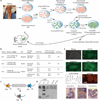
Fig. 1. Derivation and characterization of iPSCs from patients with RDEB
(A) Schematic overview of the protocol in this study. Fibroblasts and keratinocytes were derived and cultured from a skin biopsy, and iPSCs were established from both cell types. iPSCs were then either corrected in their COL7A1 loci by AAV or conventional targeting and differentiated in vitro into keratinocytes (c-iPS-KC), or left uncorrected and directly differentiated into keratinocytes (o-iPS-KC). In vitro-derived keratinocytes (from corrected and noncorrected iPSCs) were used for organotypic cultures and for in vivo skin reconstitution assays in immunocompromised mice. Red cells are uncorrected; green cells are genetically corrected. (B) The patients for which iPSC clones were derived successfully. Information on patients’ specific recessive mutations in COL7A1 locus is provided. NC1 is the immunogenic N-terminal domain of COL7A1. (C) Schematic representation of the lentiviral vector used to reprogram the patients’somatic cells. (D) Southern blot revealing the integration events of the lentiviral reprogramming cassette in somatic cells (fibroblasts and keratinocytes) and in the iPSC clones used in the study (F1-2, K3-1, and K3-4). (E) Characterization of iPSCs [clone F1-2 in (D)] revealing their bona fide undifferentiated and pluripotent state. Expression of a set of markers (OCT4, NANOG, TRA-1 −60, and SSEA3) was identified by immunofluorescence. Normal karyotype was confirmed by G-banding. Pluripotency was assessed by teratoma formation and differentiation into cell derivatives of ectoderm, mesoderm, and endoderm.
Fig. 2. HR-mediated correction of mutations in the COL7A1 locus of RDEB-derived iPSCs
(A to C) Repairing the COL7A 7 locus by conventional targeting (A), with double-negative selection and positive selection by diphtheria toxin (DT). TK, thymidine kinase; Neo, neomycin resistance; PGK, phosphoglycerate kinase I promoter. Exons carrying the mutations are in red; wild-type exons are in green. Enzymes used for Southern blot analysis as well as the location of the probes, represented by black bars, are indicated. (B) Southern blot analysis of representative neomycin-resistant clones obtained in corrected iPSCs (c-iPS) after conventional targeting (CT). KC, keratinocyte; FB, fibroblast. (C) DNA Sanger sequencing of mutant and targeted iPSCs. Mutation is shown as double peaks in the pre-correction sample, denoting the heterozygous nature of the mutations. (D to F) Repairing the COL7A1 locus by HR. (D) AAV-mediated targeting using puromycin (Pur) selection with similar coloring as in (A). (E) Southern blot analysis of representative puromycin-resistant clones. (F) DNA Sanger sequencing showing mutant and corrected sequences, as in (C). (G) Comparison of conventional and AAV-mediated targeting methods.
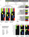
Fig. 3. Variant analysis from both whole-genome sequencing and targeted resequencing
(A) Experimental design to compare the sequence of two sets of patient somatic cells, original iPSCs (o-iPS), and corrected and looped-out iPSCs (c-iPS). (B) Heatmap of variants across keratinocytes (KC, green), fibroblasts (FB, yellow), original iPSC clones (o-iPS, blue), and corrected and Cre-excised iPSC clones (c-iPS, red) from whole-genome sequencing data. Each row represents a variant. Total number of variants for each sample is given at the bottom (with number of genes having variants in parentheses). Black indicates the variants not observed in the sample. (C) Enriched GO terms for each set of variants from whole-genome sequencing. (D) Targeted SCC-predisposing genes for resequencing. (E) Twelve new functional variants across KCs, FBs, o-iPSCs, and c-iPSCs from targeted resequencing data. Total number of variants for each sample is given at the bottom (with number of genes having variants in parentheses). Black indicates the variants not observed.
Fig. 4. Generation of pure, functional keratinocytes from patient-specific iPSCs
(A) Schematic diagram of iPSC differentiation to keratinocytes. (B) Immunofluorescence of K14, K18, p63, and Oct4 in iPS-KCs derived from patient-specific iPSCs (day 60) and in NHK. (C) Representative FACS analysis of K14, K18, and Oct4 in iPS-KCs derived from patient-specific iPSCs compared with NHKs. (D) Microarray analysis of two patient-specific iPSC– derived keratinocytes (iPS-KC1 and iPS-KC3), corresponding to patient keratinocytes AHK1 and AHK3, respectively. These cells were compared with NHKs as well as hES cells (H9). All samples were analyzed in duplicate, and differential gene expression was measured as log2 fold change relative to H9. (E) Volcano plots comparing corrected iPS-KC lines, NHK, and AHK1. Differentially expressed (DE) genes are based on adjusted P ≤ 0.01 [analysis of variance (ANOVA)] and fold change ≥2 (shown in red). (F) Venn diagram of DE genes from iPS-KCI versus NHK (DE = 2217) and from iPS-KC3 versus NHK (DE = 1844). Enriched GO terms of the common DE genes (DE = 1088).
Fig. 5. Keratinocytes derived from patient-specific iPSCs express wild-type collagen VII and stratify in vitro and in vivo
(A) Full-length collagen VII (~300 kD) was expressed in mutation-corrected keratinocytes derived from patient-specific iPSCs (c-iPS-KC1 and c-iPS-KC3) and normal keratinocytes (NHK), but not in keratinocytes from uncorrected iPS cells (o-iPS-KC1 and o-iPS-KC3). (B) Organotypic culture of corrected c-iPS-KCs on a rat type I collagen lattice. Immunofluorescence staining with antibodies to the following differentiation markers: human N-terminal collagen VII (LH7.2), K14, K10, involucrin (Inv), DSG3, E-cadherin (E-Cad), integrin α6 (ITGa6), and laminin 332 (Ln332). (C) Organotypic culture of noncorrected o-iPS-KCs on rat type I collagen lattice with antibodies as in (B). (D) Three-week xenograft of c-iPS-KCs onto nonobese diabetic severe combined immunodeficientc γ (NSG) mice. Top image: Low-power view revealing the corrected epidermis expressing human type VII collagen and K10. All other images show stratification of c-iPS-KC–derived epidermis, using the N-terminal (LH7.2) and C-terminal (LH24) type VII collagen antibodies and the indicated differentiation markers as in (B).
Similar articles
- Genetically corrected iPSCs as cell therapy for recessive dystrophic epidermolysis bullosa.
Wenzel D, Bayerl J, Nyström A, Bruckner-Tuderman L, Meixner A, Penninger JM. Wenzel D, et al. Sci Transl Med. 2014 Nov 26;6(264):264ra165. doi: 10.1126/scitranslmed.3010083. Sci Transl Med. 2014. PMID: 25429058 - Footprint-free gene mutation correction in induced pluripotent stem cell (iPSC) derived from recessive dystrophic epidermolysis bullosa (RDEB) using the CRISPR/Cas9 and piggyBac transposon system.
Itoh M, Kawagoe S, Tamai K, Nakagawa H, Asahina A, Okano HJ. Itoh M, et al. J Dermatol Sci. 2020 Jun;98(3):163-172. doi: 10.1016/j.jdermsci.2020.04.004. Epub 2020 Apr 24. J Dermatol Sci. 2020. PMID: 32376152 - Base Editor Correction of COL7A1 in Recessive Dystrophic Epidermolysis Bullosa Patient-Derived Fibroblasts and iPSCs.
Osborn MJ, Newby GA, McElroy AN, Knipping F, Nielsen SC, Riddle MJ, Xia L, Chen W, Eide CR, Webber BR, Wandall HH, Dabelsteen S, Blazar BR, Liu DR, Tolar J. Osborn MJ, et al. J Invest Dermatol. 2020 Feb;140(2):338-347.e5. doi: 10.1016/j.jid.2019.07.701. Epub 2019 Aug 19. J Invest Dermatol. 2020. PMID: 31437443 Free PMC article. - Gene therapy: pursuing restoration of dermal adhesion in recessive dystrophic epidermolysis bullosa.
Cutlar L, Greiser U, Wang W. Cutlar L, et al. Exp Dermatol. 2014 Jan;23(1):1-6. doi: 10.1111/exd.12246. Exp Dermatol. 2014. PMID: 24107073 Review. - Gene editing toward the use of autologous therapies in recessive dystrophic epidermolysis bullosa.
Perdoni C, Osborn MJ, Tolar J. Perdoni C, et al. Transl Res. 2016 Feb;168:50-58. doi: 10.1016/j.trsl.2015.05.008. Epub 2015 May 27. Transl Res. 2016. PMID: 26073463 Free PMC article. Review.
Cited by
- Context-Dependent Strategies for Enhanced Genome Editing of Genodermatoses.
March OP, Kocher T, Koller U. March OP, et al. Cells. 2020 Jan 2;9(1):112. doi: 10.3390/cells9010112. Cells. 2020. PMID: 31906492 Free PMC article. Review. - Differentiation of Human Induced Pluripotent Stem Cells into Keratinocytes.
Koch PJ, Webb S, Gugger JA, Salois MN, Koster MI. Koch PJ, et al. Curr Protoc. 2022 Apr;2(4):e408. doi: 10.1002/cpz1.408. Curr Protoc. 2022. PMID: 35384405 Free PMC article. - Direct somatic lineage conversion.
Tanabe K, Haag D, Wernig M. Tanabe K, et al. Philos Trans R Soc Lond B Biol Sci. 2015 Oct 19;370(1680):20140368. doi: 10.1098/rstb.2014.0368. Philos Trans R Soc Lond B Biol Sci. 2015. PMID: 26416679 Free PMC article. Review. - Expansion and Purification Are Critical for the Therapeutic Application of Pluripotent Stem Cell-Derived Myogenic Progenitors.
Kim J, Magli A, Chan SSK, Oliveira VKP, Wu J, Darabi R, Kyba M, Perlingeiro RCR. Kim J, et al. Stem Cell Reports. 2017 Jul 11;9(1):12-22. doi: 10.1016/j.stemcr.2017.04.022. Epub 2017 May 18. Stem Cell Reports. 2017. PMID: 28528701 Free PMC article. - Venturing into the New Science of Nucleases.
Tolarová M, McGrath JA, Tolar J. Tolarová M, et al. J Invest Dermatol. 2016 Apr;136(4):742-745. doi: 10.1016/j.jid.2016.01.021. J Invest Dermatol. 2016. PMID: 27012560 Free PMC article.
References
- Fine JD, Bruckner-Tuderman L, Eady RA, Bauer EA, Bauer JW, Has C, Heagerty A, Hintner H, Hovnanian A, Jonkman MF, Leigh I, Marinkovich MP, Martinez AE, McGrath JA, Mellerio JE, Moss C, Murrell DF, Shimizu H, Uitto J, Woodley D, Zambruno G. Inherited epidermolysis bullosa: Updated recommendations on diagnosis and classification. J. Am. Acad. Dermatol. 2014;70:1103–1126. - PubMed
- Arbuckle HA. Epidermolysis bullosa care in the United States. Dermatol. Clin. 2010;28:387–389. - PubMed
- Fine JD, Johnson LB, Suchindran C, Bauer EA, Carter M, McGuire J, Moshell A. In: Epidermolysis Bullosa. Clinical, Epidemiologic and Laboratory Advances and the Findings of the National Epidermolysis Bullosa Registry. Fine JD, Bauer EA, McGuire J, Moshell A, editors. Baltimore, MD: The Johns Hopkins University Press; 1999. pp. 175–192.
- Chen M, Kasahara N, Keene DR, Chan L, Hoeffler WK, Finlay D, Barcova M, Cannon PM, Mazurek C, Woodley DT. Restoration of type VII collagen expression and function in dystrophic epidermolysis bullosa. Nat. Genet. 2002;32:670–675. - PubMed
- Mecklenbeck S, Compton SH, Mejfa JE, Cervini R, Hovnanian A, Bruckner-Tuderman L, Barrandon Y. A microinjected COL7A 7-PAC vector restores synthesis of intact procollagen VII in a dystrophic epidermolysis bullosa keratinocyte cell line. Hum. Gene Ther. 2002;13:1655–1662. - PubMed
Publication types
MeSH terms
Substances
Grants and funding
- R01 AR055914/AR/NIAMS NIH HHS/United States
- R01 HL064274/HL/NHLBI NIH HHS/United States
- HHMI/Howard Hughes Medical Institute/United States
- K08 AR066661/AR/NIAMS NIH HHS/United States
- T32 AR007422/AR/NIAMS NIH HHS/United States
- 5R01 AR055914/AR/NIAMS NIH HHS/United States
LinkOut - more resources
Full Text Sources
Other Literature Sources


