Sodium MRI in human heart: a review - PubMed (original) (raw)
Review
Sodium MRI in human heart: a review
Paul A Bottomley. NMR Biomed. 2016 Feb.
Abstract
This paper offers a critical review of the properties, methods and potential clinical application of sodium ((23)Na) MRI in human heart. Because the tissue sodium concentration (TSC) in heart is about ~40 µmol/g wet weight, and the (23)Na gyromagnetic ratio and sensitivity are respectively about one-quarter and one-11th of that of hydrogen ((1)H), the signal-to-noise ratio of (23)Na MRI in the heart is about one-6000th of that of conventional cardiac (1)H MRI. In addition, as a quadrupolar nucleus, (23)Na exhibits ultra-short and multi-component relaxation behavior (T1 ~ 30 ms; T2 ~ 0.5-4 ms and 12-20 ms), which requires fast, specialized, ultra-short echo-time MRI sequences, especially for quantifying TSC. Cardiac (23)Na MRI studies from 1.5 to 7 T measure a volume-weighted sum of intra- and extra-cellular components present at cytosolic concentrations of 10-15 mM and 135-150 mM in healthy tissue, respectively, at a spatial resolution of about 0.1-1 ml in 10 min or so. Currently, intra- and extra-cellular sodium cannot be unambiguously resolved without the use of potentially toxic shift reagents. Nevertheless, increases in TSC attributable to an influx of intra-cellular sodium and/or increased extra-cellular volume have been demonstrated in human myocardial infarction consistent with prior animal studies, and arguably might also be seen in future studies of ischemia and cardiomyopathies--especially those involving defects in sodium transport. While technical implementation remains a hurdle, a central question for clinical use is whether cardiac (23)Na MRI can deliver useful information unobtainable by other more convenient methods, including (1)H MRI.
Keywords: MRI; T1; T2; heart; myocardial infarction; quantification; sodium; total sodium content; ultra-short echo time.
Copyright © 2015 John Wiley & Sons, Ltd.
Figures
Figure 1
(a) Partial _k_-space sampling scheme for twisted projection imaging (TPI). The maximum k forms a sphere of radius _k_max, and the projections lie on a surface of a cone whose twist is adjusted to maintain critical sample density (56). (b) The TPI pulse sequence with adiabatic half passage excitation. The hard pulse of the original TPI sequence (56) is replaced by an amplitude (amp.) and phase modulated AHP pulse, but the gradient waveforms, Gx, Gy, and Gz, are the same, and depicted for one of the projections (21). (c) Simulated point spread function for T2f =2ms and T2s =15ms signal components excited by a TPI sequence with TE=0.36ms, a 31.3 kHz bandwidth, 12 ms readout, 22cm FOV, 44×44×44 array size, and Δx =5mm nominal isotropic spatial resolution (58). The T2s component has a full-width at half-maximum (fwhm) of about 1.5Δx (7.5 mm), while the fwhm of the T2f component is 2.4Δx (12.0 mm). (Adapted from Refs. 21,56, 58).
Figure 2
Custom-built coils for human cardiac 23Na MRI at 1.5 Tesla (16.9 MHz). (a) Square 25×25 cm three-turn transmit/receive surface coil used for quantitative cardiac TSC studies employing adiabatic excitation (21). The coil has 3 embedded vials of saline gel to serve as sensitivity references, and a mount permitting exchange with a conventional cardiac 1H coil for cardiac MRI (21,22). (b) A 4-channel 23Na phased-array constructed from 15-cm, 3-turns coils for increased inductance at the lower 23Na frequency (46). The detectors were fabricated on 0.25-mm thick flexible printed circuit board, with a separate 40-cm square single-turn transmit coil for excitation.
Figure 3
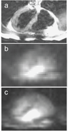
(a) Conventional 1H trans-axial image, and (b) corresponding single slice 23Na MRI of the human heart acquired with the phased-array in Fig. 2(b) using a slice-selective gradient refocused echo (GRE) sequence in 4s, and (c) 200s (array size, 32 × 32; resolution, 1 × 1 × 4 cm3; TR/TE= 33/5 ms; from Ref. 46).
Figure 4
(a) TSC values in infarcted and remote myocardium in 20 MI patients, and in adjacent tissue in 11 of the patients. Vertical bars denote means ±SD. TSC is significantly increased in MI vs remote tissue (P <0.001). (b) Trans-axial gated 1H fast spin-echo image, and (c) corresponding 23Na image from a 3D TPI data set (TR/TE=85/0.4 ms; isotropic spatial resolution =6 mm) from a 61-year-old man with a remote septal MI (arrow). Color scale is proportional to TSC and includes B1 correction; LV=left ventricle. (Adapted from Ref. 22).
Similar articles
- Sodium and quantitative hydrogen parameter changes in muscle tissue after eccentric exercise and in delayed-onset muscle soreness assessed with magnetic resonance imaging.
Höger SA, Gast LV, Marty B, Hotfiel T, Bickelhaupt S, Uder M, Heiss R, Nagel AM. Höger SA, et al. NMR Biomed. 2023 Feb;36(2):e4840. doi: 10.1002/nbm.4840. Epub 2022 Nov 5. NMR Biomed. 2023. PMID: 36196511 - Corrections of myocardial tissue sodium concentration measurements in human cardiac 23 Na MRI at 7 Tesla.
Lott J, Platt T, Niesporek SC, Paech D, G R Behl N, Niendorf T, Bachert P, Ladd ME, Nagel AM. Lott J, et al. Magn Reson Med. 2019 Jul;82(1):159-173. doi: 10.1002/mrm.27703. Epub 2019 Mar 12. Magn Reson Med. 2019. PMID: 30859615 - ECG-gated 23Na-MRI of the human heart using a 3D-radial projection technique with ultra-short echo times.
Jerecic R, Bock M, Nielles-Vallespin S, Wacker C, Bauer W, Schad LR. Jerecic R, et al. MAGMA. 2004 May;16(6):297-302. doi: 10.1007/s10334-004-0038-8. Epub 2004 May 24. MAGMA. 2004. PMID: 15160295 - Sodium MRI: a new frontier in imaging in nephrology.
Francis S, Buchanan CE, Prestwich B, Taal MW. Francis S, et al. Curr Opin Nephrol Hypertens. 2017 Nov;26(6):435-441. doi: 10.1097/MNH.0000000000000370. Curr Opin Nephrol Hypertens. 2017. PMID: 28877041 Review. - Frontiers of Sodium MRI Revisited: From Cartilage to Brain Imaging.
Zaric O, Juras V, Szomolanyi P, Schreiner M, Raudner M, Giraudo C, Trattnig S. Zaric O, et al. J Magn Reson Imaging. 2021 Jul;54(1):58-75. doi: 10.1002/jmri.27326. Epub 2020 Aug 26. J Magn Reson Imaging. 2021. PMID: 32851736 Free PMC article. Review.
Cited by
- Primary hyperaldosteronism induces congruent alterations of sodium homeostasis in different skeletal muscles: a 23Na-MRI study.
Christa M, Hahner S, Köstler H, Bauer WR, Störk S, Weng AM. Christa M, et al. Eur J Endocrinol. 2022 Mar 29;186(5):K33-K38. doi: 10.1530/EJE-22-0074. Eur J Endocrinol. 2022. PMID: 35255003 Free PMC article. - Ultra-high field cardiac MRI in large animals and humans for translational cardiovascular research.
Schreiber LM, Lohr D, Baltes S, Vogel U, Elabyad IA, Bille M, Reiter T, Kosmala A, Gassenmaier T, Stefanescu MR, Kollmann A, Aures J, Schnitter F, Pali M, Ueda Y, Williams T, Christa M, Hofmann U, Bauer W, Gerull B, Zernecke A, Ergün S, Terekhov M. Schreiber LM, et al. Front Cardiovasc Med. 2023 May 15;10:1068390. doi: 10.3389/fcvm.2023.1068390. eCollection 2023. Front Cardiovasc Med. 2023. PMID: 37255709 Free PMC article. - Quantitative sodium MR imaging: A review of its evolving role in medicine.
Thulborn KR. Thulborn KR. Neuroimage. 2018 Mar;168:250-268. doi: 10.1016/j.neuroimage.2016.11.056. Epub 2016 Nov 24. Neuroimage. 2018. PMID: 27890804 Free PMC article. Review. - Hardware and Software Setup for Quantitative 23Na Magnetic Resonance Imaging at 3T: A Phantom Study.
Giovannetti G, Flori A, Martini N, Cademartiri F, Aquaro GD, Pingitore A, Frijia F. Giovannetti G, et al. Sensors (Basel). 2024 Apr 24;24(9):2716. doi: 10.3390/s24092716. Sensors (Basel). 2024. PMID: 38732822 Free PMC article. - First in vivo fluorine-19 magnetic resonance imaging of the multiple sclerosis drug siponimod.
Starke L, Millward JM, Prinz C, Sherazi F, Waiczies H, Lippert C, Nazaré M, Paul F, Niendorf T, Waiczies S. Starke L, et al. Theranostics. 2023 Feb 5;13(4):1217-1234. doi: 10.7150/thno.77041. eCollection 2023. Theranostics. 2023. PMID: 36923535 Free PMC article.
References
- Barclay JA, Harley EJ, Houghton H. Electrolyte content of rat heart atria and ventricles. Circ Res. 1960;8:1264–1267. - PubMed
- Jennings RB, Sommers HM, Kaltenbach JP, West JJ. Electrolyte alterations in acute myocardial ischemic injury. Circ Res. 1964;14:260–264. - PubMed
- Cannon PJ, Maudsley AA, Hilal SK, Simon HE, Cassidy F. Sodium nuclear magnetic resonance imaging of myocardial tissue of dogs after coronary artery occlusion and reperfusion. J Am Coll Cardiol. 1986;7:573–579. - PubMed
Publication types
MeSH terms
Substances
LinkOut - more resources
Full Text Sources
Other Literature Sources
Medical


