Improved data analysis for the MinION nanopore sequencer - PubMed (original) (raw)
Improved data analysis for the MinION nanopore sequencer
Miten Jain et al. Nat Methods. 2015 Apr.
Abstract
Speed, single-base sensitivity and long read lengths make nanopores a promising technology for high-throughput sequencing. We evaluated and optimized the performance of the MinION nanopore sequencer using M13 genomic DNA and used expectation maximization to obtain robust maximum-likelihood estimates for insertion, deletion and substitution error rates (4.9%, 7.8% and 5.1%, respectively). Over 99% of high-quality 2D MinION reads mapped to the reference at a mean identity of 85%. We present a single-nucleotide-variant detection tool that uses maximum-likelihood parameter estimates and marginalization over many possible read alignments to achieve precision and recall of up to 99%. By pairing our high-confidence alignment strategy with long MinION reads, we resolved the copy number for a cancer-testis gene family (CT47) within an unresolved region of human chromosome Xq24.
Conflict of interest statement
COMPETING FINANCIAL INTERESTS
MA is a consultant to Oxford Nanopore Technologies.
Figures
Fig. 1
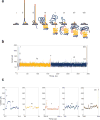
Molecular events and ionic current trace for a 2D read of a 7.25 kb M13 phage dsDNA molecule. (a) Schematic for the steps in DNA translocation through the nanopore. (i) Open channel; (ii) dsDNA with a ligated lead adaptor (blue), with a molecular motor bound to it (orange), and a hairpin adaptor (red), is captured by the nanopore. DNA translocation through the nanopore begins through the effect of an applied voltage across the membrane and the action of a molecular motor; (iii) Translocation of the lead adaptor (blue); (iv) Translocation of the template strand (gold); (v) Translocation of the hairpin adaptor (red); (vi) Translocation of the complement strand (dark blue); (vii) Translocation of the trailing adaptor (brown); (viii) Return to open channel. (b) Raw current trace for the passage of the M13 dsDNA construct through the nanopore. Regions of the ionic current trace corresponding to steps i-viii are labeled. (c) Expanded time and current scale for raw current traces corresponding to steps i–viii. Each adaptor generates a unique current signal used to aid base calling.
Fig. 2
Read length distributions and identity plots for M13. Read length histograms for mapped vs. unmapped reads across three replicate M13 experiments for (a) template; (b) complement; and (c) 2D reads. Most of the reads mapped to a known reference, with two distinct peaks at about 7.2 kb, corresponding to full-length M13, and 3.8 kb, corresponding to the phage lambda DNA (control fragment). Insets show the proportion of mappable vs. unmappable reads and the proportion of unmappable reads that found hits when compared against the NCBI NT database using BLAST (to check for contamination or missed homology). Read alignment identities for mappable reads using tuned LAST, realigned LAST, and EM trained LAST for (d) template; (e) complement; and (f) 2D reads.
Fig. 3
Maximum-likelihood (ML) alignment parameters derived using expectation-maximization (EM). The process starts from four guide alignments each generated with a different mapper using tuned parameters. (a) Insertion vs. deletion rates, expressed as events per aligned base. (b) Insertion or deletion (indel) events per aligned base vs. rate of mismatches per aligned base (see Supplement). Rates vary strongly between different guide alignments, however, EM training and realignment results in very similar rates (grey circles), regardless of the initial guide alignment. (c) Matrix for substitution emissions determined using EM reveals very low rates of A-to-T and T-to-A substitutions.
Fig. 4
M13 sequencing depth. (a) The magenta line denotes coverage by position in the genome, and the dotted blue line depicts local G/C% for that position. Ignoring sites close to the ends of the reference, which appear to be affected by adaptor trimming, the coverage drops at polymeric nucleotide runs. Coverage was calculated by binning over a sliding 5 bp window. G/C content was calculated by binning over a 50 bp sliding window. (b) Coverage depth distribution fitted with a generalized extreme value distribution.
Fig. 5
Exploring single nucleotide variant (SNV) calling with MinION reads. (a) Variant calling with substitution frequency of 1%. (b) Variant calling with substitution frequency of 5%. Dotted lines in both (a) and (b) represent variant calling using a simple transducer model, using a tuned LAST alignment and giving all substitutions equal probability. Different sampled read coverages are shown. Each curve is produced by varying the posterior base calling threshold to trade off precision for recall. Solid lines in both (a) and (b) represent variant calling using the same simple transducer model as in the dotted lines, but trained by EM and incorporating marginalization over the read to reference alignments. Results shown are averaged over three replicate M13 experiments, and for each coverage level, three samplings of the reads. ALL curve reflects all the available data for each experiment. (c) The distribution of posterior match probabilities show that there is substantial uncertainty in most matches and explain why marginalizing over the read alignments is a powerful approach.
Fig. 6
Resolution of CT47 repeat copy number estimate on human chromosome Xq24. (a) BAC end sequence alignments (RP11-482A22: AQ630638 and AZ517599) span a 247 kb region, including thirteen annotated CT47 genes (each defined within a 4.8 kb tandem repeat) and a 50 kb scaffold gap in the GRCh38/hg38 reference assembly. (b) Utilizing MinION long-reads obtained from RP11-482A22 high-molecular weight BAC DNA, nine reads span the length of the CT47-repeat region providing evidence for eight tandem copies of the CT47-repeat. Insert size estimate (170–175 kb, as determined by pulse-field gel electrophoresis) is noted as a dotted line, with flanking regions (upstream: 57 kb and downstream region: 73 kb, black line) and repeat region (37-to-42 kb, or 7.5-to-8.75 copies of the repeat, blue line). Single copy regions directly before the CT47 repeats are shown in orange (6.6 kb) and green (2.6 kb), repeat copies are labeled in blue, and grey lines describe read alignment into flanking region. The size of the repeat region are provided on the left (range 36 kb ’ 42 kb). (c) Shearing the BAC DNA to increase sequence coverage provided copy number estimates by read depth. All bases not included in the CT47 repeat unit are labeled as flanking region (grey distribution, mean: 46.2 base coverage). Base coverage across the CT47 repeats are summarized over one copy of the repeat to provide an estimate of the combined number (dark blue distribution, mean: 329.3 base coverage), and are similar to single copy estimates when normalized for eight copies (light blue distribution, mean: 41.15 base coverage).
Comment in
- Successful test launch for nanopore sequencing.
Loman NJ, Watson M. Loman NJ, et al. Nat Methods. 2015 Apr;12(4):303-4. doi: 10.1038/nmeth.3327. Nat Methods. 2015. PMID: 25825834 No abstract available.
Similar articles
- Oxford Nanopore MinION Sequencing and Genome Assembly.
Lu H, Giordano F, Ning Z. Lu H, et al. Genomics Proteomics Bioinformatics. 2016 Oct;14(5):265-279. doi: 10.1016/j.gpb.2016.05.004. Epub 2016 Sep 17. Genomics Proteomics Bioinformatics. 2016. PMID: 27646134 Free PMC article. Review. - MinION™ nanopore sequencing of environmental metagenomes: a synthetic approach.
Brown BL, Watson M, Minot SS, Rivera MC, Franklin RB. Brown BL, et al. Gigascience. 2017 Mar 1;6(3):1-10. doi: 10.1093/gigascience/gix007. Gigascience. 2017. PMID: 28327976 Free PMC article. - ECNano: A cost-effective workflow for target enrichment sequencing and accurate variant calling on 4800 clinically significant genes using a single MinION flowcell.
Leung AW, Leung HC, Wong CL, Zheng ZX, Lui WW, Luk HM, Lo IF, Luo R, Lam TW. Leung AW, et al. BMC Med Genomics. 2022 Mar 4;15(1):43. doi: 10.1186/s12920-022-01190-3. BMC Med Genomics. 2022. PMID: 35246132 Free PMC article. - Full-Length HLA Class I Genotyping with the MinION Nanopore Sequencer.
Lang K, Surendranath V, Quenzel P, Schöfl G, Schmidt AH, Lange V. Lang K, et al. Methods Mol Biol. 2018;1802:155-162. doi: 10.1007/978-1-4939-8546-3_10. Methods Mol Biol. 2018. PMID: 29858807 - On-Site MinION Sequencing.
Runtuwene LR, Tuda JSB, Mongan AE, Suzuki Y. Runtuwene LR, et al. Adv Exp Med Biol. 2019;1129:143-150. doi: 10.1007/978-981-13-6037-4_10. Adv Exp Med Biol. 2019. PMID: 30968366 Review.
Cited by
- Oxford Nanopore MinION Sequencing and Genome Assembly.
Lu H, Giordano F, Ning Z. Lu H, et al. Genomics Proteomics Bioinformatics. 2016 Oct;14(5):265-279. doi: 10.1016/j.gpb.2016.05.004. Epub 2016 Sep 17. Genomics Proteomics Bioinformatics. 2016. PMID: 27646134 Free PMC article. Review. - Using Nanopore Sequencing to Obtain Complete Bacterial Genomes from Saliva Samples.
Baker JL. Baker JL. mSystems. 2022 Oct 26;7(5):e0049122. doi: 10.1128/msystems.00491-22. Epub 2022 Aug 22. mSystems. 2022. PMID: 35993719 Free PMC article. - A long road/read to rapid high-resolution HLA typing: The nanopore perspective.
Liu C. Liu C. Hum Immunol. 2021 Jul;82(7):488-495. doi: 10.1016/j.humimm.2020.04.009. Epub 2020 May 1. Hum Immunol. 2021. PMID: 32386782 Free PMC article. Review. - De Novo Long-Read Whole-Genome Assemblies and the Comparative Pan-Genome Analysis of Ascochyta Blight Pathogens Affecting Field Pea.
Ogaji YO, Lee RC, Sawbridge TI, Cocks BG, Daetwyler HD, Kaur S. Ogaji YO, et al. J Fungi (Basel). 2022 Aug 22;8(8):884. doi: 10.3390/jof8080884. J Fungi (Basel). 2022. PMID: 36012871 Free PMC article. - Long-Read Sequencing Emerging in Medical Genetics.
Mantere T, Kersten S, Hoischen A. Mantere T, et al. Front Genet. 2019 May 7;10:426. doi: 10.3389/fgene.2019.00426. eCollection 2019. Front Genet. 2019. PMID: 31134132 Free PMC article. Review.
References
- Li H. Aligning sequence reads, clone sequences and assembly contigs with BWA-MEM. 2013;00:3.
- Harris RS. Ph.D. thesis. The Pennsylvania State University; 2007. Improved pairwise alignment of genomic DNA.
Publication types
MeSH terms
Grants and funding
- HG006321/HG/NHGRI NIH HHS/United States
- R01 HG006321/HG/NHGRI NIH HHS/United States
- U54HG007990/HG/NHGRI NIH HHS/United States
- HG007827/HG/NHGRI NIH HHS/United States
- U54 HG007990/HG/NHGRI NIH HHS/United States
- R01 HG007827/HG/NHGRI NIH HHS/United States
LinkOut - more resources
Full Text Sources
Other Literature Sources


