Upregulation of cytosolic phosphoenolpyruvate carboxykinase is a critical metabolic event in melanoma cells that repopulate tumors - PubMed (original) (raw)
. 2015 Apr 1;75(7):1191-6.
doi: 10.1158/0008-5472.CAN-14-2615. Epub 2015 Feb 24.
Shunqun Luo 1, Ruihua Ma 1, Jing Liu 1, Pingwei Xu 1, Huafeng Zhang 2, Ke Tang 2, Jingwei Ma 2, Yi Zhang 2, Xiaoyu Liang 2, Yanling Sun 1, Tiantian Ji 1, Ning Wang 3, Bo Huang 4
Affiliations
- PMID: 25712344
- PMCID: PMC4629827
- DOI: 10.1158/0008-5472.CAN-14-2615
Upregulation of cytosolic phosphoenolpyruvate carboxykinase is a critical metabolic event in melanoma cells that repopulate tumors
Yong Li et al. Cancer Res. 2015.
Abstract
Although metabolic defects have been investigated extensively in differentiated tumor cells, much less attention has been directed to the metabolic properties of stem-like cells that repopulate tumors [tumor-repopulating cells (TRC)]. Here, we show that melanoma TRCs cultured in three-dimensional soft fibrin gels reprogram glucose metabolism by hijacking the cytosolic enzyme phosphoenolpyruvate carboxykinase (PCK1), a key player in gluconeogenesis. Surprisingly, upregulated PCK1 in TRCs did not mediate gluconeogenesis but promoted glucose side-branch metabolism, including in the serine and glycerol-3-phosphate pathways. Moreover, this retrograde glucose carbon flow strengthened rather than antagonized glycolysis and glucose consumption. Silencing PCK1 or inhibiting its enzymatic activity slowed the growth of TRCs in vitro and impeded tumorigenesis in vivo. Overall, our work unveiled metabolic features of TRCs in melanoma that have implications for targeting a unique aspect of this disease.
©2015 American Association for Cancer Research.
Conflict of interest statement
Disclosure of Potential Conflicts of Interest
B. Huang was supported by Soundny (Sheng-Qi-An) Biotech. All other authors declare no competing financial interests.
Figures
Figure 1
The expression of PCK1 is upregulated in tumor-repopulating cells (TRCs). A, The expression of PCK1 in H22 hepatocarcinoma, B16 melanoma and EL4 lymphoma TRCs as well as murine embryonic stem cells (mESCs) and mesenchymal stem cells (mMSCs) was analyzed by RT-PCR. Tumor cells cultured in 2D rigid dish were used as control. Data shown are representative of three independent experiments. B, The expression of PCK1 in H22, B16 and EL4 TRCs was analyzed by real-time PCR. Tumor cells cultured in 2D rigid dish were used as control. Results represent means ± SEM from three independent experiences, *, p<0.05; ***, p<0.001. C, PCK1 promoter-EGFP-expressing B16 cells were cultured in 3D soft fibrin gels for five days to form TRC spheroids and the fluorescence of spheroids was measured. Then, these cultured TRCs were seeded in conventional rigid dish for further culture. The fluorescence was measured at different time points. Scale bar = 20 μm. Data shown are representative of three independent experiments. D, The expression of PCK1 was analyzed by western blot. Tumor cells cultured in 2D rigid dish were used as control. Data shown are representative of three independent experiments.
Figure 2
PCK1 regulates carbon flow of glucose in B16 TRCs. A, B16 TRCs and normal B16 cells (105 each) were cultured in conventional rigid plate with the same culture medium. The glucose concentration in the supernatant was measured at the beginning and after 12h culture. The glucose consumption rate was calculated correspondingly (left). In parallel, B16 TRCs were transfected with two PCK1 siRNAs or control siRNA and cultured in rigid plates. Similarly, glucose consumption rate was measured (right). Results were normalized as relative consumption against negative control siRNA. B, B16 TRCs and normal B16 cells (105 each) were cultured in conventional rigid plate. The lactate concentration in the supernatant was measured at the beginning and after 12h culture. The production of lactate was calculated correspondingly (left). Lactate production by B16 TRCs after PCK1 knockdown was calculated (right). Results were normalized as relative production against control siRNA. C, TRCs were transfected with two PCK1 siRNAs or control siRNA and cultured in soft 3D fibrin gels for 36 hours. The intracellular L-serine (left) and glycine (right) were analyzed by HLPC. Results were normalized as relative levels against control siRNA. D, detection of intracellular glycerol-3-phosphate (G-3-P) levels in B16 TRCs and control normal B16 cells (left). TRCs were transfected with PCK1 or control siRNA and cultured in soft 3D fibrin gels for 36 hours. G-3-P levels in TRCs were analyzed (right). Results were normalized as relative levels against control siRNA. All above data represent means ± SEM from four independent experiences, *, p<0.05; **, p<0.01; ***, p<0.001.
Figure 3
PCK1 promotes B16 TRC growth in vitro and to form a tumor in vivo. A, PCK1 knockdown inhibited TRC growth. B16 TRCs were transfected with two PCK1 siRNAs or control siRNA and cultured in soft 3D fibrin gels. 5 days later, the colony size (left) and colony number (right) of TRCs were measured. Data are mean ± SEM from three separate experiments. *, p<0.05; ***, p<0.001. B, PCK1-overexpressing (PCK1-OE) and mock control B16 cells were cultured in soft 3D fibrin gels. 5 days later, the colony size (left) and colony number (right) of TRCs were measured. 1st generation: cells were culture up to Day5 in 3D fibrin gels .2nd generation: the cells from 1st generation were harvested and cultured for another 5 days in soft gels. Data are mean ± SEM from three separate experiments. *, p<0.05; ***, p<0.001. C, Inhibition of PCK1 activity resulted in TRC enrichment. Bulk B16 cells were continually treated with 0.05 mM 3MPA. The cells (1,250) from different time points were seeded back to 3D soft fibrin gels. The colony number was counted. Data are mean ± SEM from three separate experiments. *, p<0.05; ***, p<0.001. D, knockdown of PCK1 impaired tumor-repopulating ability of B16 TRCs. 5×10² PCK1 or control siRNA-transfected B16 TRCs were i.v. or s.c. injected to mice. 21 days later, the lung metastasis was shown (left) and the number of metastatic nodules was counted (middle). The volume of skin melanoma was measured (right). Data shown are representative of three independent experiments. Error bars represent means ± SEM, *, p<0.05; **, p<0.01.
Figure 4
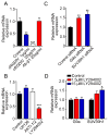
The expression of PCK1 in B16 TRCs is regulated by αvβ3/PI3K signaling pathway. A, B16 TRCs were cultured in the presence of αvβ3 integrin-antagonizing oligopeptide cyclo-(Arg-Gly-Asp-D-Phe-Val, cRGDfV) or β1 blocking antibody. The expression of PCK1 was detected by real time PCR. B, B16 TRCs were treated with 20 μM U0126 (ERK1/2 inhibitor), 5 mM AKTi1/2 (AKT kinase1/2 inhibitor) or 15 μM LY294002 (PI3K inhibitor) for 12 hours. The expression of PCK1 was detected by real time PCR. C, B16 TRCs were transfected with G9a, SUV39h1 or control siRNA and the expression of PCK1 was detected by real time PCR. D, B16 TRCs were treated with LY294002 and the expression of G9a and SUV39h1 mRNA was detected by real-time PCR. All data represent means ± SEM from three independent experiences, *, p<0.05; **, p<0.01; ***, p<0.001.
Similar articles
- Downregulation of PCK2 remodels tricarboxylic acid cycle in tumor-repopulating cells of melanoma.
Luo S, Li Y, Ma R, Liu J, Xu P, Zhang H, Tang K, Ma J, Liu N, Zhang Y, Sun Y, Ji T, Liang X, Yin X, Liu Y, Tong W, Niu Y, Wang N, Wang X, Huang B. Luo S, et al. Oncogene. 2017 Jun 22;36(25):3609-3617. doi: 10.1038/onc.2016.520. Epub 2017 Feb 6. Oncogene. 2017. PMID: 28166201 - Hypoxia Promotes Breast Cancer Cell Growth by Activating a Glycogen Metabolic Program.
Tang K, Zhu L, Chen J, Wang D, Zeng L, Chen C, Tang L, Zhou L, Wei K, Zhou Y, Lv J, Liu Y, Zhang H, Ma J, Huang B. Tang K, et al. Cancer Res. 2021 Oct 1;81(19):4949-4963. doi: 10.1158/0008-5472.CAN-21-0753. Epub 2021 Aug 4. Cancer Res. 2021. PMID: 34348966 - Low Levels of Sox2 are required for Melanoma Tumor-Repopulating Cell Dormancy.
Jia Q, Yang F, Huang W, Zhang Y, Bao B, Li K, Wei F, Zhang C, Jia H. Jia Q, et al. Theranostics. 2019 Jan 1;9(2):424-435. doi: 10.7150/thno.29698. eCollection 2019. Theranostics. 2019. PMID: 30809284 Free PMC article. - Gluconeogenesis in cancer cells - Repurposing of a starvation-induced metabolic pathway?
Grasmann G, Smolle E, Olschewski H, Leithner K. Grasmann G, et al. Biochim Biophys Acta Rev Cancer. 2019 Aug;1872(1):24-36. doi: 10.1016/j.bbcan.2019.05.006. Epub 2019 May 30. Biochim Biophys Acta Rev Cancer. 2019. PMID: 31152822 Free PMC article. Review. - What is the metabolic role of phosphoenolpyruvate carboxykinase?
Yang J, Kalhan SC, Hanson RW. Yang J, et al. J Biol Chem. 2009 Oct 2;284(40):27025-9. doi: 10.1074/jbc.R109.040543. Epub 2009 Jul 27. J Biol Chem. 2009. PMID: 19636077 Free PMC article. Review. No abstract available.
Cited by
- Research progress of tumor-derived extracellular vesicles in the treatment of malignant pleural effusion.
Zhang S, Chen L, Zong Y, Li Q, Zhu K, Li Z, Meng R. Zhang S, et al. Cancer Med. 2023 Jan;12(2):983-994. doi: 10.1002/cam4.5005. Epub 2022 Jul 21. Cancer Med. 2023. PMID: 35861052 Free PMC article. Review. - Dysregulation of metabolic enzymes in tumor and stromal cells: Role in oncogenesis and therapeutic opportunities.
Khan MA, Zubair H, Anand S, Srivastava SK, Singh S, Singh AP. Khan MA, et al. Cancer Lett. 2020 Mar 31;473:176-185. doi: 10.1016/j.canlet.2020.01.003. Epub 2020 Jan 7. Cancer Lett. 2020. PMID: 31923436 Free PMC article. Review. - Pancreatic Ductal Adenocarcinoma Cortical Mechanics and Clinical Implications.
Angstadt S, Zhu Q, Jaffee EM, Robinson DN, Anders RA. Angstadt S, et al. Front Oncol. 2022 Jan 31;12:809179. doi: 10.3389/fonc.2022.809179. eCollection 2022. Front Oncol. 2022. PMID: 35174086 Free PMC article. Review. - O-GlcNAc modified-TIP60/KAT5 is required for PCK1 deficiency-induced HCC metastasis.
Liu R, Gou D, Xiang J, Pan X, Gao Q, Zhou P, Liu Y, Hu J, Wang K, Tang N. Liu R, et al. Oncogene. 2021 Dec;40(50):6707-6719. doi: 10.1038/s41388-021-02058-z. Epub 2021 Oct 14. Oncogene. 2021. PMID: 34650217 Free PMC article. - Downregulation of PCK2 remodels tricarboxylic acid cycle in tumor-repopulating cells of melanoma.
Luo S, Li Y, Ma R, Liu J, Xu P, Zhang H, Tang K, Ma J, Liu N, Zhang Y, Sun Y, Ji T, Liang X, Yin X, Liu Y, Tong W, Niu Y, Wang N, Wang X, Huang B. Luo S, et al. Oncogene. 2017 Jun 22;36(25):3609-3617. doi: 10.1038/onc.2016.520. Epub 2017 Feb 6. Oncogene. 2017. PMID: 28166201
References
- Piccirillo SG, Reynolds BA, Zanetti N, et al. Bone morphogenetic proteins inhibit the tumorigenic potential of human brain tumour-initiating cells. Nature. 2006;444:761–5. - PubMed
- Schepers AG, Snippert HJ, Stange DE, et al. Lineage tracing reveals Lgr5+ stem cell activity in mouse intestinal adenomas. Science. 2012;337:730–5. - PubMed
Publication types
MeSH terms
Substances
LinkOut - more resources
Full Text Sources
Other Literature Sources


