Imaging Chronic Tuberculous Lesions Using Sodium [(18)F]Fluoride Positron Emission Tomography in Mice - PubMed (original) (raw)
Imaging Chronic Tuberculous Lesions Using Sodium [(18)F]Fluoride Positron Emission Tomography in Mice
Alvaro A Ordonez et al. Mol Imaging Biol. 2015 Oct.
Abstract
Purpose: Calcification is a hallmark of chronic tuberculosis (TB) in humans, often noted years to decades (after the initial infection) on chest radiography, but not visualized well with traditional positron emission tomography (PET). We hypothesized that sodium [(18)F]fluoride (Na[(18)F]F) PET could be used to detect microcalcifications in a chronically Mycobacterium tuberculosis-infected murine model.
Procedures: C3HeB/FeJ mice, which develop necrotic and hypoxic TB lesions, were aerosol-infected with M. tuberculosis and imaged with Na[(18)F]F PET.
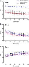
Results: Pulmonary TB lesions from chronically infected mice demonstrated significantly higher Na[(18)F]F uptake compared with acutely infected or uninfected animals (P < 0.01), while no differences were noted in the blood or bone compartments (P > 0.08). Ex vivo biodistribution studies confirmed the imaging findings, and tissue histology demonstrated microcalcifications in TB lesions from chronically infected mice, which has not been demonstrated previously in a murine model.
Conclusion: Na[(18)F]F PET can be used for the detection of chronic TB lesions and could prove to be a useful noninvasive biomarker for TB studies.
Keywords: Chronic; Microcalcification; Na[18F]F; PET; Tuberculosis.
Conflict of interest statement
Conflict of Interest. None of the authors report any financial or potential conflicts of interest.
Figures
Fig. 1
Transverse, coronal, and sagittal sections from Na[18F]F PET/CT imaging performed on a chronically _M. tuberculosis_-infected, b acutely _M. tuberculosis_-infected, and c uninfected mice, 40 min post tracer injection. Radio-densities demonstrating TB lesions are clearly visible in the lungs of infected mice (a and b), but Na[18F]F PET signal is only noted in the chronically infected mice (arrows). Na[18F]F PET signal is also noted in bones and the urinary bladder (Bl). H heart.
Fig. 2
Dynamic Na[18F]F PET imaging demonstrates significantly higher signal in a pulmonary lesions from chronically infected mice (blue) compared with acutely infected (red) or uninfected (dotted black) animals. No difference is evident in b the blood or c bone compartments amongst the three different groups. Three animals were imaged for each group. Data is represented as median and interquartile range.
Fig. 3
Ex vivo biodistribution studies performed after completion of imaging. Na[18F]F uptake is significantly higher in the chronically infected lung tissues, while uptake is similar amongst the three different groups in other tissue compartments. Three animals were used for each group. Data is represented as median and interquartile range.
Fig. 4
Histology sections from chronic (left panels) and acutely infected (middle panels) mice and uninfected (right panels) controls are shown. H&E staining (a), von Kossa staining for calcium phosphate deposits in black (b and c), Alizarin Red with calcium deposits visualized in red (d), and acid-fast staining for M. tuberculosis bacilli (e). Well-defined necrotic granulomas are noted on H&E staining in the _M. tuberculosis_-infected (acute and chronic) tissues. Von Kossa and Alizarin Red staining demonstrate deposits only in TB lesions from chronically infected tissues (b–d). AFB staining demonstrates large numbers of bacilli in infected (acute and chronic) tissues (e).
Fig. 5
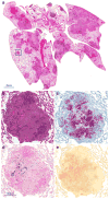
a Histology demonstrating pulmonary lesions in a _M. tuberculosis_-infected mouse with chronic infection. Higher magnification of a TB lesion (inset) demonstrates a classic granulomatous lesion with central necrosis (b) and a large number of M. tuberculosis bacilli seen with AFB staining (c). Microcalcifications represented as black and red deposits are evident on von Kossa (d) and Alizarin Red staining (e), respectively.
Similar articles
- Noninvasive pulmonary [18F]-2-fluoro-deoxy-D-glucose positron emission tomography correlates with bactericidal activity of tuberculosis drug treatment.
Davis SL, Nuermberger EL, Um PK, Vidal C, Jedynak B, Pomper MG, Bishai WR, Jain SK. Davis SL, et al. Antimicrob Agents Chemother. 2009 Nov;53(11):4879-84. doi: 10.1128/AAC.00789-09. Epub 2009 Sep 8. Antimicrob Agents Chemother. 2009. PMID: 19738022 Free PMC article. - Identifying active vascular microcalcification by (18)F-sodium fluoride positron emission tomography.
Irkle A, Vesey AT, Lewis DY, Skepper JN, Bird JL, Dweck MR, Joshi FR, Gallagher FA, Warburton EA, Bennett MR, Brindle KM, Newby DE, Rudd JH, Davenport AP. Irkle A, et al. Nat Commun. 2015 Jul 7;6:7495. doi: 10.1038/ncomms8495. Nat Commun. 2015. PMID: 26151378 Free PMC article. - Molecular imaging in oncology: (18)F-sodium fluoride PET imaging of osseous metastatic disease.
Mick CG, James T, Hill JD, Williams P, Perry M. Mick CG, et al. AJR Am J Roentgenol. 2014 Aug;203(2):263-71. doi: 10.2214/AJR.13.12158. AJR Am J Roentgenol. 2014. PMID: 25055258 Review. - [18F] Sodium Fluoride PET Kinetic Parameters in Bone Imaging.
Puri T, Frost ML, Cook GJ, Blake GM. Puri T, et al. Tomography. 2021 Dec 1;7(4):843-854. doi: 10.3390/tomography7040071. Tomography. 2021. PMID: 34941643 Free PMC article. Review.
Cited by
- Molecular Imaging of Pulmonary Inflammation and Infection.
Giraudo C, Evangelista L, Fraia AS, Lupi A, Quaia E, Cecchin D, Casali M. Giraudo C, et al. Int J Mol Sci. 2020 Jan 30;21(3):894. doi: 10.3390/ijms21030894. Int J Mol Sci. 2020. PMID: 32019142 Free PMC article. Review. - Determination of [11C]rifampin pharmacokinetics within Mycobacterium tuberculosis-infected mice by using dynamic positron emission tomography bioimaging.
DeMarco VP, Ordonez AA, Klunk M, Prideaux B, Wang H, Zhuo Z, Tonge PJ, Dannals RF, Holt DP, Lee CK, Weinstein EA, Dartois V, Dooley KE, Jain SK. DeMarco VP, et al. Antimicrob Agents Chemother. 2015 Sep;59(9):5768-74. doi: 10.1128/AAC.01146-15. Epub 2015 Jul 13. Antimicrob Agents Chemother. 2015. PMID: 26169396 Free PMC article. - New methods to image unstable atherosclerotic plaques.
Andrews JPM, Fayad ZA, Dweck MR. Andrews JPM, et al. Atherosclerosis. 2018 May;272:118-128. doi: 10.1016/j.atherosclerosis.2018.03.021. Epub 2018 Mar 14. Atherosclerosis. 2018. PMID: 29602139 Free PMC article. Review. - Mouse models of human TB pathology: roles in the analysis of necrosis and the development of host-directed therapies.
Kramnik I, Beamer G. Kramnik I, et al. Semin Immunopathol. 2016 Mar;38(2):221-37. doi: 10.1007/s00281-015-0538-9. Epub 2015 Nov 5. Semin Immunopathol. 2016. PMID: 26542392 Free PMC article. Review. - Visualizing the dynamics of tuberculosis pathology using molecular imaging.
Ordonez AA, Tucker EW, Anderson CJ, Carter CL, Ganatra S, Kaushal D, Kramnik I, Lin PL, Madigan CA, Mendez S, Rao J, Savic RM, Tobin DM, Walzl G, Wilkinson RJ, Lacourciere KA, Via LE, Jain SK. Ordonez AA, et al. J Clin Invest. 2021 Mar 1;131(5):e145107. doi: 10.1172/JCI145107. J Clin Invest. 2021. PMID: 33645551 Free PMC article. Review.
References
- WHO Global tuberculosis report. [Accessed 21 November];2014 http://www.who.int/tb/publications/global_report/gtbr14_executive_summar....
- Robbins SL, Kumar V. Robbins and Cotran pathologic basis of disease. 8th. Saunders/Elsevier; Philadelphia: 2010.
- Sathekge M, Maes A, Kgomo M, et al. Use of 18F-FDG PET to predict response to first-line tuberculostatics in HIV-associated tuberculosis. J Nucl Med. 2011;52:880–885. - PubMed
Publication types
MeSH terms
Substances
Grants and funding
- DP2 OD006492/OD/NIH HHS/United States
- R01-HL116316/HL/NHLBI NIH HHS/United States
- R01 EB020539/EB/NIBIB NIH HHS/United States
- R01-EB020539/EB/NIBIB NIH HHS/United States
- DP2-OD006492/OD/NIH HHS/United States
- R01 HL116316/HL/NHLBI NIH HHS/United States
LinkOut - more resources
Full Text Sources
Other Literature Sources
Medical
Molecular Biology Databases


