TGFβ Is a Master Regulator of Radiation Therapy-Induced Antitumor Immunity - PubMed (original) (raw)
TGFβ Is a Master Regulator of Radiation Therapy-Induced Antitumor Immunity
Claire Vanpouille-Box et al. Cancer Res. 2015.
Abstract
T cells directed to endogenous tumor antigens are powerful mediators of tumor regression. Recent immunotherapy advances have identified effective interventions to unleash tumor-specific T-cell activity in patients who naturally develop them. Eliciting T-cell responses to a patient's individual tumor remains a major challenge. Radiation therapy can induce immune responses to model antigens expressed by tumors, but it remains unclear whether it can effectively prime T cells specific for endogenous antigens expressed by poorly immunogenic tumors. We hypothesized that TGFβ activity is a major obstacle hindering the ability of radiation to generate an in situ tumor vaccine. Here, we show that antibody-mediated TGFβ neutralization during radiation therapy effectively generates CD8(+) T-cell responses to multiple endogenous tumor antigens in poorly immunogenic mouse carcinomas. Generated T cells were effective at causing regression of irradiated tumors and nonirradiated lung metastases or synchronous tumors (abscopal effect). Gene signatures associated with IFNγ and immune-mediated rejection were detected in tumors treated with radiation therapy and TGFβ blockade in combination but not as single agents. Upregulation of programmed death (PD) ligand-1 and -2 in neoplastic and myeloid cells and PD-1 on intratumoral T cells limited tumor rejection, resulting in rapid recurrence. Addition of anti-PD-1 antibodies extended survival achieved with radiation and TGFβ blockade. Thus, TGFβ is a fundamental regulator of radiation therapy's ability to generate an in situ tumor vaccine. The combination of local radiation therapy with TGFβ neutralization offers a novel individualized strategy for vaccinating patients against their tumors.
©2015 American Association for Cancer Research.
Figures
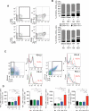
Figure 1. TGFβ blockade with RT enhances TIDC activation and induces CD8+ T cells responses to endogenous tumor antigens
(A) Treatment schema. (B-C) Analysis of 4T1 TIDC at day 22 (n=9/group). To obtain sufficient material, 3 tumors were pooled to obtain 3 independent samples for each group. Viable cells were gated on CD45+CD11c+IAd+ TIDC and analyzed for expression of activation markers CD40 and CD70. (B) Representative dot plots. (C) Bar graphs showing significant increase in mean percentage of TIDC expressing CD40 and CD70 in tumors of mice treated with RT+1D11 (P<0.005 compared to all other groups). (D-H) IFNγ production by CD8+ T cells from LN draining 4T1 (D-G) or TSA (H) tumors in response to peptides derived from survivin (D and E, closed circles), Twist (F, closed squares,), and gp70 (AH1A5) (G and H, closed triangles) or irrelevant peptide (open symbols). Each symbol represents one animal. Horizontal lines indicate the mean of antigen-specific (solid lines) or control (dashed lines) IFNγ concentration. Data are representative of three independent experiments. **p<0.005; ***p<0.0005; ****p<0.00005.
Figure 2. TGFβ blockade with RT inhibits irradiated tumor and non-irradiated metastases
(A) Tumor models and treatment schema. (B, C) Tumor volume overtime (B) and lung metastases quantified at day 28 (C) in 4T1 tumor-bearing mice treated with Sham+Isotype (n=9, open squares), Sham+1D11 (n=8, grey circles), RT+Isotype (n=9, open circles) and RT+1D11 (n=9, black squares). Data are representative of three independent experiments. (D, E) Tumor volume overtime of primary (right flank) (D) and secondary (left flank) (E) TSA tumors in mice treated with Sham+Isotype (n=21, open squares), Sham+1D11 (n=21, grey circles), RT (primary only) +Isotype (n=20, open circles) and RT (primary only) +1D11 (n=21, black squares). Data are representative of two experiments. *p<0.05; **p<0.005; ***p<0.0005.
Figure 3. TGFβ blockade with RT activates immune-mediated tissue rejection pathways
(A) Experimental schema. Genome-wide analysis of RNA from 4T1 tumor of mice treated as indicated in Figure 1 and harvested at day 22 (n=3/group). (B) Heat map showing the top 500 genes selectively upregulated in RT+1D11 tumors. (C) Top 3 self-organizing networks according to IPA representing schematic relationship among genes upregulated in RT+1D11 tumors. Upregulated genes and gene complexes are in red, while no color fill designates genes that are part of the network but not of the gene list. Bold lines indicate direct interaction. Dotted lines indicate indirect interaction.
Figure 4. The therapeutic effect of local RT and TGFβ blockade is T cell-dependent
4T1 primary (A, B) and TSA secondary (C) tumors of mice treated as indicated in Figure 1 were harvested at day 22 (n=3/group) and stained for T cell markers. (A) Representative fields (x200) showing CD8+ T cells (red). Nuclei were stained with DAPI (blue). Mean number ± SD of CD4+ (white bars) and CD8+ (black bars) cells/field in 3 mice/group in 4T1 (B) and TSA (C) tumors. (D) Treatment schema for depletion of CD4+ or CD8+ T cells in 4T1 tumor-bearing mice treated with RT and TGFβ blockade. (E) Tumor volume and (F) lung metastases at day 28 in mice treated with Sham+Isotype (open squares, n=7), Sham+1D11 (grey circles, n=7), RT+Isotype (open circles, dashed black line), RT+1D11 (red circle, red line, n=7), RT+1D11+CD4 depletion (red triangles, dashed red line, n=7) and RT+1D11+CD8 depletion (open red diamond, dashed and dotted red line, n=7). Data are representative of two independent experiments. (G) Representative photographs of lungs. *p<0.05; **p<0.005; ***p<0.0005; ****p<0.00005.
Figure 5. Enhanced expression of PD-1 and its ligands in tumors treated with TGFβ blockade and RT
4T1 tumors of mice treated as indicated in Figure 1 were harvested at day 22 (n=9/group). To obtain sufficient material, 3 tumors were pooled to obtain 3 independent samples for each group. (A-B) Expression of CD69 and PD-1 was analyzed in CD4+ and CD8+ T cells. Representative dot plots. (B) Bar graphs show mean percentage of T cells expressing one or both markers, as indicated. (C) Representative dot plots showing the gating strategy for 4T1 and myeloid cells, and expression of PD-1 ligands. (D) Bar graphs show PD-L2 and PD-L1 mean fluorescence intensity (MFI) ± SD on gated Epcam+CD45− 4T1 cells and CD45+CD11b+ myeloid cells. *p<0.05; **p<0.005; ***p<0.0005.
Figure 6. Blocking PD-1 in mice treated with RT and TGFβ blockade improves tumor rejection and survival
(A) Treatment schema. (B) Mean tumor volume in each group up to day 30, and individual mice tumor growth curves for groups receiving RT. (C) Survival of mice treated with Sham+Isotype (dashed black line, n=16), Sham+1D11 (dashed green line, n=16), Sham+anti-PD-1 (grey line, n=16), Sham+1D11+anti-PD-1 (orange line, n=16), RT+Isotype (blue line, n=15), RT+1D11 (red line, n=16), RT+ anti-PD-1 (dotted black line, n=16) and RT+1D11+anti-PD-1 (dashed purple line, n=15). Data are representative of two independent experiments. (D) IFNγ production by CD8+ T cells from LN draining 4T1 tumors in response to AH1A5 peptide (closed triangles, n=3) or irrelevant peptide (n=3, open triangles). Each symbol represents one animal. Horizontal lines indicate the mean of antigen-specific (solid lines) or control (dashed lines) IFNγ concentration. Data are representative of two experiments. *p<0.05; **p<0.005; ***p<0.005.
Similar articles
- TGFβ Blockade Enhances Radiotherapy Abscopal Efficacy Effects in Combination with Anti-PD1 and Anti-CD137 Immunostimulatory Monoclonal Antibodies.
Rodríguez-Ruiz ME, Rodríguez I, Mayorga L, Labiano T, Barbes B, Etxeberria I, Ponz-Sarvise M, Azpilikueta A, Bolaños E, Sanmamed MF, Berraondo P, Calvo FA, Barcelos-Hoff MH, Perez-Gracia JL, Melero I. Rodríguez-Ruiz ME, et al. Mol Cancer Ther. 2019 Mar;18(3):621-631. doi: 10.1158/1535-7163.MCT-18-0558. Epub 2019 Jan 25. Mol Cancer Ther. 2019. PMID: 30683810 - Activin A Promotes Regulatory T-cell-Mediated Immunosuppression in Irradiated Breast Cancer.
De Martino M, Daviaud C, Diamond JM, Kraynak J, Alard A, Formenti SC, Miller LD, Demaria S, Vanpouille-Box C. De Martino M, et al. Cancer Immunol Res. 2021 Jan;9(1):89-102. doi: 10.1158/2326-6066.CIR-19-0305. Epub 2020 Oct 22. Cancer Immunol Res. 2021. PMID: 33093219 Free PMC article. - Systemic Tolerance Mediated by Melanoma Brain Tumors Is Reversible by Radiotherapy and Vaccination.
Jackson CM, Kochel CM, Nirschl CJ, Durham NM, Ruzevick J, Alme A, Francica BJ, Elias J, Daniels A, Dubensky TW Jr, Lauer P, Brockstedt DG, Baxi EG, Calabresi PA, Taube JM, Pardo CA, Brem H, Pardoll DM, Lim M, Drake CG. Jackson CM, et al. Clin Cancer Res. 2016 Mar 1;22(5):1161-72. doi: 10.1158/1078-0432.CCR-15-1516. Epub 2015 Oct 21. Clin Cancer Res. 2016. PMID: 26490306 Free PMC article. - Recent Advances in Lung Cancer Immunotherapy: Input of T-Cell Epitopes Associated With Impaired Peptide Processing.
Leclerc M, Mezquita L, Guillebot De Nerville G, Tihy I, Malenica I, Chouaib S, Mami-Chouaib F. Leclerc M, et al. Front Immunol. 2019 Jul 3;10:1505. doi: 10.3389/fimmu.2019.01505. eCollection 2019. Front Immunol. 2019. PMID: 31333652 Free PMC article. Review. - Immune checkpoint inhibitors with radiotherapy and locoregional treatment: synergism and potential clinical implications.
Esposito A, Criscitiello C, Curigliano G. Esposito A, et al. Curr Opin Oncol. 2015 Nov;27(6):445-51. doi: 10.1097/CCO.0000000000000225. Curr Opin Oncol. 2015. PMID: 26447875 Review.
Cited by
- Tumor Microenvironment as a Regulator of Radiation Therapy: New Insights into Stromal-Mediated Radioresistance.
Krisnawan VE, Stanley JA, Schwarz JK, DeNardo DG. Krisnawan VE, et al. Cancers (Basel). 2020 Oct 11;12(10):2916. doi: 10.3390/cancers12102916. Cancers (Basel). 2020. PMID: 33050580 Free PMC article. Review. - Rationale and evidence to combine radiation therapy and immunotherapy for cancer treatment.
Ishihara D, Pop L, Takeshima T, Iyengar P, Hannan R. Ishihara D, et al. Cancer Immunol Immunother. 2017 Mar;66(3):281-298. doi: 10.1007/s00262-016-1914-6. Epub 2016 Oct 14. Cancer Immunol Immunother. 2017. PMID: 27743027 Free PMC article. Review. - The Effects of Gynecological Tumor Irradiation on the Immune System.
Romero Fernandez J, Cordoba Largo S, Benlloch Rodriguez R, Gil Haro B. Romero Fernandez J, et al. Cancers (Basel). 2024 Aug 9;16(16):2804. doi: 10.3390/cancers16162804. Cancers (Basel). 2024. PMID: 39199577 Free PMC article. Review. - Special Conference on Tumor Immunology and Immunotherapy: A New Chapter.
Byrne KT, Vonderheide RH, Jaffee EM, Armstrong TD. Byrne KT, et al. Cancer Immunol Res. 2015 Jun;3(6):590-597. doi: 10.1158/2326-6066.CIR-15-0106. Epub 2015 May 12. Cancer Immunol Res. 2015. PMID: 25968457 Free PMC article. - Advances in Combining Radiation and Immunotherapy in Breast Cancer.
Nguyen AT, Shiao SL, McArthur HL. Nguyen AT, et al. Clin Breast Cancer. 2021 Apr;21(2):143-152. doi: 10.1016/j.clbc.2021.03.007. Epub 2021 Mar 16. Clin Breast Cancer. 2021. PMID: 33810972 Free PMC article. Review.
References
Publication types
MeSH terms
Substances
LinkOut - more resources
Full Text Sources
Other Literature Sources
Medical
Molecular Biology Databases
Research Materials


