Unraveling the 3D genome: genomics tools for multiscale exploration - PubMed (original) (raw)
Review
Unraveling the 3D genome: genomics tools for multiscale exploration
Viviana I Risca et al. Trends Genet. 2015 Jul.
Abstract
A decade of rapid method development has begun to yield exciting insights into the 3D architecture of the metazoan genome and the roles it may play in regulating transcription. Here we review core methods and new tools in the modern genomicist's toolbox at three length scales, ranging from single base pairs to megabase-scale chromosomal domains, and discuss the emerging picture of the 3D genome that these tools have revealed. Blind spots remain, especially at intermediate length scales spanning a few nucleosomes, but thanks in part to new technologies that permit targeted alteration of chromatin states and time-resolved studies, the next decade holds great promise for hypothesis-driven research into the mechanisms that drive genome architecture and transcriptional regulation.
Keywords: chromatin structure; genome architecture; genomics methods; transcriptional regulation.
Copyright © 2015 Elsevier Ltd. All rights reserved.
Figures
Figure 1. Overview of chromatin structure and assays at three scales
Chromatin structure is divided into three size scales, by analogy to protein structure. Primary structure encompasses DNA methylation (pink) and sequence features, DNA-bound factors (blue), nucleosome position and modifications (multi-colored), and DNA accessibility. Secondary structure encompasses local structures formed by nucleosome-nucleosome interactions, and although several models are shown here, the lack of methods that can probe this organizational scale of chromatin means that sequence-resolved in vivo architecture at this scale is not fully understood. Tertiary structure encompasses promoter-enhancer looping (on the order of several kb to several hundreds of kb) and megabase-scale chromosome domains. Many methods exist for assaying the primary and tertiary scales of chromatin structure for both architecture and the identity of DNA-bound trans factors, but no genomics methods directly assay secondary structure.
Figure 2. Methods that assay the primary structure of chromatin
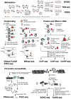
(A) The most widely used assays for cytosine methylation with base-pair accuracy rely on sodium bisulfite, which converts unprotected cytosines to uracils. In a sequencing reaction, methylated and hydroxymethylated sites are read as cytosines, while all other cytosines read as thymines. Tet-assisted bisulfite sequencing uses two additional enzymatic steps to selectively protect hydroxymethylcytosines from bisulfite conversion, and reduced-representation bisulfite sequencing uses a methylation-sensitive restriction enzyme to cleave near methylated CpGs, ensuring that they are read during sequencing. (B) DNA footprinting can be done with either endonucleases or transposases, which cleave unprotected DNA, and with MNase, which has hybrid endonuclease and exonuclease activity and “nibbles” free DNA until it reaches an obstacle, such as a TF or a nucleosome. DNA fragments are purified, size selected and sequenced. DNase-seq focuses on TF footprints from short fragments, whereas DNase-FLASH isolates multiple size classes to assay both nucleosome and TF footprints. (C) Fragmented chromatin can be assayed to identify the sequence of DNA bound to protein (ChIP) or RNA (ChIRP). The fragmentation methods vary, from sonication of crosslinked chromatin to MNase digestion of uncrosslinked chromatin (called native ChIP). Target proteins are pulled down with antibodies, whereas RNAs of interest are pulled down with biotinylated antisense oligos. ChIP-exo employs lambda exonuclease to digest away excess free DNA that is pulled down, increasing the resolution of the method. (D) DNA accessibility, a useful indicator of active regulatory regions, can be assayed by DNase I cleavage (DNase-seq/DHS-seq), Tn5 transposase insertion of sequencing adapters (ATAC-seq), or fragmentation by sonication (SONO-seq), or the depletion of protein-bound fragments (FAIRE-seq). A 30-nm chromatin fiber cartoon has been used to depict the contrast between open and closed chromatin in this panel.
Figure 3. Methods that assay the tertiary structure of chromatin
(A) DamID relies on expression of a fusion between the bacterial adenine methylase Dam and the protein of interest in cells. Dam methylates (yellow) TCGA motifs on any genomic DNA it contacts, and genomic DNA is enzymatically cleaved at methylated restriction sites, the sites are then ligated to adapter oligos (gray) that are complementary to PCR primers. Another restriction digestion cleaves non-methylated sites. The amplification step selects for densely methylated areas, in which methylated restriction sites are adjacent. (B) The chromosome conformation capture family of methods is based on a restriction digest of crosslinked chromatin followed by proximity ligation of the fragment ends in dilute solution. Proximity ligation creates junctions between fragments from genomic regions that interact in 3D. Combining this approach with different strategies for fragmentation, enrichment, and readout of the ligation junctions yields a diverse set of methods. In 3C, PCR with primers on either side of the ligation junction of interest assay a single site. In 4C, the library is cleaved with a second RE and recircularized. PCR primers oriented outward from the test (bait) locus are then used to amplify the unknown regions with which the bait interacts. In 5C, oligos are designed to bridge ligation junctions and are then hybridized to and ligated across the junctions to create “carbon copies” of the junctions that can be PCR-amplified with common primers. Capture-C involves fragmentation of a 3C library then oligo hybridization capture to enrich for all sequence fragments (junctions and non-junctions) from a subset of the genome. Hi-C is a very high-throughput version of 3C that assays all interactions between all genomic loci. Fragment ends are biotinylated (light blue squares) before proximity ligation to facilitate their enrichment for sequencing library construction. Refinements of Hi-C include proximity ligation within crosslinked nuclei rather than in dilute solution, and the use of DNase I fragmentation to replace restriction digest. Because DNase I generates a variety of end types, ends must be blunted and ligated to biotinylated linkers (pink with light blue square) to replace the biotinylation of sticky ends from RE digest. ChIA-PET assays DNA-DNA contacts involving a particular protein by incorporating a ChIP step in the workflow before proximity ligation.
References
- Lander ES, et al. Initial sequencing and analysis of the human genome. Nature. 2001;409:860–921. - PubMed
- International Human Genome Sequencing Consortium. Finishing the euchromatic sequence of the human genome. Nature. 2004;431:931–945. - PubMed
- Lenhard B, et al. Metazoan promoters: emerging characteristics and insights into transcriptional regulation. Nat Rev Genet. 2012;13:233–245. - PubMed
Publication types
MeSH terms
Substances
Grants and funding
- P50 HG007735/HG/NHGRI NIH HHS/United States
- R21 HG007726/HG/NHGRI NIH HHS/United States
- P50HG007735/HG/NHGRI NIH HHS/United States
- R21HG007726/HG/NHGRI NIH HHS/United States
LinkOut - more resources
Full Text Sources
Other Literature Sources


