Effects of diurnal variation of gut microbes and high-fat feeding on host circadian clock function and metabolism - PubMed (original) (raw)
. 2015 May 13;17(5):681-9.
doi: 10.1016/j.chom.2015.03.006. Epub 2015 Apr 16.
Sean M Gibbons 2, Kristina Martinez 1, Alan L Hutchison 3, Edmond Y Huang 1, Candace M Cham 1, Joseph F Pierre 1, Aaron F Heneghan 4, Anuradha Nadimpalli 1, Nathaniel Hubert 1, Elizabeth Zale 1, Yunwei Wang 1, Yong Huang 1, Betty Theriault 5, Aaron R Dinner 6, Mark W Musch 1, Kenneth A Kudsk 4, Brian J Prendergast 7, Jack A Gilbert 8, Eugene B Chang 9
Affiliations
- PMID: 25891358
- PMCID: PMC4433408
- DOI: 10.1016/j.chom.2015.03.006
Effects of diurnal variation of gut microbes and high-fat feeding on host circadian clock function and metabolism
Vanessa Leone et al. Cell Host Microbe. 2015.
Abstract
Circadian clocks and metabolism are inextricably intertwined, where central and hepatic circadian clocks coordinate metabolic events in response to light-dark and sleep-wake cycles. We reveal an additional key element involved in maintaining host circadian rhythms, the gut microbiome. Despite persistence of light-dark signals, germ-free mice fed low or high-fat diets exhibit markedly impaired central and hepatic circadian clock gene expression and do not gain weight compared to conventionally raised counterparts. Examination of gut microbiota in conventionally raised mice showed differential diurnal variation in microbial structure and function dependent upon dietary composition. Additionally, specific microbial metabolites induced under low- or high-fat feeding, particularly short-chain fatty acids, but not hydrogen sulfide, directly modulate circadian clock gene expression within hepatocytes. These results underscore the ability of microbially derived metabolites to regulate or modify central and hepatic circadian rhythm and host metabolic function, the latter following intake of a Westernized diet.
Copyright © 2015 Elsevier Inc. All rights reserved.
Figures
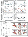
Figure 1. Germ-free mice exhibit differential canonical liver gene transcriptomic signatures and are resistant to diet-induced obesity
GF and CONV C57Bl/6 liver transcriptomes generated via Affymetrix Mouse Genome 430 2.0 array (_n_=3 per group). Canonical pathways (A) enriched in GF versus CONV identified by Significant Analysis of Microarray software (FC >1.5 and FDR <5%) followed by submission to Ingenuity Pathway Analysis software. Significant pathways were determined by one-tail Fisher’s Exact test (p-value <0.05, i.e. −log(p-value) >1.3, (red line)). Weight gain (B) and food consumption (C) of SPF and GF mice (_n_=17–18 per group) fed RC or HF. Data represent mean ± s.e.m. ***p<0.001; **p<0.01; *p<0.05 via one-way ANOVA followed by Dunnett’s post-test relative to SPF-RC control where star color represents treatment exhibiting significance. Diurnal circadian gene expression patterns relative to GAPDH in mediobasal hypothalamus (D) and liver (E) in GF and SPF mice fed RC or HF. ***p<0.001; **p<0.01; *p<0.05 via one-way ANOVA followed by Dunnett’s post-test relative to SPF-RC control - star color indicates treatment exhibiting significance. See also Figure S1A–F and Table S2.
Figure 2. Diurnal gut microbe community structure is altered by high fat feeding
Principal Coordinate Analysis (PCoA) of weighted UniFrac distances from 16S rRNA amplicon sequences colored by diet in fecal pellets (A) collected via repeat sampling over 48h (_n_=3 mice/treatment) and in cecal contents (B) collected over 24h (n **=**2–3 mice/time point) from SPF C57Bl/6 mice fed HF or RC. (C) PCoA analysis of cecal contents colored by Zeitgeber (ZT) time (PC=principle coordinate). (D) 16S rRNA abundance determined via qPCR using universal primers in cecal contents. Data represent mean ± s.e.m. **p<0.01; *p<0.05 via unpaired t-test at each time point. (E) Relative abundance of oscillating 16S rRNA OTUs determined via eJTK_CYCLE in fecal samples collected every 6h over 48h and in cecal contents (F) collected every 4h over 24h at sacrifice. Data represent mean ± s.e.m. See also Figure S2A–E, Table S3 and S5.
Figure 3. High fat diet shifts diurnal microbial function and metabolite production
Principle Coordinate Analysis (PCoA) of Biolog™ spectrophotometric analysis of cecal contents from mice fed RC and HF incubated anaerobically and colored by diet (A) and Zeitgeber (ZT) time (B). Percent oscillating vs. non-oscillating substrate and sensitivity chemicals in fecal pellets (C) from HF or RC collected over 48h (_n_=3/time point). Diurnal cecal butyrate concentration (D, top) and H2S production (bottom) in cecal contents collected from RC or HF (expressed as μmol/g of content) (_n_=2–3/time point). Data represents mean ± s.e.m. *p<0.05,**p<0.01 determined via unpaired t-test at each time point. (E) Rosburia butyryl-CoA: acetate CoA-transferase (BUT Ros_Eub, top) and dissimilatory sulfite reductase (dsrAB, bottom) gene abundance (right axis, normalized to 16S abundance) in cecal contents from mice fed RC or HF. Butyrate and H2S concentration are presented on the left axis. (F) Fecal butyrate concentration (top) and H2S production (bottom) collected via repeat sampling over 48h from mice fed RC and HF (expressed as μmol/g of content) (_n_=3/time point. (G) BUT Ros_Eub (top) and dsrAB (bottom) gene abundance (right axis, normalized to 16S abundance) in fecal pellets over 48h. Data represents mean ± s.e.m. #p<0.1, *p<0.05, **p<0.01 determined via unpaired t-test at each time point. See also Figure S3A–D, Table S4 and S5.
Figure 4. In vitro and in vivo exposure to diet-induced microbial metabolites alters host circadian gene expression
Expression of per2 (A) and bmal1 (B) relative to GAPDH following addition of 5mM sodium acetate, 5mM sodium butyrate, or 1mM sodium hydrosulfide (NaHS) in hepanoids after serum-shock. Data is represented as mean ± s.e.m (_n_=3 replicates/treatment/time point). ***p<0.001; **p<0.01; and *p<0.05 determined via unpaired t-test compared to no trt within time point. See also Table S5. Ratio of per2:bmal1 mRNA (C) in MBH and liver of GF mice treated with saline or butyrate (But) at ZT2 or ZT14 for 5 days. Treatments are saline(ZT2)-saline(ZT14), saline(ZT2)-But(ZT14), But(ZT2)-saline(ZT14). Data represent mean ± s.e.m (_n_=4/treatment). *p<0.05; NS, not significant determined via unpaired t-test. Proposed experimental model (D) diet-induced change in gut microbe metabolic oscillatory patterns alters the balance between food consumption, the central CC, and hepatic regulatory networks of metabolism promoting DIO.
Comment in
- Message in a biota: gut microbes signal to the circadian clock.
Marcinkevicius EV, Shirasu-Hiza MM. Marcinkevicius EV, et al. Cell Host Microbe. 2015 May 13;17(5):541-3. doi: 10.1016/j.chom.2015.04.013. Cell Host Microbe. 2015. PMID: 25974294
Similar articles
- Message in a biota: gut microbes signal to the circadian clock.
Marcinkevicius EV, Shirasu-Hiza MM. Marcinkevicius EV, et al. Cell Host Microbe. 2015 May 13;17(5):541-3. doi: 10.1016/j.chom.2015.04.013. Cell Host Microbe. 2015. PMID: 25974294 - Administration of Exogenous Melatonin Improves the Diurnal Rhythms of the Gut Microbiota in Mice Fed a High-Fat Diet.
Yin J, Li Y, Han H, Ma J, Liu G, Wu X, Huang X, Fang R, Baba K, Bin P, Zhu G, Ren W, Tan B, Tosini G, He X, Li T, Yin Y. Yin J, et al. mSystems. 2020 May 19;5(3):e00002-20. doi: 10.1128/mSystems.00002-20. mSystems. 2020. PMID: 32430404 Free PMC article. - Oat Fiber Modulates Hepatic Circadian Clock via Promoting Gut Microbiota-Derived Short Chain Fatty Acids.
Han S, Gao H, Song R, Zhang W, Li Y, Zhang J. Han S, et al. J Agric Food Chem. 2021 Dec 29;69(51):15624-15635. doi: 10.1021/acs.jafc.1c06130. Epub 2021 Dec 20. J Agric Food Chem. 2021. PMID: 34928598 - Rhythm and bugs: circadian clocks, gut microbiota, and enteric infections.
Rosselot AE, Hong CI, Moore SR. Rosselot AE, et al. Curr Opin Gastroenterol. 2016 Jan;32(1):7-11. doi: 10.1097/MOG.0000000000000227. Curr Opin Gastroenterol. 2016. PMID: 26628099 Free PMC article. Review. - Gut microbiota as a transducer of dietary cues to regulate host circadian rhythms and metabolism.
Choi H, Rao MC, Chang EB. Choi H, et al. Nat Rev Gastroenterol Hepatol. 2021 Oct;18(10):679-689. doi: 10.1038/s41575-021-00452-2. Epub 2021 May 17. Nat Rev Gastroenterol Hepatol. 2021. PMID: 34002082 Free PMC article. Review.
Cited by
- Microbiota Composition and Probiotics Supplementations on Sleep Quality-A Systematic Review and Meta-Analysis.
Santi D, Debbi V, Costantino F, Spaggiari G, Simoni M, Greco C, Casarini L. Santi D, et al. Clocks Sleep. 2023 Dec 13;5(4):770-792. doi: 10.3390/clockssleep5040050. Clocks Sleep. 2023. PMID: 38131749 Free PMC article. Review. - Gut microbiota-astrocyte axis: new insights into age-related cognitive decline.
Zhang L, Wei J, Liu X, Li D, Pang X, Chen F, Cao H, Lei P. Zhang L, et al. Neural Regen Res. 2025 Apr 1;20(4):990-1008. doi: 10.4103/NRR.NRR-D-23-01776. Epub 2024 Apr 16. Neural Regen Res. 2025. PMID: 38989933 Free PMC article. - How the AHR Became Important in Intestinal Homeostasis-A Diurnal FICZ/AHR/CYP1A1 Feedback Controls Both Immunity and Immunopathology.
Rannug A. Rannug A. Int J Mol Sci. 2020 Aug 8;21(16):5681. doi: 10.3390/ijms21165681. Int J Mol Sci. 2020. PMID: 32784381 Free PMC article. Review. - Invasions of Host-Associated Microbiome Networks.
Murall CL, Abbate JL, Touzel MP, Allen-Vercoe E, Alizon S, Froissart R, McCann K. Murall CL, et al. Adv Ecol Res. 2017;57:201-281. doi: 10.1016/bs.aecr.2016.11.002. Adv Ecol Res. 2017. PMID: 39404686 Free PMC article. - The Role of the Gut Microbiota in Lipid and Lipoprotein Metabolism.
Yu Y, Raka F, Adeli K. Yu Y, et al. J Clin Med. 2019 Dec 17;8(12):2227. doi: 10.3390/jcm8122227. J Clin Med. 2019. PMID: 31861086 Free PMC article. Review.
References
- Balsalobre A, Damiola F, Schibler U. A serum shock induces circadian gene expression in mammalian tissue culture cells. Cell. 1998;93:929–937. - PubMed
- Bass J, Turek FW. Sleepless in America: a pathway to obesity and the metabolic syndrome? Arch Intern Med. 2005;165:15–16. - PubMed
Publication types
MeSH terms
Grants and funding
- UL1 TR000430/TR/NCATS NIH HHS/United States
- P30 DK042086/DK/NIDDK NIH HHS/United States
- R01 DK038510/DK/NIDDK NIH HHS/United States
- NIDDK P30DK42086/PHS HHS/United States
- UH3 DK083993/DK/NIDDK NIH HHS/United States
- T32 GM007281/GM/NIGMS NIH HHS/United States
- DK42086/DK/NIDDK NIH HHS/United States
- T32 DK007074/DK/NIDDK NIH HHS/United States
- R37 DK047722/DK/NIDDK NIH HHS/United States
- UH3DK083993/DK/NIDDK NIH HHS/United States
- T32 EB009412/EB/NIBIB NIH HHS/United States
- DK097268/DK/NIDDK NIH HHS/United States
- DK47722/DK/NIDDK NIH HHS/United States
- NIGMS T32GM07281/PHS HHS/United States
- R01 DK097268/DK/NIDDK NIH HHS/United States
- R01 DK047722/DK/NIDDK NIH HHS/United States
LinkOut - more resources
Full Text Sources
Other Literature Sources
Molecular Biology Databases


