The development of innate lymphoid cells requires TOX-dependent generation of a common innate lymphoid cell progenitor - PubMed (original) (raw)
. 2015 Jun;16(6):599-608.
doi: 10.1038/ni.3168. Epub 2015 Apr 27.
Affiliations
- PMID: 25915732
- PMCID: PMC4439271
- DOI: 10.1038/ni.3168
The development of innate lymphoid cells requires TOX-dependent generation of a common innate lymphoid cell progenitor
Corey R Seehus et al. Nat Immunol. 2015 Jun.
Abstract
Diverse innate lymphoid cell (ILC) subtypes have been defined on the basis of effector function and transcription factor expression. ILCs derive from common lymphoid progenitors, although the transcriptional pathways that lead to ILC-lineage specification remain poorly characterized. Here we found that the transcriptional regulator TOX was required for the in vivo differentiation of common lymphoid progenitors into ILC lineage-restricted cells. In vitro modeling demonstrated that TOX deficiency resulted in early defects in the survival or proliferation of progenitor cells, as well as ILC differentiation at a later stage. In addition, comparative transcriptome analysis of bone marrow progenitors revealed that TOX-deficient cells failed to upregulate many genes of the ILC program, including genes that are targets of Notch, which indicated that TOX is a key determinant of early specification to the ILC lineage.
Conflict of interest statement
COMPETING FINANCIAL INTERESTS
The authors declare no competing financial interests.
Figures
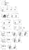
Figure 1
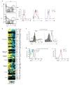
TOX is expressed in ILC progenitors and mature ILC lineages. (a) Deletion of the neomycin cassette was accomplished by breeding to FLPase recombinase expressing mice. FLPase was removed in subsequent breeding to generate _Tox_TOM reporter mice. Restriction enzyme sites (P=PstI, S=StuI, R=EcoRI) are shown in surrounding non-coding sequences only. ToxTOM mice were also bred to _Id2_GFP mice to generate a dual reporter strain used in many experiments. (b) Flow cytometry of Lin−CD122+ cells from BM and spleen for TOM and GFP in NKp, _i_NK, and mNK populations (_n_=3). Numbers refer to frequency of gated population here and in subsequent figures. (c) Expression of TOM and GFP in BM CLP reporter mice as indicated (_n_=3). (d) TOM and GFP expression in BM Lin−α4β7+CD127+ cells subgated by CD25 (_n_=3). (e) Lung ILC2s from WT and ToxTOMId2GFP mice were analyzed for reporter expression (_n_=3). (f) Expression of Tox and Gata3 by qRT-PCR in IL-33-expanded ILC2s, in vitro generated TH2 cells, and CD19+B220+ splenic B cells (_n_=2–3). (g, h) Expression of TOM in WT and ToxTOM lamina propria cells gated as shown (_n_=3). (i, j) Expression of TOM in splenic subpopulations (n=3) as in (g). *P <0.05, NS= non-significant, _P_ >0.05 (Student’s _t_-test). Data (± s.d.) are representative of three (b–e, g–j) or two (f) independent experiments.
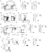
Figure 2
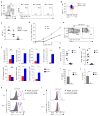
TOX is required for the development of ILC progenitors. (a) Staining for BM CLP from WT and Tox−/− mice (_n_=6). (b, c) Compiled data of the frequency (b) and number (c) of Flt3+Sca-1int cells within the Lin−CD45+ cell population from WT and Tox−/− BM. (d) Lin−α4β7+CD127+ cells from BM of WT and Tox−/− animals (_n_=3). (e) Gated Lin−α4β7+CD127+ cells from WT and Tox−/− animals were analyzed for CD25. Shown is the frequency of CD25− cells (_n_=3). (f) Number of Lin−α4β7+CD127+CD25− cells in WT and Tox−/− animals. (g) Lin−CD45+NK1.1+NKp46+ cells were analyzed for cNK (CD27+CD127−) and ILC1s (CD27+CD127+) from WT and Tox−/− mice in BM. The lack of Lin−CD45+NK1.1+NKp46+ cells from Tox−/− mice precluded any additional subset analysis. (h, i) Compiled data of the frequency (h) and number (i) of NK1.1+NKp46+ cells within the Lin−CD45+ cell population from BM. (j) Staining for BM ILC2p from WT and Tox−/− mice. (k, l) Compiled frequency (k) and absolute numbers (l) of BM ILC2p. **P <0.01, _***P_ <0.001, NS= non-significant, _P_ >0.05 (Student’s _t_-test). Symbols (b, c, f, h, I, k, l) represent individual mice analyzed independently; horizontal lines denote the mean.
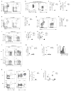
Figure 3
TOX is required for the development of mature ILCs. (a) Comparison of lung NK1.1+NKp46+ cells within the Lin−CD45+ population from WT and Tox−/− mice (_n_=3). (b) Intracellular staining of WT, lung Lin−CD45+NK1.1+NKp46+ population as shown in (a) including cNK and ILC1s (_n_=3). (c, d) Compiled frequency (c) and absolute numbers (d) of lung NK1.1+NKp46+ population. (e, g) Colon (e) and small intestine (g) lamina propria NK1.1+NKp46+ cells from WT and Tox−/− mice (_n_=3). (f, h) Frequency of colon (f) and small intestine (h) NK1.1+NKp46+ population. (i) Comparison of lung ILC2s from WT and Tox−/− mice (_n_=10). (j, k) Compiled frequency (j) and absolute numbers (k) of lung ILC2s. (l) Staining for lung ILC2 in WT and Tox−/− animals treated with IL-33 or PBS (_n_=3–5). (m, n) Frequency (m) and absolute numbers (n) of ILC2s from WT and Tox−/− lung, treated as indicated. (o) Comparison of ST2 expression on IL-33-expanded lung Lin−CD45+Thy-1+ cells isolated from WT or Tox−/− mice (_n_=3–5). (p) Comparison of splenic Lin−CD45+CD127+c-Kit+ cells from WT and Tox−/− mice gated as shown (_n_=4). (q) Absolute numbers of total spleen Lin−CD45+CD127+c-Kit+. (r) Absolute numbers of RORγt+CD4− and CD4+ cells as gated in (p). *P <0.05, _**P_ <0.01, _***P_ <0.001, NS= non-significant, _P_ >0.05 (Student’s _t_-test). Symbols (c, d, f, h, j, k, m, n, q, r) represent individual mice analyzed independently; horizontal lines denote the mean.
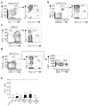
Figure 4
TOX regulates the development of ILCs by a cell-intrinsic mechanism. (a–d) Donor-derived (CD45.2+) cells in (a) BM (_n_=4), (b, c) lung (_n_=3), and (d) spleen (_n_=3) were analyzed for allelic markers for WT (Thy-1.1+) or Tox−/− (Thy-1.2+) genotypes. (e) Ratio of _Tox_−/− to WT cells in the indicated populations. ***P <0.001 (Student’s _t_-test). Data (± s.d.) are compiled from four (a) or three (b, c, d) experiments, pooling two animals per experiment.
Figure 5
Whole transcriptome analysis of Tox−/− α4β7+ progenitors reveals a block in induction of the ILC gene program. (a) Gating strategy used for isolation of Lin−CD127+α4β7+CD25−Flt3− cells from WT and Tox−/− mice (_n_=4). (b) Heat map of select gene expression. PE, poorly expressed (average FPKM of both genotypes ≤ 2), NS, not significant (Q value ≥ 0.05) (Student’s _t_-test, Benjamini and Hochberg correction for multiple tests). (c) Expression of ICOS or isotype control (filled histograms) in CHILP from Id2GFP reporter mice (_n_=2). (d) WT progenitor cells as gated in (a) were compared to ILC2p and CLP for Notch1 surface expression (_n_=2). (e) CLP from WT and _Tox−/−_mice were analyzed for Notch1 (_n_=2). Data are compiled from four (b) independent experiments, pooling 2–3 animals per experiment per genotype.
Figure 6
In vitro ILC specification is blocked early in the absence of TOX. (a) Subpopulations at day six of Lin− cells subgated on Thy-1 and CD25 were analyzed for TOM expression (_n_=3). (b) Calculated fold expansion over number of input WT or Tox−/− CLP at day six. (c) Differentiation of WT and Tox−/− CLP at day six was assessed by expression of Thy-1 and CD25 (_n_=6). (d, e) Frequency (d) and number (e) of Lin−Thy-1hi cells (% of Lin−) gated as in (c). (f) Lin−Thy-1hiCD25−ICOS+ WT and Tox−/− cells at day six in culture (_n_=6). (g) Lin−Thy-1lo and Lin−Thy-1hi cell populations were isolated by cell sorting and expression of indicated genes was determined by qRT-PCR. Data was normalized to Hprt expression and presented relative to in vivo, IL-33-expanded ILC2s. *P <0.05, _**P_ <0.01, _***P_ <0.001, NS= non-significant, _P_ >0.05 (Student’s _t_-test). Symbols (b, d, e) represent individual mice analyzed independently; horizontal lines denote the mean. Data (± s.d.) are compiled from four mice (g), using CLP pooled from two animals per experiment.
Figure 7
In the absence of TOX, there is a cell-intrinsic differentiation defect in the generation of ILC1s and functional ILC2s. (a) Frequency of WT (CD45.2+) and Tox−/− (CD45.2−) in vitro differentiated non-ILCs (ICOS−NK1.1−), ILC2s (ICOS+NK1.1−T-bet−GATA-3+) and ILC1s (ICOS+NK1.1+T-bet+GATA-3−) (_n_=4). (b) Transcription factor staining for WT ILC1s and ILC2s (_n_=4). (c) WT to Tox−/− ratio of populations in (a). (d) CLPs were isolated from WT and Tox−/− BM, cultured for six, 12, or 18 days, and numbers of Lin−Thy-1hi cells determined. Data are calculated based on input number of CLP. (e) Analysis of day 18 cultures as shown (_n_=7). (f) Gene expression in day 18 Lin−Thy-1hiICOS− or ICOS+ cells, determined as in Fig. 6. (g, h) Frequency (% of Lin−Thy-1hi) (g) and cell numbers (h) of WT culture-generated ICOS+ cells gated as in (e). Cell numbers are expressed as total cells recovered at day 18 per 5,000 cells plated at day 12. (i, j) Quantification of secreted IL-5 (i) or IL-13 (j) from in vitro differentiated CLP. (k, l) Expression of α4β7 (k) and PLZF (l) on Lin−Thy-1hi cells. Histograms display results from isotype control, ICOS− and ICOS+ cells (_n_=3). *P <0.05, _**P_ <0.01, _***P_ <0.001, NS= non-significant, _P_ >0.05 (Student’s _t_-test). Symbols (c, g, h, I, j) represent individual mice analyzed independently; horizontal lines denote the mean. Data are compiled from seven (d) or three (f) independent experiments using two animals pooled per experiment.
Comment in
- TOX sets the stage for innate lymphoid cells.
Spits H. Spits H. Nat Immunol. 2015 Jun;16(6):594-5. doi: 10.1038/ni.3177. Nat Immunol. 2015. PMID: 25988899 No abstract available.
Similar articles
- Polychromic Reporter Mice Reveal Unappreciated Innate Lymphoid Cell Progenitor Heterogeneity and Elusive ILC3 Progenitors in Bone Marrow.
Walker JA, Clark PA, Crisp A, Barlow JL, Szeto A, Ferreira ACF, Rana BMJ, Jolin HE, Rodriguez-Rodriguez N, Sivasubramaniam M, Pannell R, Cruickshank J, Daly M, Haim-Vilmovsky L, Teichmann SA, McKenzie ANJ. Walker JA, et al. Immunity. 2019 Jul 16;51(1):104-118.e7. doi: 10.1016/j.immuni.2019.05.002. Epub 2019 May 22. Immunity. 2019. PMID: 31128961 Free PMC article. - TOX sets the stage for innate lymphoid cells.
Spits H. Spits H. Nat Immunol. 2015 Jun;16(6):594-5. doi: 10.1038/ni.3177. Nat Immunol. 2015. PMID: 25988899 No abstract available. - Single-cell RNA-seq identifies a PD-1hi ILC progenitor and defines its development pathway.
Yu Y, Tsang JC, Wang C, Clare S, Wang J, Chen X, Brandt C, Kane L, Campos LS, Lu L, Belz GT, McKenzie AN, Teichmann SA, Dougan G, Liu P. Yu Y, et al. Nature. 2016 Nov 3;539(7627):102-106. doi: 10.1038/nature20105. Epub 2016 Sep 29. Nature. 2016. PMID: 27749818 - The Role of TOX in the Development of Innate Lymphoid Cells.
Seehus CR, Kaye J. Seehus CR, et al. Mediators Inflamm. 2015;2015:243868. doi: 10.1155/2015/243868. Epub 2015 Oct 18. Mediators Inflamm. 2015. PMID: 26556952 Free PMC article. Review. - Transcriptional regulation of innate and adaptive lymphocyte lineages.
De Obaldia ME, Bhandoola A. De Obaldia ME, et al. Annu Rev Immunol. 2015;33:607-42. doi: 10.1146/annurev-immunol-032414-112032. Epub 2015 Feb 4. Annu Rev Immunol. 2015. PMID: 25665079 Review.
Cited by
- Stage-specific GATA3 induction promotes ILC2 development after lineage commitment.
Furuya H, Toda Y, Iwata A, Kanai M, Kato K, Kumagai T, Kageyama T, Tanaka S, Fujimura L, Sakamoto A, Hatano M, Suto A, Suzuki K, Nakajima H. Furuya H, et al. Nat Commun. 2024 Jul 5;15(1):5610. doi: 10.1038/s41467-024-49881-y. Nat Commun. 2024. PMID: 38969652 Free PMC article. - Transcriptomic diversity of innate lymphoid cells in human lymph nodes compared to BM and spleen.
Hashemi E, McCarthy C, Rao S, Malarkannan S. Hashemi E, et al. Commun Biol. 2024 Jun 25;7(1):769. doi: 10.1038/s42003-024-06450-9. Commun Biol. 2024. PMID: 38918571 Free PMC article. - The TOX-RAGE axis mediates inflammatory activation and lung injury in severe pulmonary infectious diseases.
Kim H, Park HH, Kim HN, Seo D, Hong KS, Jang JG, Seo EU, Kim IY, Jeon SY, Son B, Cho SW, Kim W, Ahn JH, Lee W. Kim H, et al. Proc Natl Acad Sci U S A. 2024 Jun 25;121(26):e2319322121. doi: 10.1073/pnas.2319322121. Epub 2024 Jun 20. Proc Natl Acad Sci U S A. 2024. PMID: 38900789 Free PMC article. - RAG suppresses group 2 innate lymphoid cells.
Ver Heul AM, Mack M, Zamidar L, Tamari M, Yang TL, Trier AM, Kim DH, Janzen-Meza H, Van Dyken SJ, Hsieh CS, Karo JM, Sun JC, Kim BS. Ver Heul AM, et al. bioRxiv [Preprint]. 2024 Apr 28:2024.04.23.590767. doi: 10.1101/2024.04.23.590767. bioRxiv. 2024. PMID: 38712036 Free PMC article. Preprint. - Transcription factor Tox2 is required for metabolic adaptation and tissue residency of ILC3 in the gut.
Das A, Martinez-Ruiz GU, Bouladoux N, Stacy A, Moraly J, Vega-Sendino M, Zhao Y, Lavaert M, Ding Y, Morales-Sanchez A, Harly C, Seedhom MO, Chari R, Awasthi P, Ikeuchi T, Wang Y, Zhu J, Moutsopoulos NM, Chen W, Yewdell JW, Shapiro VS, Ruiz S, Taylor N, Belkaid Y, Bhandoola A. Das A, et al. Immunity. 2024 May 14;57(5):1019-1036.e9. doi: 10.1016/j.immuni.2024.04.001. Epub 2024 Apr 26. Immunity. 2024. PMID: 38677292
References
- Artis D, Spits H. The biology of innate lymphoid cells. Nature. 2015;517:293–301. - PubMed
- Klose CS, et al. Differentiation of type 1 ILCs from a common progenitor to all helper-like innate lymphoid cell lineages. Cell. 2014;157:340–356. - PubMed
Publication types
MeSH terms
Substances
Grants and funding
- R00 DK098310/DK/NIDDK NIH HHS/United States
- R01 AI054977/AI/NIAID NIH HHS/United States
- HHSN272201300006C/AI/NIAID NIH HHS/United States
- 5R01AI054977/AI/NIAID NIH HHS/United States
- K99 DK098310/DK/NIDDK NIH HHS/United States
- DK098310/DK/NIDDK NIH HHS/United States
LinkOut - more resources
Full Text Sources
Other Literature Sources
Molecular Biology Databases
