Fibroblast-Derived Exosomes Contribute to Chemoresistance through Priming Cancer Stem Cells in Colorectal Cancer - PubMed (original) (raw)
Fibroblast-Derived Exosomes Contribute to Chemoresistance through Priming Cancer Stem Cells in Colorectal Cancer
Yibing Hu et al. PLoS One. 2015.
Abstract
Chemotherapy resistance observed in patients with colorectal cancer (CRC) may be related to the presence of cancer stem cells (CSCs), but the underlying mechanism(s) remain unclear. Carcinoma-associated fibroblasts (CAFs) are intimately involved in tumor recurrence, and targeting them increases chemo-sensitivity. We investigated whether fibroblasts might increase CSCs thus mediating chemotherapy resistance. CSCs were isolated from either patient-derived xenografts or CRC cell lines based on expression of CD133. First, CSCs were found to be inherently resistant to cell death induced by chemotherapy. In addition, fibroblast-derived conditioned medium (CM) promoted percentage, clonogenicity and tumor growth of CSCs (i.e., CD133+ and TOP-GFP+) upon treatment with 5-fluorouracil (5-Fu) or oxaliplatin (OXA). Further investigations exhibited that exosomes, isolated from CM, similarly took the above effects. Inhibition of exosome secretion decreased the percentage, clonogenicity and tumor growth of CSCs. Altogether, our findings suggest that, besides targeting CSCs, new therapeutic strategies blocking CAFs secretion even before chemotherapy shall be developed to gain better clinical benefits in advanced CRCs.
Conflict of interest statement
Competing Interests: The authors have declared that no competing interests exist.
Figures
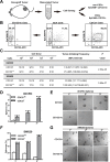
Fig 1. CD133 identifies CSCs in colorectal cancer.
(A)Schematic of CD133+ and CD133-/lo tumor cells sorting from dissociated colorectal xenograft tumor by FACS. (B) A representative example of post-sorting analysis of the sorted CD133+ and CD133-/lo XhCRC cells.(C) Tumor-initiating frequency of CD133+ and CD133-/lo CRC cells in immunodeficient mice (D-G)Serial sphere formation assays for purified CD133+ and CD133-/loCRC cells (i.e., XhCRC and SW620). Spheres were enumerated (D, F) and representative images are shown (E, G).Scale bars, 100μm.***P< 0.001.
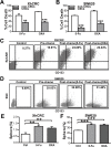
Fig 2. CSCs display cell-autonomous resistance to chemotherapy.
(A, B) Cell death analysis of CSCs and non-CSCs from XhCRC or SW620 cells was assessed by CCK-8 activity assay upon chemotherapeutic treatment (5-Fu or OXA).**P< 0.01, ***P< 0.001. (C, D) Enrichment of CSCs in bulk cells from XhCRC (C) and SW620 cells (D) was assessed by FACS analysis based on CD133 expression upon chemotherapy. (E, F) Sphere-forming capacity of bulk cells (XhCRC or SW620 cells) pre-treated by chemotherapeutic agents or DMSO (Ctrl). **P< 0.01, ***P< 0.001.
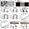
Fig 3. 18Co cells prime SW620 CSCs to be more drug resistance via paracrine pathway.
(A) Sphere-forming capacity of SW620 CSCs treated with 18Co-derived CM during chemotherapy (5-Fu or OXA). Representative microscopic images are shown. Scale bars, 100μm. (B, C) 18Co-derived conditioned medium affected tumor growth of SW620 CSCs in female Balb/c-nu mice treated with OXA. Tumor growth curves are shown in C, Shown in D are tumor weights and images at the end of experiments. Data are presented as mean ± SD; *P< 0.1; **P< 0.05; ***P< 0.001.
Fig 4. CAFs prime XhCRC CSCs to be more drug resistance through paracrine pathway.
(A) CAFs derived from patient specimen were validated by positive immunostaining for CAF markers (α-SMA,Vimentin and FAP) and negative immunostaining for an epithelial marker (EpCAM). Scale bars, 30μm. (B) XhCRC CSCs were validated by positive immunostaining for epithelial marker (EpCAM) and CSC marker (CD133), Scale bars, 10μm. (C) Sphere-forming capacity of XhCRC CSCs in CAFs-derived conditioned media (e.g., 5-FU, OXA, DMSO-treated CAFs). (D) Sphere-forming capacity of XhCRC CSCs treated with CAF-derived CM during chemotherapy (5-Fu or OXA). Representative microscopic images are shown. Scale bars, 50μm. (E, F) CAF-derived CM affected on tumor growth of XhCRC CSCs in female NOD/SCID mice treated with OXA. Tumor growth curves are shown in E, Shown in F are tumor weights and images at the end of experiments. Data are presented as mean ± SD; *P< 0.1; **P< 0.05. (G)CAF-derived CM affected on tumor growth of XhCRC CSCs transplanted in female NOD/SCID mice upon treatment with 5-Fu.
Fig 5. Fibroblast-derived exosomes prime CSCs to be more chemoresistance.
(A) Transmission electron microscopic image of the exosomes derived from 18Co cells and CAFs. Scale bars, 100nm. (B) Western blotting of CD81 in exosomes. (C) Representative microscopy of SW620 cells exposed to DiI-labeled exosomes for 12h. (D, E) Sphere-forming capacity of SW620 or XhCRC CSCs treated with 18Co/CAF-derived exosomes during chemotherapy (5-Fu or OXA). (F) Sphere-forming capacity of SW620 CSCs treated with ultracentrifugation-purified 18Co-derived exosomes during chemotherapy (5-Fu or OXA). (G, H)Fibroblasts were treated with 10mM GW4869 (dissolves in DMSO) for 24h. The CMs derived from GW4869/DMSO-pretreated fibroblasts were added to CSCs. (I-L) CAF-derived exosomes affected on tumor growth of XhCRC CSCs in female NOD/SCID mice treated with 5-Fu or OXA. Tumor growth curves are shown in I(5-Fu) and K(OXA), and tumor weights and images at the end of experiments are shown in L(5-Fu) and J(OXA). Data are presented as mean ± SD; *P< 0.1; **P< 0.05.
Fig 6. Exosomes prime CSCs through mediating Wnt activity.
(A) TOP–GFP levels are associated with CSC maker expression. (B) Sphere-forming efficiency and representative spheres (insert) of TOP-GFP+ and TOP-GFP-/lo SW620 cells. *** P<0.001. (C)Representative flow cytometry and quantification (insert) of TOP-GFP expression of CSCs treated with 18Co-derived exosomes (green line).Gray line represents the control group.(D, E) Representative flow cytometry and quantification (insert) of TOP-GFP expression of CSCs treated with 18Co-derived exosomes (magenta line) or GW4869 (black line)/DMSO (orange line)-pretreated 18Co-CM upon chemotherapy (5-Fu or OXA). Gray line represents the control group. (F) Fibroblast-derived exosomes were purified by ultracentrifugation. The pellet, supernatant and fibroblast cell lysate were subjected to western blotting for exosome maker CD81 and Wnt3a.
Similar articles
- Exosomal Wnt-induced dedifferentiation of colorectal cancer cells contributes to chemotherapy resistance.
Hu YB, Yan C, Mu L, Mi YL, Zhao H, Hu H, Li XL, Tao DD, Wu YQ, Gong JP, Qin JC. Hu YB, et al. Oncogene. 2019 Mar;38(11):1951-1965. doi: 10.1038/s41388-018-0557-9. Epub 2018 Nov 2. Oncogene. 2019. PMID: 30390075 Free PMC article. - Cancer associated fibroblasts-derived exosomes contribute to radioresistance through promoting colorectal cancer stem cells phenotype.
Liu L, Zhang Z, Zhou L, Hu L, Yin C, Qing D, Huang S, Cai X, Chen Y. Liu L, et al. Exp Cell Res. 2020 Jun 15;391(2):111956. doi: 10.1016/j.yexcr.2020.111956. Epub 2020 Mar 10. Exp Cell Res. 2020. PMID: 32169425 - Colorectal cancer stem cell and chemoresistant colorectal cancer cell phenotypes and increased sensitivity to Notch pathway inhibitor.
Huang R, Wang G, Song Y, Tang Q, You Q, Liu Z, Chen Y, Zhang Q, Li J, Muhammand S, Wang X. Huang R, et al. Mol Med Rep. 2015 Aug;12(2):2417-24. doi: 10.3892/mmr.2015.3694. Epub 2015 Apr 28. Mol Med Rep. 2015. PMID: 25936357 Free PMC article. - Neuroblastoma stem cells - mechanisms of chemoresistance and histone deacetylase inhibitors.
Khalil MA, Hrabeta J, Cipro S, Stiborova M, Vicha A, Eckschlager T. Khalil MA, et al. Neoplasma. 2012;59(6):737-46. doi: 10.4149/neo_2012_093. Neoplasma. 2012. PMID: 22862175 Review. - CD133 as a target for colon cancer.
Catalano V, Di Franco S, Iovino F, Dieli F, Stassi G, Todaro M. Catalano V, et al. Expert Opin Ther Targets. 2012 Mar;16(3):259-67. doi: 10.1517/14728222.2012.667404. Epub 2012 Mar 3. Expert Opin Ther Targets. 2012. PMID: 22385077 Review.
Cited by
- Therapeutic combinations of exosomes alongside cancer stem cells (CSCs) and of CSC-derived exosomes (CSCEXs) in cancer therapy.
Zabeti Touchaei A, Norollahi SE, Najafizadeh A, Babaei K, Bakhshalipour E, Vahidi S, Samadani AA. Zabeti Touchaei A, et al. Cancer Cell Int. 2024 Oct 5;24(1):334. doi: 10.1186/s12935-024-03514-y. Cancer Cell Int. 2024. PMID: 39369258 Free PMC article. Review. - Extracellular vesicles and particles impact the systemic landscape of cancer.
Lucotti S, Kenific CM, Zhang H, Lyden D. Lucotti S, et al. EMBO J. 2022 Sep 15;41(18):e109288. doi: 10.15252/embj.2021109288. Epub 2022 Sep 2. EMBO J. 2022. PMID: 36052513 Free PMC article. Review. - Tumour immune microenvironment biomarkers predicting cytotoxic chemotherapy efficacy in colorectal cancer.
Wilkinson K, Ng W, Roberts TL, Becker TM, Lim SH, Chua W, Lee CS. Wilkinson K, et al. J Clin Pathol. 2021 Oct;74(10):625-634. doi: 10.1136/jclinpath-2020-207309. Epub 2021 Mar 22. J Clin Pathol. 2021. PMID: 33753562 Free PMC article. Review. - The Role of Exosomes in Stemness and Neurodegenerative Diseases-Chemoresistant-Cancer Therapeutics and Phytochemicals.
Beeraka NM, Doreswamy SH, Sadhu SP, Srinivasan A, Pragada RR, Madhunapantula SV, Aliev G. Beeraka NM, et al. Int J Mol Sci. 2020 Sep 17;21(18):6818. doi: 10.3390/ijms21186818. Int J Mol Sci. 2020. PMID: 32957534 Free PMC article. Review. - Knockdown of miR-92a suppresses the stemness of colorectal cancer cells via mediating SOCS3.
Li L, Zhang J, Peng H, Jiang X, Liu Z, Tian H, Hou S, Xie X, Peng Q, Zhou T. Li L, et al. Bioengineered. 2022 Mar;13(3):5613-5624. doi: 10.1080/21655979.2021.2022267. Bioengineered. 2022. PMID: 35184640 Free PMC article.
References
Publication types
MeSH terms
Substances
Grants and funding
This study was supported by grants from the National Natural Science Foundation of China (Nos. 81172065, 81272660), Program for New Century Excellent Talents in University (No. NCET-12-0208), Scientific Research Foundation for the Returned Overseas Chinese Scholars, State Education Ministry (No. JYBHG201002), the Fundamental Research Funds for the Central Universities (HUST, No.01-18-540005), Tongji Hospital Funds for the Returned Overseas Scientists and Outstanding Young Scientists (2012yq004) (all to JCQ). The funders had no role in study design, data collection and analysis, decision to publish, or preparation of the manuscript.
LinkOut - more resources
Full Text Sources
Other Literature Sources
Medical
Research Materials


