Coma and cerebral imaging - PubMed (original) (raw)
Coma and cerebral imaging
Walter F Haupt et al. Springerplus. 2015.
Abstract
The clinical sign of coma is a common feature in critical care medicine. However, little information has been put forth on the correlations between coma and cerebral imaging methods. The purpose of the article is to compile the available information derived from various imaging methods and placing it in a context of clinical knowledge of coma and related states. The definition of coma and the cerebral structures responsible for consciousness are described; the mechanisms of cerebral lesions leading to impaired consciousness and coma are explained. Cerebral imaging methods provide a large array of information on the structural changes of brain tissue in the various diseases leading to coma. Circumscript lesions produce space-occupying masses that displace the brain, ultimately leading to various types of herniation. Generalized disease of the brain usually leads to diffuse brain swelling which also can cause herniation. Epileptic states, however, rarely are detectable by imaging methods and mandate EEG examinations. Another important aspect of imaging in coma is the increasing use of functional imaging methods, which can detect the function of loss of function in various areas of the brain and render information on the extent and severity of brain damage as well as on the prognosis of disease. The MRI methods of (1)H-spectroscopy and diffusion tensor imaging may provide more functional information in the future.
Keywords: Brain death; Brain disease; Coma; Computed tomography (CT); Functional Magnetic Resonance Imaging (fMRI); Magnetic Resonance Imaging (MRI).
Figures
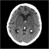
Figure 1
68 year- old patient with bilateral embolic thalamic infarctions causing coma.
Figure 2
90 year-old patient with thrombosis of inner veins, causing bilateral thalamic and basal ganglia edema, resulting in coma.
Figure 3
45 year-old patient with coma and lymphocytic pleocytosis in CSF, caused by immune-mediated encephalitis.
Figure 4
26 y/o patient with traumatic brain injury. A. CT scan only shows impression fracture and mild swelling. B. MRI shows severe contusion of right temporal lobe and bilateral mesencephalic lesions causing coma.
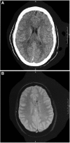
Figure 5
16 y/o patient with traumatic brain injury. A. CT shows no lesion. B. MRI shows multiple disseminated microbleeds in the T2* sequences.
Similar articles
- Magnetic Resonance Imaging Application in the Area of Mild and Acute Traumatic Brain Injury: Implications for Diagnostic Markers?
Toth A. Toth A. In: Kobeissy FH, editor. Brain Neurotrauma: Molecular, Neuropsychological, and Rehabilitation Aspects. Boca Raton (FL): CRC Press/Taylor & Francis; 2015. Chapter 24. In: Kobeissy FH, editor. Brain Neurotrauma: Molecular, Neuropsychological, and Rehabilitation Aspects. Boca Raton (FL): CRC Press/Taylor & Francis; 2015. Chapter 24. PMID: 26269902 Free Books & Documents. Review. - Exploring Serum Biomarkers for Mild Traumatic Brain Injury.
Papa L, Edwards D, Ramia M. Papa L, et al. In: Kobeissy FH, editor. Brain Neurotrauma: Molecular, Neuropsychological, and Rehabilitation Aspects. Boca Raton (FL): CRC Press/Taylor & Francis; 2015. Chapter 22. In: Kobeissy FH, editor. Brain Neurotrauma: Molecular, Neuropsychological, and Rehabilitation Aspects. Boca Raton (FL): CRC Press/Taylor & Francis; 2015. Chapter 22. PMID: 26269900 Free Books & Documents. Review. - Imaging assessment of traumatic brain injury.
Currie S, Saleem N, Straiton JA, Macmullen-Price J, Warren DJ, Craven IJ. Currie S, et al. Postgrad Med J. 2016 Jan;92(1083):41-50. doi: 10.1136/postgradmedj-2014-133211. Epub 2015 Nov 30. Postgrad Med J. 2016. PMID: 26621823 Review. - Neuropathology of Mild Traumatic Brain Injury: Correlation to Neurocognitive and Neurobehavioral Findings.
Bigler ED. Bigler ED. In: Kobeissy FH, editor. Brain Neurotrauma: Molecular, Neuropsychological, and Rehabilitation Aspects. Boca Raton (FL): CRC Press/Taylor & Francis; 2015. Chapter 31. In: Kobeissy FH, editor. Brain Neurotrauma: Molecular, Neuropsychological, and Rehabilitation Aspects. Boca Raton (FL): CRC Press/Taylor & Francis; 2015. Chapter 31. PMID: 26269912 Free Books & Documents. Review. - Early detection of consciousness in patients with acute severe traumatic brain injury.
Edlow BL, Chatelle C, Spencer CA, Chu CJ, Bodien YG, O'Connor KL, Hirschberg RE, Hochberg LR, Giacino JT, Rosenthal ES, Wu O. Edlow BL, et al. Brain. 2017 Sep 1;140(9):2399-2414. doi: 10.1093/brain/awx176. Brain. 2017. PMID: 29050383 Free PMC article.
Cited by
- Fractional Amplitude of Low-Frequency Fluctuations and Functional Connectivity in Comatose Patients Subjected to Resting-State Functional Magnetic Resonance Imaging.
Huang L, Zheng Y, Zeng Z, Li M, Zhang L, Gao Y. Huang L, et al. Ann Indian Acad Neurol. 2019 Apr-Jun;22(2):203-209. doi: 10.4103/aian.AIAN_420_17. Ann Indian Acad Neurol. 2019. PMID: 31007434 Free PMC article.
References
- Bollinger O. Internationale Beiträge zur wissenschaftlichen Medizin. Berlin Hirschwald: Festschrift Rudolf Virchow; 1891.
- Brandt T, Hohlfeld R, Noth J, Reichmann H. Klinische Neurologie. Stuttgart: Kohlhammer; 2009.
LinkOut - more resources
Full Text Sources
Other Literature Sources


