Micro-CT Study of Rhynchonkos stovalli (Lepospondyli, Recumbirostra), with Description of Two New Genera - PubMed (original) (raw)
Micro-CT Study of Rhynchonkos stovalli (Lepospondyli, Recumbirostra), with Description of Two New Genera
Matt Szostakiwskyj et al. PLoS One. 2015.
Abstract
The Early Permian recumbirostran lepospondyl Rhynchonkos stovalli has been identified as a possible close relative of caecilians due to general similarities in skull shape as well as similar robustness of the braincase, a hypothesis that implies the polyphyly of extant lissamphibians. In order to better assess this phylogenetic hypothesis, we studied the morphology of the holotype and three specimens previously attributed to R. stovalli. With the use of micro-computed x-ray tomography (μCT) we are able to completely describe the external and internal cranial morphology of these specimens, dramatically revising our knowledge of R. stovalli and recognizing two new taxa, Aletrimyti gaskillae gen et sp. n. and Dvellacanus carrolli gen et sp. n. The braincases of R. stovalli, A. gaskillae, and D. carrolli are described in detail, demonstrating detailed braincase morphology and new information on the recumbirostran supraoccipital bone. All three taxa show fossorial adaptations in the braincase, sutural articulations of skull roof bones, and in the lower jaw, but variation in cranial morphology between these three taxa may reflect different modes of head-first burrowing behaviors and capabilities. We revisit the homology of the supraoccipital, median anterior bone, and temporal bone of recumbirostrans, and discuss implications of alternate interpretations of the homology of these elements. Finally, we evaluate the characteristics previously used to unite Rhynchonkos stovalli with caecilians in light of these new data. These proposed similarities are more ambiguous than previous descriptions suggest, and result from the composite nature of previous descriptions, ambiguities in external morphology, and functional convergence between recumbirostrans and caecilians for head-first burrowing.
Conflict of interest statement
Competing Interests: The authors have declared that no competing interests exist.
Figures
Fig 1. Volume renderings and volumizations of Rhynchonkos stovalli (FM-UR 1039).
Shown in dorsal view (A, B) and right lateral view (C, D). Volume renderings (A, C) depict the specimen after being submitted to HRXCT. Volumizations (B, D) show the bony elements after they have been isolated. Scale bar equals 1mm. Abbreviations: a, angular; art, articular; bo, basioccipital; d, dentary; ect, ectopterygoid; eo, exoccipital; fro, frontal; jug, jugal; lac, lacrimal; max, maxilla; n, nasal; opi, opisthotic; par, parietal; po, postorbital; pof, postfrontal; pp, postparietal; ppo, palpebral ossification; prf, prefrontal; prm, premaxilla; pte, pterygoid; sa, surangular; sm, septomaxilla; so, supraoccipital; sq, squamosal; tab, tabular; v, vomer.
Fig 2. Ventral skull roof reconstructions and interpretive drawings of the descending flange of the prefrontals, frontals, and parietals.
Rhynchonkos stovalli (FM-UR 1039); (A, B) and Aletrimyti gaskillae, gen. et sp. nov. (FM-UR 1040); (C, D), in ventral view, based on select micro-CT images. Bold line indicates the descending flange of the prefrontal, frontal, and parietal. Grey line indicates a depression in the bone. Not shown to scale. Abbreviations: f, frontal; p, parietal; pin, pineal foramen; prf, prefrontal; V1, foramen for the ophthalmic branch of the trigeminal nerve.
Fig 3. Isolated elements of the palate and neighboring elements in ventral view.
A, Rhynchonkos stovalli (FM-UR 1039); B, Aletrimyti gaskillae, gen. et sp. nov. (FM-UR 1040); C, Dvellecanus carrolli, gen. et sp. nov. (UCMP 202940). Reconstructed from HRXCT images. Scale bar equals 1mm. Abbreviations: ect, ectopterygoid; epi, epipterygoid; max, maxilla; pal, palatine; prm, premaxilla; ps, parasphenoid; pte, pterygoid; q, quadrate; v, vomer.
Fig 4. Interpretive drawings of Rhynchonkos stovalli (FM-UR 1039).
A, medial view; B, right lateral view. Lower jaw has been removed to better reveal palate and cheek elements. Scale bar equals 1mm. Abbreviations: bo, basioccipital; eo, exoccipital; epi, epipterygoid; fro, frontal; jug, jugal; lac, lacrimal; mab, ‘median anterior braincase bone’; max, maxilla; n, nasal; opi, opisthotic; par, parietal; pls, pleurosphenoid; po, postorbital; pof, postfrontal; pp, postparietal; ppo, palpebral ossification; prf, prefrontal; prm, premaxilla; pro, prootic; ps, parasphenoid; pte, pterygoid; se, sphenethmoid; sm, septomaxilla; so, supraoccipital; sq, squamosal; sta, stapes; tab, tabular; v, vomer.
Fig 5. Isolated elements of the lower jaw and quadrate.
Shown in right lateral (A, C), left lateral (B), and medial (D, E, F) views. A, D, Rhynchonkos stovalli (FM-UR 1039); B, E, Aletrimyti gaskillae, gen. et sp. nov. (FM-UR 1040); C, F, Dvellecanus carrolli, gen. et sp. nov. (UCLA-VP 2940). Reconstructed from HRXCT images. Scale bar equals 1mm. Abbreviations: a, angular; art, articular; co1, anterior coronoid; co2, posterior coronoid; d, dentary; pa, prearticular; sa, surangular; sp, splenial; spp, post-splenial.
Fig 6. Isolated braincase elements of Rhynchonkos stovalli (FM-UR 1039).
Shown in right lateral (A), dorsal (B), right anterolateral oblique (C), and left posterolateral oblique (D) views. Surrounding elements have been removed to expose braincase. Scale bars equal 1mm. Abbreviations: bo, basioccipital; eo, exoccipital; mab, ‘median anterior braincase bone’; opi, opisthotic; pls, pleurosphenoid; pro, prootic; ps, parasphenoid; se, sphenethmoid; si, ossification of the subiculum infundibulum; so, supraoccipital; sta, stapes; II, optic nerve foramen; III, oculomotor nerve foramen; IV, trochlear nerve foramen; V, fenestra prootica; X, foramen for the vagus nerve and jugular vein; XII, hypoglossal nerve foramen.
Fig 7. Volume renderings and volumizations of Aletrimyti gaskillae, gen. et sp. nov. (FM-UR 1040).
Shown in dorsal view (A, B) and left lateral view (C, D). Volume renderings (A, C) depict the specimen after being submitted to HRXCT. Volumizations (B, D) show the bony elements after they have been isolated. Scale bar equals 1mm. Abbreviations: a, angular; bo, basioccipital; d, dentary; ect, ectopterygoid; eo, exoccipital; fro, frontal; jug, jugal; lac, lacrimal; max, maxilla; n, nasal; opi, opisthotic; pal, palatine; par, parietal; po, postorbital; pof, postfrontal; pp, postparietal; ppo, palpebral ossification; prf, prefrontal; prm, premaxilla; pte, pterygoid; q, quadrate; qj, quadratojugal; sa, surangular; sm, septomaxilla; so, supraoccipital; sq, squamosal; sta, stapes; tab, tabular; v, vomer.
Fig 8. Interpretive drawings of Aletrimyti gaskillae, gen. et sp. nov. (FM-UR 1040).
A, medial view; B, right lateral view. Lower jaw has been removed to better reveal palate and cheek elements. Scale bar equals 1mm. Abbreviations: bo, basioccipital; eo, exoccipital; fro, frontal; jug, jugal; lac, lacrimal; mab, ‘median anterior braincase bone’; max, maxilla; n, nasal; opi, opisthotic; par, parietal; pls, pleurosphenoid; po, postorbital; pof, postfrontal; pp, postparietal; ppo, palpebral ossification; prf, prefrontal; prm, premaxilla; pro, prootic; ps, parasphenoid; pte, pterygoid; q, quadrate; se, sphenethmoid; sm, septomaxilla; so, supraoccipital; sq, squamosal; sta, stapes; tab, tabular; v, vomer.
Fig 9. Isolated braincase elements of Aletrimyti gaskillae, gen. et sp. nov. (FM-UR 1040).
Shown in right lateral (A), dorsal (B), right anterolateral oblique (C), and right posterolateral oblique (D) views. Surrounding elements have been removed to expose braincase. Scale bars equal 1mm. Abbreviations: bo, basioccipital; eo, exoccipital; mab, ‘median anterior braincase bone’; opi, opisthotic; pls, pleurosphenoid; pro, prootic; ps, parasphenoid; se, sphenethmoid; si, ossification of the subiculum infundibulum; so, supraoccipital; sta, stapes; II, optic nerve foramen; III, oculomotor nerve foramen; IV, trochlear nerve foramen; V, fenestra prootica; X, foramen for the vagus nerve and jugular vein; XII, hypoglossal nerve foramen.
Fig 10. Volume renderings and volumizations of Dvellecanus carrolli, gen. et sp. nov. (UCMP 202940).
Shown in dorsal view (A, B) and right lateral view (C, D). Volume renderings (A, C) depict the specimen after being submitted to HRXCT. Volumizations (B, D) show the bony elements after they have been isolated. Scale bar equals 1mm. Abbreviations: a, angular; d, dentary; fro, frontal; jug, jugal; lac, lacrimal; max, maxilla; n, nasal; occ, occipital complex; opi, opisthotic; pal, palatine; par, parietal; po, postorbital; pof, postfrontal; pp, postparietal; ppo, palpebral ossification; prf, prefrontal; prm, premaxilla; pte, pterygoid; q, quadrate; qj, quadratojugal; sa, surangular; sm, septomaxilla; so, supraoccipital; sq, squamosal; sta, stapes; tab, tabular.
Fig 11. Interpretive drawings of Dvellecanus carrolli, gen. et sp. nov. (UCMP 202940).
A, medial view; B, right lateral view. Lower jaw has been removed to better reveal palate and cheek elements. Hashed line indicates presumed ventral border of the squamosal. Scale bar equals 1mm. Abbreviations: ect, ectopterygoid; fro, frontal; jug, jugal; lac, lacrimal; mab, ‘median anterior braincase bone’; max, maxilla; n, nasal; occ, occipital complex; opi, opisthotic; pal, palatine; par, parietal; pls, pleurosphenoid; po, postorbital; pof, postfrontal; pp, postparietal; ppo, palpebral ossification; prf, prefrontal; prm, premaxilla; pro, prootic; ps, parasphenoid; pte, pterygoid; q, quadrate; se, sphenethmoid; sm, septomaxilla; so, supraoccipital; sq, squamosal; sta, stapes; tab, tabular; v, vomer.
Fig 12. Isolated braincase elements of Dvellecanus carrolli, gen. et sp. nov. (UCMP 202940).
Shown in right lateral (A), dorsal (B), left anterolateral oblique (C), and left posterolateral oblique (D) views. Surrounding elements have been removed to expose braincase. Scale bars equal 1mm. Abbreviations: occ, occipital complex; opi, opisthotic; pls, pleurosphenoid; pro, prootic; ps, parasphenoid; se, sphenethmoid; si, ossification of the subiculum infundibulum; so, supraoccipital; sta, stapes; II, optic nerve foramen; III, oculomotor nerve foramen; IV, trochlear nerve foramen; V, fenestra prootica; X, foramen for the vagus nerve and jugular vein.
Fig 13. Isolated elements of the right otic capsule of Dvellecanus carrolli, gen. et sp. nov. (UCMP 202940).
Scan (A) and interpretive line drawing (B) in ventromedial oblique view, with the anterior oriented to the left. Surrounding braincase elements have been removed to expose the otic capsule. Scale bar equals 1mm. Abbreviations: ant semi, groove for the anterior semicircular canal; cr inf, crista interfenestralis; fen vest, fenestra vestibuli; hor semi, groove for the horizontal semicircular canal; lag cr, lagenar crest; lag rec, lagenar recess; opi, opisthotic; pro, prootic; rec scala tymp, recessus scala tympani; V, fenestra prootica; X, foramen for the vagus nerve and jugular vein.
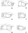
Fig 14. Reconstructions of the sphenethmoid and the descending flange of the frontal and parietal.
Specimens previously attributed to Rhynchonkos (A, C, E) and select microsaurs (B, D, F) in right lateral view. A, Aletrimyti gaskillae, gen. et sp. nov. (FM-UR 1040); B, Nannaroter mckinziei (OMNH 73107); C, Rhynchonkos stovalli (FM-UR 1039); D, Micraroter erythrogeios (FM-UR 2311); E, Dvellecanus carrolli, gen. et sp. nov. (UCMP 202940); F, Huskerpeton englehorni (UNSM 32144). Dashed line indicates the bony margins of the orbit. Scale bars equal 1mm. Abbreviations: fro, frontal; par, parietal; ps, parasphenoid; se, sphenethmoid; II, optic nerve foramen.
Fig 15. Posterior braincases of selected tetrapods in dorsal view, showing ossifications of the synotic tectum.
Interpretive line drawings (A-E) and volumizations (F-I). A, Limnoscelis palustris, after [42]; B, Aerosaurus cf. A. wellesi, after [43]; C, Captorhinus laticeps, after [44]; D, Petrolacosaurus kansensis, after [45]; E, Youngina capensis, after [46]; F, Huskerpeton englehorni (UNSM 32144); G, Rhynchonkos stovalli (FM-UR 1039); H, Aletrimyti (FM-UR 1040); I, Dvellecanus (UCMP 202940). Images not to scale. Abbreviations: eo, exoccipital; lap, lateral ascending process; map, median ascending process; opi, opisthotic; ot, otic; pop, posterior process; pro, prootic; so, supraoccipital; sta, stapes.
Similar articles
- Cranial Morphology of the Brachystelechid 'Microsaur' Quasicaecilia texana Carroll Provides New Insights into the Diversity and Evolution of Braincase Morphology in Recumbirostran 'Microsaurs'.
Pardo JD, Szostakiwskyj M, Anderson JS. Pardo JD, et al. PLoS One. 2015 Jun 24;10(6):e0130359. doi: 10.1371/journal.pone.0130359. eCollection 2015. PLoS One. 2015. PMID: 26107260 Free PMC article. - Cranial Morphology of the Carboniferous-Permian Tetrapod Brachydectes newberryi (Lepospondyli, Lysorophia): New Data from µCT.
Pardo JD, Anderson JS. Pardo JD, et al. PLoS One. 2016 Aug 26;11(8):e0161823. doi: 10.1371/journal.pone.0161823. eCollection 2016. PLoS One. 2016. PMID: 27563722 Free PMC article. - New material of the 'microsaur' Llistrofus from the cave deposits of Richards Spur, Oklahoma and the paleoecology of the Hapsidopareiidae.
Gee BM, Bevitt JJ, Garbe U, Reisz RR. Gee BM, et al. PeerJ. 2019 Jan 25;7:e6327. doi: 10.7717/peerj.6327. eCollection 2019. PeerJ. 2019. PMID: 30701139 Free PMC article. - A new fossil from the Jurassic of Patagonia reveals the early basicranial evolution and the origins of Crocodyliformes.
Pol D, Rauhut OW, Lecuona A, Leardi JM, Xu X, Clark JM. Pol D, et al. Biol Rev Camb Philos Soc. 2013 Nov;88(4):862-72. doi: 10.1111/brv.12030. Epub 2013 Feb 28. Biol Rev Camb Philos Soc. 2013. PMID: 23445256 Review.
Cited by
- A new recumbirostran 'microsaur' from the lower Permian Bromacker locality, Thuringia, Germany, and its fossorial adaptations.
MacDougall MJ, Jannel A, Henrici AC, Berman DS, Sumida SS, Martens T, Fröbisch NB, Fröbisch J. MacDougall MJ, et al. Sci Rep. 2024 Feb 20;14(1):4200. doi: 10.1038/s41598-023-46581-3. Sci Rep. 2024. PMID: 38378723 Free PMC article. - Neurosensory anatomy of Varanopidae and its implications for early synapsid evolution.
Bazzana KD, Evans DC, Bevitt JJ, Reisz RR. Bazzana KD, et al. J Anat. 2022 May;240(5):833-849. doi: 10.1111/joa.13593. Epub 2021 Nov 14. J Anat. 2022. PMID: 34775594 Free PMC article. - Joermungandr bolti, an exceptionally preserved 'microsaur' from the Mazon Creek Lagerstätte reveals patterns of integumentary evolution in Recumbirostra.
Mann A, Calthorpe AS, Maddin HC. Mann A, et al. R Soc Open Sci. 2021 Jul 21;8(7):210319. doi: 10.1098/rsos.210319. eCollection 2021 Jul. R Soc Open Sci. 2021. PMID: 34295525 Free PMC article. - Can We Reliably Calibrate Deep Nodes in the Tetrapod Tree? Case Studies in Deep Tetrapod Divergences.
Pardo JD, Lennie K, Anderson JS. Pardo JD, et al. Front Genet. 2020 Oct 16;11:506749. doi: 10.3389/fgene.2020.506749. eCollection 2020. Front Genet. 2020. PMID: 33193596 Free PMC article. Review. - Mandibular musculature constrains brain-endocast disparity between sarcopterygians.
Challands TJ, Pardo JD, Clement AM. Challands TJ, et al. R Soc Open Sci. 2020 Sep 23;7(9):200933. doi: 10.1098/rsos.200933. eCollection 2020 Sep. R Soc Open Sci. 2020. PMID: 33047053 Free PMC article.
References
- Anderson JS (2008) Focal review: the origin(s) of modern amphibians. Evol Biol 35: 231–347. - PubMed
- Sigurdsen T, Green DM (2011) The origin of modern amphibians: a re-evaluation. Zool J Linn Soc 162: 457–469.
- Jenkins FA, Walsh DM (1993) An Early Jurassic caecilian with limbs. Nature 365:246–250.
- Jenkins FA, Walsh DM, Carroll RL (2007) Anatomy of Eocaecilia micropodia, a limbed caecilian of the Early Jurassic. Bull Mus Comp Zool 158: 285–366.
Publication types
MeSH terms
Grants and funding
This research was supported by Natural Sciences and Engineering Research Council of Canada (NSERC) Discovery Grant 327756-2011 held by Jason S Anderson. The funder had no role in study design, data collection and analysis, decision to publish, or preparation of the manuscript.
LinkOut - more resources
Full Text Sources
Other Literature Sources


