Exosome-delivered microRNAs modulate the inflammatory response to endotoxin - PubMed (original) (raw)
Exosome-delivered microRNAs modulate the inflammatory response to endotoxin
Margaret Alexander et al. Nat Commun. 2015.
Abstract
MicroRNAs regulate gene expression posttranscriptionally and function within the cells in which they are transcribed. However, recent evidence suggests that microRNAs can be transferred between cells and mediate target gene repression. We find that endogenous miR-155 and miR-146a, two critical microRNAs that regulate inflammation, are released from dendritic cells within exosomes and are subsequently taken up by recipient dendritic cells. Following uptake, exogenous microRNAs mediate target gene repression and can reprogramme the cellular response to endotoxin, where exosome-delivered miR-155 enhances while miR-146a reduces inflammatory gene expression. We also find that miR-155 and miR-146a are present in exosomes and pass between immune cells in vivo, as well as demonstrate that exosomal miR-146a inhibits while miR-155 promotes endotoxin-induced inflammation in mice. Together, our findings provide strong evidence that endogenous microRNAs undergo a functional transfer between immune cells and constitute a mechanism of regulating the inflammatory response.
Figures
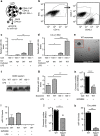
Figure 1. miR-155 is transferred between BMDCs and is present in exosomes.
(a) A schematic of the co-culture experiment. (b) Representative FACS plots where co-cultured CD45.1+ Wt and CD45.2 + miR-155−/− CD11c+ BMDCs were separated (_n_=4). (c) Relative miR-155 levels were quantified via qRT–PCR from isolated miR-155−/− BMDCs that had been cultured alone or with Wt BMDCs in the presence or the absence of LPS for 24 h (_n_=4). (d) Relative miR-155 levels were measured via qRT–PCR in miR-155−/− BMDCs either cultured alone or with Wt BMDCs separated by a 0.4-μm filter for 24 h with or without LPS (_n_=3). (e) Cryo-EM of exosomes isolated from Wt BMDCs. Scale bar, 200 nm. Red box is enlarged in the upper left corner. (f) CD63 protein levels in the exosomal pellet from Wt and miR-155−/− BMDCs treated with or without LPS. (g) Relative levels of miR-155 in exosomes derived from Wt or miR-155−/− BMDCs treated with or without LPS (_n_=3). (h) Exosome quantification of Wt BMDCs treated with or without GW4869 (_n_=3). Limit of detection is 2 × 10⁷ exosomes. (i) Relative levels of miR-155 were measured in the exosomal pellet from Wt BMDCs treated with or without LPS and GW4869 as quantified by qRT–PCR (_n_=2). (j) Exosome quantification of Wt and Rab27 DKO BMDC-derived exosomes (_n_=2). Limit of detection is 2 × 10⁷ exosomes. (k) miR-155 levels in exosome pellets from Wt and Rab27 DKO BMDC-conditioned medium (_n_=2). Data represent two independent experiments and are presented as the mean±s.d. (error bars). *P<0.05; **P<0.01, Student's _t_-test.
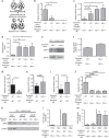
Figure 2. Functional transfer of miR-155 via exosomes in vitro.
(a) Schematic of the exosome transfer experiment. (b) qRT–PCR was used to measure relative miR-155 levels in miR-155−/− BMDCs that received either Wt or miR-155−/− exosomes derived from BMDCs treated with or without GW4869 (_n_=5). (c,d) mRNA levels of miR-155 targets, BACH1 and SHIP1, from the same experiment shown in b as measured by qRT–PCR (_n_=5). (e) Representative western blottings of SHIP1 and β-actin in miR-155−/− BMDCs given either Wt or miR-155−/− exosomes. (f) Protein levels of SHIP1 were quantified using ImageJ software (_n_=2). (g) Relative miR-155 levels in Wt BMDCs given either Wt or miR-155−/− exosomes as quantified by qRT–PCR (_n_=6). (h,i) BACH1 and SHIP1 mRNA levels were measured in the same experiment shown in g as quantified by qRT–PCR (_n_=6). (j) qRT–PCR was used to quantify HO1 mRNA levels during the experiment in b (_n_=5). (k) Western blotting for AGO2 and β-actin from miR-155−/− BMDCs given Wt or miR-155−/− exosomes. On the left is the input (whole-cell lysate), the middle is from the pan-AGO pulldown where one-third of input was used and the right is the IgG pulldown where one-third of the input was used. (l) Relative miR-155 levels were quantified via qRT–PCR in the same experiment shown in k. (m) miR-146a levels were quantified using qRT–PCR during the experiment in k. Levels in l,m are plotted as Ago:IgG. Dotted line separates input from pull-down groups. Data represent two independent experiments and are presented as the mean±s.d. (error bars). *P<0.05; **P<0.01, Student's _t_-test.
Figure 3. Functional transfer of miR-146a via exosomes in vitro.
(a) Levels of miR-146a in the exosomal pellet derived from BMDCs that were treated with or without GW4869 and LPS (_n_=2). (b) miR-146a levels in Wt and Rab27 DKO BMDC-derived exosomal pellets (_n_=2). (c) Schematic of miR-146a exosome-transfer experiment where Wt or miR-146a−/− exosomes were isolated from BMDCs and transferred to recipient miR-146a−/− BMDCs. RNA was isolated after 24 h and the presence of miR-146a was assayed via qRT–PCR. (d) Relative levels of miR-146a in miR-146a−/− BMDCs given exosomes derived from Wt or miR-146a−/− BMDCs (_n_=4). (e) mRNA levels of miR-146a target, IRAK1, were measured from the same cells as in d via qRT–PCR (_n_=4). (f) Representative western blottings of IRAK1 and β-actin from miR-146a−/− cells given either Wt or miR-146a−/− exosomes. (g) IRAK1 protein levels were quantified using ImageJ software (_n_=2). (h) mRNA levels of miR-146a target, TRAF6, were measured in the same cells as in d via qRT–PCR. (i) Western blottings for TRAF6 and β-actin from miR-146a−/− BMDCs given either Wt or miR-146a−/− exosomes (_n_=2). (j) Western blotting results are quantified with ImageJ software. (k) Copy number of miR-146a in Wt and miR-146a−/− exosomes (_n_=3). Copy number is calculated based on a standard curve where a known amount of synthetic miR-146a was spiked into miR-146a−/− BMDC-derived exosome pellet followed by RNA isolation and qRT–PCR. (l) Copy number of miR-146a was measured via qRT–PCR in miR-146a−/− recipient BMDCs that received either Wt or miR-146a−/− exosomes (146a−/− BMDC+Wt exos and 146a−/− BMDC+146a−/− exos), as well as in Wt and miR-146a−/− donor BMDCs (_n_=3). Average copy number is displayed above. Copy number is calculated based on a standard curve where a known amount of synthetic miR-146a was spiked into miR-146a−/− BMDC pellet followed by RNA isolation and qRT–PCR. Data represent two independent experiments and are presented as the mean±s.d. (error bars). *P<0.05, Student's _t_-test.
Figure 4. Seed-dependent repression of miRNA targets by exosome-delivered miR-155 and miR-146a.
(a) Schematic for mimic experiment. (b,c) Relative mRNA levels of the miR-155 targets SHIP1 and BACH1 were measured via qRT–PCR in recipient cells that received exosomes with no mimics (No) (_n_=7), miR-155 seed mutant mimics (Seed) (_n_=4), with scrambled mimics (Scram) (_n_=3), or with miR-155-mimics (Mimic) (_n_=7). (d,e) qRT–PCR was preformed to assay the mRNA levels of the miR-146a targets, IRAK1 and TRAF6, following treatment with exosomes containing miR-146a mimics and controls as in b,c. Results are reported normalized to exosomes with no mimics added, which is set as 1. (f) Protein levels of TRAF6, IRAK1 and β-actin were determined via western blotting using lysates from miR-146a−/− BMDCs that received exosomes containing no mimics, seed mutant mimics or Wt mimics. Numbers below the blot represent relative protein levels with no mimics set as 1 following normalization to β-actin. (g) Schematic for 3′-UTR luciferase reporter assays in h,i. (h) Results from 3′-UTR luciferase reporter assays where miR-155−/− BMDCs were transfected with a pmiReport control vector, a BACH1 3′-UTR vector (BACH1), a BACH1 miR-155-binding site (bs) mutant vector (BACH1 mutant), or a 2mer-positive control vector. Transfected BMDCs were treated 6 h later with or without Wt exosomes and per cent change in luciferase activity of exosome treated BMDCs compared with no exosome treatment was calculated after 24 h (_n_=4). (i) Results from 3′-UTR luciferase reporter assay where miR-146a−/− BMDCs were transfected with a pmiReport control vector, a TRAF6 3′-UTR vector (TRAF6) or TRAF6 miR-146a bs mutant vector (TRAF6 mutant). Six hours later, the BMDCs were treated with or without Wt exosomes and per cent repression of luciferase activity was calculated 24 h after exosome transfer (_n_=4). Results represent two independent experiments. All data are presented as the mean±s.d. (error bars). *P<0.05; **P<0.01, ****P<0.0001; Student's _t_-test.
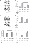
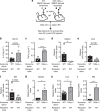
Figure 5. Exosomal transfer of miR-155 and miR-146a programme the response to LPS in vitro
(a) A schematic of the experimental design for b. (b) Exosomes were isolated from Wt or miR-155−/− BMDCs and given to miR-155−/− BMDCs for 24 h. Cells were then treated with or without LPS and media was taken after 6 h for an IL-6 enzyme-linked immunosorbent assay. Relative IL-6 protein levels are shown (_n_=4). (c) Schematic for experiments in d–f. (d–f) qRT–PCR was used to quantify mRNA levels of IL-10, IL-6 and IL-12 p40 in miR-146a−/− BMDCs given exosomes from Wt or miR-146a−/− BMDCs for 24 h followed by stimulation with or without LPS for 6 h (_n_=4). Data represent two independent experiments. All data are presented as the mean±s.d. (error bars). *P<0.05; Student's _t_-test.
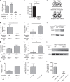
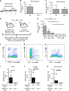
Figure 6. Transfer of endogenous miR-155 between haematopoietic cells in vivo.
(a) CD63 western blotting using exosomes isolated directly from the BM of Wt or miR-155−/− mice. 1 and 2 stand for two biological replicates. (b,c) Levels of miR-155 and miR-146a in exosomes isolated form Wt and miR-155−/− mouse BM as measured by qRT–PCR (_n_=2). (d) Schematic of the in-vivo experiment. (e) qRT–PCR was used to quantify levels of miR-155 in miR-155−/− CD45.2+ BM cells that were B220+, CD3+, or CD11b+ from miR-155−/− mice that were either reconstituted with Wt (CD45.1+) and miR-155−/− BM, or miR-155−/− BM alone as indicated (_n_=5). (f–h) Representative FACS plots of the cell types in isolated BM shown in e (_n_=5). (i–k) miR-155−/− mice were i.p. injected multiple times over a week with either Wt or miR-155−/− exosomes. CD3+ T cells, B220+ B cells and CD11b+ myeloid cells were sorted from mouse spleens and qRT–PCR was preformed to analyse the delivery of miR-155 to each cell type (_n_=5). All data are presented as the mean±s.d. (error bars). *P<0.05; **P<0.01, ***P<0.001, Student's _t_-test.
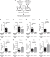
Figure 7. miR-155-containing exosomes promote a heightened response to LPS in miR-155−/− mice.
(a) Schematic of the experimental design where miR-155−/− mice were i.p. injected with either Wt or miR-155−/− BMDC-derived exosomes and then challenged with LPS 24 h later. Blood was taken 2 h post LPS injection and the spleen, liver and BM were harvested 24 h post injection. (b,c) Serum TNFα and IL-6 concentrations were analysed via enzyme-linked immunosorbent assay 2 h after injection of LPS in miR-155−/− mice that had been pretreated with either Wt or miR-155−/− exosomes (_n_=5). (d–f) qRT–PCR was preformed using RNA isolated from the spleen, liver and BM, to assay the relative levels of exosomally delivered miR-155 (_n_=5). (g–i) mRNA levels of the miR-155 targets SHIP1 and BACH1 were measured in the spleen, liver and/or the BM using qRT–PCR (_n_=5). All data are presented as the mean±s.d. (error bars). *P<0.05; **P<0.01, Student's _t_-test.
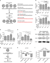
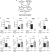
Figure 8. miR-146a-containing exosomes reduce inflammatory responses to LPS in miR-146a−/− mice.
(a) Schematic of the experimental design where miR-146a−/− mice were i.p. injected with either Wt or miR-146a−/− BMDC-derived exosomes and then challenged with LPS 24 h later. Blood was taken 2 h post LPS injection and the spleen, liver and BM were harvested 24 h post injection. (b,c) Serum TNFα and IL-6 were analysed via enzyme-linked immunosorbent assay 2 h after injection of LPS (_n_=5). (d–f) qRT–PCR was preformed using RNA isolated from the spleen, liver and BM, to assay the relative levels of exosomally delivered miR-146a (_n_=5). (g–i) mRNA levels of the miR-146a targets TRAF6 and IRAK1 were measured in the spleen, liver and/or the BM using qRT–PCR (_n_=5). All data are presented as the mean±s.d. (error bars). *P<0.05; **P<0.01, Student's _t_-test.
Figure 9. miR-146a-containing exosomes reduce inflammatory response to LPS in Wt mice.
(a) Schematic of the experimental design where Wt mice were i.p. injected with either Wt or miR-146a−/− BMDC-derived exosomes and then challenged with LPS 24 h later. Blood was taken 2 h post LPS injection and the spleen, liver and BM were harvested 24 h post injection. (b,c) Serum TNFα and IL-6 were analysed via enzyme-linked immunosorbent assay 2 h after injection of LPS (_n_=5). (d–f) qRT–PCR using RNA isolated from the spleen, liver and BM was performed to assay the relative levels of exosomally delivered miR-146a (_n_=5). (g–i) mRNA levels of the miR-146a targets TRAF6 and IRAK1 were measured in the spleen, liver and/or the BM using qRT–PCR (_n_=5). Results represent two independent experiments. All data are presented as the mean±s.d. (error bars). *P<0.05; ****P<0.0001, Student's _t_-test.
Similar articles
- Exosome-mediated miR-146a transfer suppresses type I interferon response and facilitates EV71 infection.
Fu Y, Zhang L, Zhang F, Tang T, Zhou Q, Feng C, Jin Y, Wu Z. Fu Y, et al. PLoS Pathog. 2017 Sep 14;13(9):e1006611. doi: 10.1371/journal.ppat.1006611. eCollection 2017 Sep. PLoS Pathog. 2017. PMID: 28910400 Free PMC article. - Exosomal miR-146a Contributes to the Enhanced Therapeutic Efficacy of Interleukin-1β-Primed Mesenchymal Stem Cells Against Sepsis.
Song Y, Dou H, Li X, Zhao X, Li Y, Liu D, Ji J, Liu F, Ding L, Ni Y, Hou Y. Song Y, et al. Stem Cells. 2017 May;35(5):1208-1221. doi: 10.1002/stem.2564. Epub 2017 Feb 5. Stem Cells. 2017. PMID: 28090688 - BM-MSCs-derived microvesicles promote allogeneic kidney graft survival through enhancing micro-146a expression of dendritic cells.
Wu XQ, Yan TZ, Wang ZW, Wu X, Cao GH, Zhang C. Wu XQ, et al. Immunol Lett. 2017 Nov;191:55-62. doi: 10.1016/j.imlet.2017.09.010. Epub 2017 Sep 28. Immunol Lett. 2017. PMID: 28963073 - Exosome-Encapsulated microRNAs as Potential Circulating Biomarkers in Colon Cancer.
Hosseini M, Khatamianfar S, Hassanian SM, Nedaeinia R, Shafiee M, Maftouh M, Ghayour-Mobarhan M, ShahidSales S, Avan A. Hosseini M, et al. Curr Pharm Des. 2017;23(11):1705-1709. doi: 10.2174/1381612822666161201144634. Curr Pharm Des. 2017. PMID: 27908272 Review. - Mesenchymal stem cells-derived exosomal microRNAs contribute to wound inflammation.
Ti D, Hao H, Fu X, Han W. Ti D, et al. Sci China Life Sci. 2016 Dec;59(12):1305-1312. doi: 10.1007/s11427-016-0240-4. Epub 2016 Nov 18. Sci China Life Sci. 2016. PMID: 27864711 Review.
Cited by
- Exosomes derived from synovial fibroblasts under hypoxia aggravate rheumatoid arthritis by regulating Treg/Th17 balance.
Ding Y, Wang L, Wu H, Zhao Q, Wu S. Ding Y, et al. Exp Biol Med (Maywood). 2020 Aug;245(14):1177-1186. doi: 10.1177/1535370220934736. Epub 2020 Jul 2. Exp Biol Med (Maywood). 2020. PMID: 32615822 Free PMC article. - Inhibition of miR-155 Protects Against LPS-induced Cardiac Dysfunction and Apoptosis in Mice.
Wang H, Bei Y, Huang P, Zhou Q, Shi J, Sun Q, Zhong J, Li X, Kong X, Xiao J. Wang H, et al. Mol Ther Nucleic Acids. 2016 Oct 11;5(10):e374. doi: 10.1038/mtna.2016.80. Mol Ther Nucleic Acids. 2016. PMID: 27727247 Free PMC article. - Extracellular vesicle therapeutics from plasma and adipose tissue.
Iannotta D, Yang M, Celia C, Di Marzio L, Wolfram J. Iannotta D, et al. Nano Today. 2021 Aug;39:101159. doi: 10.1016/j.nantod.2021.101159. Epub 2021 Apr 27. Nano Today. 2021. PMID: 33968157 Free PMC article. - Aging and Metabolic Reprogramming of Adipose-Derived Stem Cells Affect Molecular Mechanisms Related to Cardiovascular Diseases.
Holvoet P. Holvoet P. Cells. 2023 Dec 7;12(24):2785. doi: 10.3390/cells12242785. Cells. 2023. PMID: 38132104 Free PMC article. Review. - Small RNA Sequencing in Cells and Exosomes Identifies eQTLs and 14q32 as a Region of Active Export.
Tsang EK, Abell NS, Li X, Anaya V, Karczewski KJ, Knowles DA, Sierra RG, Smith KS, Montgomery SB. Tsang EK, et al. G3 (Bethesda). 2017 Jan 5;7(1):31-39. doi: 10.1534/g3.116.036137. G3 (Bethesda). 2017. PMID: 27799337 Free PMC article.
References
- Tian T., Wang Y., Wang H., Zhu Z. & Xiao Z. Visualizing of the cellular uptake and intracellular trafficking of exosomes by live-cell microscopy. J. Cell. Biochem. 111, 488–496 (2010) . - PubMed
- Théry C., Zitvogel L. & Amigorena S. Exosomes: composition, biogenesis and function. Nat. Rev. Immunol. 2, 569–579 (2002) . - PubMed
- Valadi H. et al. Exosome-mediated transfer of mRNAs and microRNAs is a novel mechanism of genetic exchange between cells. Nat. Cell Biol. 9, 654–659 (2007) . - PubMed
Publication types
MeSH terms
Substances
LinkOut - more resources
Full Text Sources
Other Literature Sources
Molecular Biology Databases
