Regulation of dendrite morphogenesis by extrinsic cues - PubMed (original) (raw)
Review
Regulation of dendrite morphogenesis by extrinsic cues
Pamela Valnegri et al. Trends Neurosci. 2015 Jul.
Abstract
Dendrites play a central role in the integration and flow of information in the nervous system. The morphogenesis and maturation of dendrites is hence an essential step in the establishment of neuronal connectivity. Recent studies have uncovered crucial functions for extrinsic cues in the development of dendrites. We review the contribution of secreted polypeptide growth factors, contact-mediated proteins, and neuronal activity in distinct phases of dendrite development. We also highlight how extrinsic cues influence local and global intracellular mechanisms of dendrite morphogenesis. Finally, we discuss how these studies have advanced our understanding of neuronal connectivity and have shed light on the pathogenesis of neurodevelopmental disorders.
Keywords: calcium signaling; contact-mediated regulators; dendrite morphogenesis; neuronal activity; secreted polypeptide growth factors.
Copyright © 2015 Elsevier Ltd. All rights reserved.
Figures
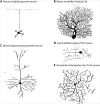
Figure 1. Diverse patterns of dendrite branching in different type of neurons
(A) Mouse cerebellar granule neuron have only four to five dendrites, each of which ends with a dendritic claw that harbors postsynaptic dendritic specializations. (B) Elaborate dendritic tree in a mouse Purkinje cell. (C) Mouse hippocampal pyramidal neuron characterized by two distinct dendritic trees, the basal and apical dendrites. (D) Dendritic tree in Caenorhabditis elegans PVD neuron. (E) Multidendritic class IV da neuron in Drosophila melanogaster.
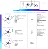
Figure 2. Cell-extrinsic regulators of dendrite morphogenesis
Summary of molecules regulating different stages of dendrite development, as described in the text. *Molecules that mediate repulsion between sister dendrites.
Figure 3. Effects of calcium signaling on dendrite morphogenesis
Calcium influx from voltage-gated calcium channels VGCCs or NMDA receptors (NMDAR) activates several CaMK family members and MAPKs which in turn regulate dendrite growth and elaboration. VGCC are also responsible to generate compartmentalized calcium transients to trigger dendrite pruning. In later stage of dendrite development, CaMKIIβ activated by calcium influx from TRPC5 channel drives dendrite pruning.
Similar articles
- Mapping CRMP3 domains involved in dendrite morphogenesis and voltage-gated calcium channel regulation.
Quach TT, Wilson SM, Rogemond V, Chounlamountri N, Kolattukudy PE, Martinez S, Khanna M, Belin MF, Khanna R, Honnorat J, Duchemin AM. Quach TT, et al. J Cell Sci. 2013 Sep 15;126(Pt 18):4262-73. doi: 10.1242/jcs.131409. Epub 2013 Jul 18. J Cell Sci. 2013. PMID: 23868973 - Mechanisms of dendritic maturation.
Libersat F, Duch C. Libersat F, et al. Mol Neurobiol. 2004 Jun;29(3):303-20. doi: 10.1385/MN:29:3:303. Mol Neurobiol. 2004. PMID: 15181241 Review. - [Intrinsic and extrinsic mechanisms regulating neuronal dendrite morphogenesis].
Zhao W, Zou W. Zhao W, et al. Zhejiang Da Xue Xue Bao Yi Xue Ban. 2020 May 25;49(1):90-99. doi: 10.3785/j.issn.1008-9292.2020.02.09. Zhejiang Da Xue Xue Bao Yi Xue Ban. 2020. PMID: 32621417 Free PMC article. Review. Chinese. - Molecular mechanisms that mediate dendrite morphogenesis.
Lefebvre JL. Lefebvre JL. Curr Top Dev Biol. 2021;142:233-282. doi: 10.1016/bs.ctdb.2020.12.008. Epub 2021 Feb 17. Curr Top Dev Biol. 2021. PMID: 33706919 Review. - Dendrite morphogenesis from birth to adulthood.
Prigge CL, Kay JN. Prigge CL, et al. Curr Opin Neurobiol. 2018 Dec;53:139-145. doi: 10.1016/j.conb.2018.07.007. Epub 2018 Aug 6. Curr Opin Neurobiol. 2018. PMID: 30092409 Free PMC article. Review.
Cited by
- Interplay between axonal Wnt5-Vang and dendritic Wnt5-Drl/Ryk signaling controls glomerular patterning in the Drosophila antennal lobe.
Hing H, Reger N, Snyder J, Fradkin LG. Hing H, et al. PLoS Genet. 2020 May 1;16(5):e1008767. doi: 10.1371/journal.pgen.1008767. eCollection 2020 May. PLoS Genet. 2020. PMID: 32357156 Free PMC article. - Reelin-Nrp1 Interaction Regulates Neocortical Dendrite Development in a Context-Specific Manner.
Kohno T, Ishii K, Hirota Y, Honda T, Makino M, Kawasaki T, Nakajima K, Hattori M. Kohno T, et al. J Neurosci. 2020 Oct 21;40(43):8248-8261. doi: 10.1523/JNEUROSCI.1907-20.2020. Epub 2020 Oct 2. J Neurosci. 2020. PMID: 33009002 Free PMC article. - The Zinc-BED Transcription Factor Bedwarfed Promotes Proportional Dendritic Growth and Branching through Transcriptional and Translational Regulation in Drosophila.
Bhattacharjee S, Iyer EPR, Iyer SC, Nanda S, Rubaharan M, Ascoli GA, Cox DN. Bhattacharjee S, et al. Int J Mol Sci. 2023 Mar 28;24(7):6344. doi: 10.3390/ijms24076344. Int J Mol Sci. 2023. PMID: 37047316 Free PMC article. - Mechanisms regulating dendritic arbor patterning.
Ledda F, Paratcha G. Ledda F, et al. Cell Mol Life Sci. 2017 Dec;74(24):4511-4537. doi: 10.1007/s00018-017-2588-8. Epub 2017 Jul 22. Cell Mol Life Sci. 2017. PMID: 28735442 Free PMC article. Review. - The Zinc-BED transcription factor Bedwarfed promotes proportional dendritic growth and branching through transcriptional and translational regulation in Drosophila.
Bhattacharjee S, Iyer EPR, Iyer SC, Nanda S, Rubaharan M, Ascoli GA, Cox DN. Bhattacharjee S, et al. bioRxiv [Preprint]. 2023 Feb 15:2023.02.15.528686. doi: 10.1101/2023.02.15.528686. bioRxiv. 2023. PMID: 36824896 Free PMC article. Updated. Preprint.
References
- Goldberg JL. Intrinsic neuronal regulation of axon and dendrite growth. Curr Opin Neurobiol. 2004 Oct;14(5):551–7. doi:S0959-4388(04)00126-6 [pii] 10.1016/j.conb.2004.08.012. - PubMed
- Huang EJ, Reichardt LF. Trk receptors: roles in neuronal signal transduction. Annu Rev Biochem. 2003;72:609–42. doi:10.1146/annurev.biochem.72.121801.161629121801.161629 [pii] - PubMed
- McAllister AK, Lo DC, Katz LC. Neurotrophins regulate dendritic growth in developing visual cortex. Neuron. 1995 Oct;15(4):791–803. doi:0896-6273(95)90171-X [pii] - PubMed
Publication types
MeSH terms
Substances
LinkOut - more resources
Full Text Sources
Other Literature Sources


