Genome-Wide DNA Methylation Analysis Identifies Novel Hypomethylated Non-Pericentromeric Genes with Potential Clinical Implications in ICF Syndrome - PubMed (original) (raw)
Genome-Wide DNA Methylation Analysis Identifies Novel Hypomethylated Non-Pericentromeric Genes with Potential Clinical Implications in ICF Syndrome
L Simo-Riudalbas et al. PLoS One. 2015.
Abstract
Introduction and results: Immunodeficiency, centromeric instability and facial anomalies syndrome (ICF) is a rare autosomal recessive disease, characterized by severe hypomethylation in pericentromeric regions of chromosomes (1, 16 and 9), marked immunodeficiency and facial anomalies. The majority of ICF patients present mutations in the DNMT3B gene, affecting the DNA methyltransferase activity of the protein. In the present study, we have used the Infinium 450K DNA methylation array to evaluate the methylation level of 450,000 CpGs in lymphoblastoid cell lines and untrasformed fibroblasts derived from ICF patients and healthy donors. Our results demonstrate that ICF-specific DNMT3B variants A603T/STP807ins and V699G/R54X cause global DNA hypomethylation compared to wild-type protein. We identified 181 novel differentially methylated positions (DMPs) including subtelomeric and intrachromosomic regions, outside the classical ICF-related pericentromeric hypomethylated positions. Interestingly, these sites were mainly located in intergenic regions and inside the CpG islands. Among the identified hypomethylated CpG-island associated genes, we confirmed the overexpression of three selected genes, BOLL, SYCP2 and NCRNA00221, in ICF compared to healthy controls, which are supposed to be expressed in germ line and silenced in somatic tissues.
Conclusions: In conclusion, this study contributes in clarifying the direct relationship between DNA methylation defect and gene expression impairment in ICF syndrome, identifying novel direct target genes of DNMT3B. A high percentage of the DMPs are located in the subtelomeric regions, indicating a specific role of DNMT3B in methylating these chromosomal sites. Therefore, we provide further evidence that hypomethylation in specific non-pericentromeric regions of chromosomes might be involved in the molecular pathogenesis of ICF syndrome. The detection of DNA hypomethylation at BOLL, SYCP2 and NCRNA00221 may pave the way for the development of specific clinical biomarkers with the aim to facilitate the identification of ICF patients.
Conflict of interest statement
Competing Interests: The authors have declared that no competing interests exist.
Figures
Fig 1. Genome-wide DNA methylation profiles in ICF patients and control samples.
(A) Histograms shows bimodal distribution pattern of DNA methylation profiles in ICF patients and normal donors. The frequency of CpGs according to DNA methylation levels are depicted in the graph. (B) Table showing number of average poorly methylated (methylation levels beta<0.33) and average highly methylated (methylation levels Beta>0.66). (C) Scatter plot represents comparison of DNA methylation levels of total CpG sites using the Infinium 450K DNA methylation assay. Green triangle selects hypomethylated area for ICF patients compared to controls. (D) Box plot displaying the distribution of Beta-values of total CpG sites of ICF versus healthy control donors. Normality was tested using the Shapiro-Wilk test and significance was evaluated with the Mann-Whitney U test and is indicated by three asterisks *** (p<0.001).
Fig 2. Identification of Differentially methylated CpGs.
(A) Unsupervised hierarchical clustering and heatmap of four cord blood donors (purple), three unrelated healthy donors (blue) and two ICF patients (orange) using 5000 random selected CpGs. DNA Methylation levels scale is shown. Each column represents patients and each row represents the different CpGs. (B) Supervised cluster and heatmap representing the distinctive 181 CpGs corresponding to the comparison between ICF patients (orange) and all control samples (dark blue).
Fig 3. Schematic representation of chromosomes and CpG localization.
Gene names and intergenic CpGs are represented and localized by blue lines.
Fig 4. Genomic distribution and gene features of the 53 differentially methylated CpGs.
(A) Chromosomal sub-localization classified in different groups: subtelomeric, pericentromeric and intrachromosomal. (B) Associated RNA transcription classified in: coding, non-coding and intergenic. (C) CpG context and neighborhood classified in: island, shore, shelf and open sea/others. (D) Functional genomic distribution classified in: promoter (TSS1500, TSS200, 5´UTR), genic (1stexon, body and 3´UTR) and intergenic.
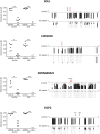
Fig 5. Validation of DNA methylation of four representative genes (BOLL, SYCP2, LDHALD6 and NCRNA00221) levels by bisulfite genomic sequencing analysis.
Left panel depicts DNA methylation values (Beta) from ICF and healthy donors using both methodologies bisulfite genomic sequencing (BSG) and 450K array. Right panel shows bisulfite genomic sequencing analysis of genes in one representative control and ICF patient. CpG dinucleotides are shown in vertical lines. Multiple single clones are represented for each sample. Presence of unmethylated or methylated CpGs is indicated by white or black squares, respectively. Red arrows mark the localization of the differentially methylated CpGs by 450K array. The distance to transcription start site (bp) is also indicated.
Fig 6. Gene expression analysis for the selected 4 CpG island-promoter associated genes BOLL, SYCP2, LDHALD6 and NCRNA00221.
Fold change values of the differentially DNA methylated genes in lymphoblastoid ICF patients and healthy donors were evaluated by qRT-PCR. In parallel, Fold change values were also tested in untransformed fibroblast form an ICF patient and a healthy donor. Values were determined at least in triplicate. Statistic analysis was evaluated using student t test and significance symbols correspond to (* p<0.05; ** p<0.01 and *** p<0.001).
Similar articles
- Satellite 2 methylation patterns in normal and ICF syndrome cells and association of hypomethylation with advanced replication.
Hassan KM, Norwood T, Gimelli G, Gartler SM, Hansen RS. Hassan KM, et al. Hum Genet. 2001 Oct;109(4):452-62. doi: 10.1007/s004390100590. Hum Genet. 2001. PMID: 11702227 - DNA methyltransferase 3B (DNMT3B) mutations in ICF syndrome lead to altered epigenetic modifications and aberrant expression of genes regulating development, neurogenesis and immune function.
Jin B, Tao Q, Peng J, Soo HM, Wu W, Ying J, Fields CR, Delmas AL, Liu X, Qiu J, Robertson KD. Jin B, et al. Hum Mol Genet. 2008 Mar 1;17(5):690-709. doi: 10.1093/hmg/ddm341. Epub 2007 Nov 20. Hum Mol Genet. 2008. PMID: 18029387 - Whole-genome methylation scan in ICF syndrome: hypomethylation of non-satellite DNA repeats D4Z4 and NBL2.
Kondo T, Bobek MP, Kuick R, Lamb B, Zhu X, Narayan A, Bourc'his D, Viegas-Péquignot E, Ehrlich M, Hanash SM. Kondo T, et al. Hum Mol Genet. 2000 Mar 1;9(4):597-604. doi: 10.1093/hmg/9.4.597. Hum Mol Genet. 2000. PMID: 10699183 - ICF syndrome cells as a model system for studying X chromosome inactivation.
Gartler SM, Hansen RS. Gartler SM, et al. Cytogenet Genome Res. 2002;99(1-4):25-9. doi: 10.1159/000071571. Cytogenet Genome Res. 2002. PMID: 12900541 Review. - The gene mutations and subtelomeric DNA methylation in immunodeficiency, centromeric instability and facial anomalies syndrome.
Hu H, Chen C, Shi S, Li B, Duan S. Hu H, et al. Autoimmunity. 2019 Aug-Sep;52(5-6):192-198. doi: 10.1080/08916934.2019.1657846. Epub 2019 Sep 2. Autoimmunity. 2019. PMID: 31476899 Review.
Cited by
- Enhanced insulin-regulated phagocytic activities support extreme health span and longevity in multiple populations.
Wu D, Bi X, Li P, Xu D, Qiu J, Li K, Zheng S, Chow KH. Wu D, et al. Aging Cell. 2023 May;22(5):e13810. doi: 10.1111/acel.13810. Epub 2023 Mar 8. Aging Cell. 2023. PMID: 36883688 Free PMC article. - The Alteration of Subtelomeric DNA Methylation in Aging-Related Diseases.
Hu H, Li B, Duan S. Hu H, et al. Front Genet. 2019 Jan 9;9:697. doi: 10.3389/fgene.2018.00697. eCollection 2018. Front Genet. 2019. PMID: 30687384 Free PMC article. Review. - Epigenetic aging of human hematopoietic cells is not accelerated upon transplantation into mice.
Frobel J, Rahmig S, Franzen J, Waskow C, Wagner W. Frobel J, et al. Clin Epigenetics. 2018 May 22;10:67. doi: 10.1186/s13148-018-0499-7. eCollection 2018. Clin Epigenetics. 2018. PMID: 29796118 Free PMC article. - DNA methylation in mammalian development and disease.
Smith ZD, Hetzel S, Meissner A. Smith ZD, et al. Nat Rev Genet. 2025 Jan;26(1):7-30. doi: 10.1038/s41576-024-00760-8. Epub 2024 Aug 12. Nat Rev Genet. 2025. PMID: 39134824 Review. - Losing DNA methylation at repetitive elements and breaking bad.
Pappalardo XG, Barra V. Pappalardo XG, et al. Epigenetics Chromatin. 2021 Jun 3;14(1):25. doi: 10.1186/s13072-021-00400-z. Epigenetics Chromatin. 2021. PMID: 34082816 Free PMC article. Review.
References
- Kondo T, Bobek MP, Kuick R, Lamb B, Zhu X, Narayan A et al. (2000) Whole-genome methylation scan in ICF syndrome: hypomethylation of non-satellite DNA repeats D4Z4 and NBL2. Hum Mol Genet. 9(4):597–604. - PubMed
- Ehrlich M, Buchanan KL, Tsien F, Jiang G, Sun B, Uicker W et al. (2001) DNA methyltransferase 3B mutations linked to the ICF syndrome cause dysregulation of lymphogenesis genes. Hum Mol Genet 10(25):2917–31. - PubMed
Publication types
MeSH terms
Substances
LinkOut - more resources
Full Text Sources
Other Literature Sources
Molecular Biology Databases


