Low Back Pain: Current Surgical Approaches - PubMed (original) (raw)
Review
Low Back Pain: Current Surgical Approaches
Santosh Baliga et al. Asian Spine J. 2015 Aug.
Abstract
Low back pain (LBP) is a worldwide phenomenon. The UK studies place LBP as the largest single cause of absence from work; up to 80% of the population will experience LBP at least once in their lifetime. Most individuals do not seek medical care and are not disabled by their pain once it is managed by nonoperative measures. However, around 10% of patients go on to develop chronic pain. This review outlines the basics of the traditional approach to spinal surgery for chronic LBP secondary to osteoarthritis of the lumbar spine as well as explains the novel concepts and terminology of back pain surgery. Traditionally, the stepwise approach to surgery starts with local anaesthetic and steroid injection followed by spinal fusion. Fusion aims to alleviate pain by preventing movement between affected spinal segments; this commonly involves open surgery, which requires large soft tissue dissection and there is a possibility of blood loss and prolonged recovery time. Established minimally invasive spine surgery techniques (MISS) aim to reduce all of these complications and they include laparoscopic anterior lumbar interbody fusion and MISS posterior instrumentation with pedicle screws and rods. Newer MISS techniques include extreme lateral interbody fusion and axial interbody fusion. The main problem of fusion is the disruption of the biomechanics of the rest of the spine; leading to adjacent level disease. Theoretically, this can be prevented by performing motion-preserving surgeries such as total disc replacement, facet arthroplasty, and non fusion stabilisation. We outline the basic concepts of the procedures mentioned above as well as explore some of the novel surgical therapies available for chronic LBP.
Keywords: Intervertebral disc degeneration; Low back pain; Minimally invasive surgical procedures; Pedicle screws, Zygapophyseal joints; Spinal fusion.
Conflict of interest statement
Conflict of Interest: No potential conflict of interest relevant to this article was reported.
Figures
Fig. 1. In the normal spine, the discs have high water content (left). As the disc degenerates, it dehydrates, losing height or collapse (right). This puts pressure on the facet joints and may result in arthritis of these joints. Both diagrams show a spinal segment; two adjacent vertebrae with a pair of facet joints and the intervertebral disc.
Fig. 2. The classic versus new treatment ladder of back pain. Reprinted from Bertagnoli [21] with permission from North American Spine Society.
Fig. 3. A picture of interspinous spacer (SMS Q Spine) implanted between the spinous process of two lumbar vertebrae.
Fig. 4. Radiographs of an L5-S1 total disc replacement and a photograph of the prosthesis. Reprinted image of the Freedom Lumbar Disc with permission from AxioMed Spine Co., Cleveland, OH, USA.
Fig. 5. Schematic diagram of the total facet arthroplasty system. Reprinted with permission from Globus Medical Inc., Phoenixville, PA, USA.
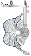
Fig. 6. Approaches to lumbar spine can be broadly divided into anterior (red arrow) and posterior (blue arrow). Axial magnetic resonance imaging also demonstrating extreme lateral access (black arrow) and transforaminal (green arrow) approaches.
Fig. 7. Pedicle screws are inserted posteriorly through the pedicles into the vertebral body. Rods are used to stabilise the two vertebrae by linking them to the pedicle screws. If indicated, several levels can be fused at the same time.
Fig. 8. Schematic drawing showing the extreme lateral interbody fusion procedure; the retractor is inserted into the retroperitoneal space, penetrating the psoas muscle, and positioned directly on the lateral intervertebral disc space.
Fig. 9. Radiograph showing interbody fusion after the extreme lateral interbody fusion procedure supplemented by posterior instrumentation, which can also be achieved by minimally invasive spine surgery; useful in correcting the spinal deformity.
Fig. 10. The axial interbody fusion rod is implanted into the L5-S1 disc space, making it a stable construct containing bone graft. Reprinted with permission from Surgi-C Ltd., Birmingham, UK.
Fig. 11. (A) Computer tomography image of the axial interbody fusion with the formation of a bridging callus between the sacrum and the L5 vertebra. (B) Schematic showing AxiaLIF Rod inserted between S1 and L5 vertebrae. Reprinted from Tobler et al. [62] with permission of Wolters Kluwer Health Inc.
Similar articles
- Minimally invasive lateral interbody fusion for the treatment of rostral adjacent-segment lumbar degenerative stenosis without supplemental pedicle screw fixation.
Wang MY, Vasudevan R, Mindea SA. Wang MY, et al. J Neurosurg Spine. 2014 Dec;21(6):861-6. doi: 10.3171/2014.8.SPINE13841. Epub 2014 Oct 10. J Neurosurg Spine. 2014. PMID: 25303619 - Minimally invasive spine surgery in chronic low back pain patients.
Spoor AB, Öner FC. Spoor AB, et al. J Neurosurg Sci. 2013 Sep;57(3):203-18. J Neurosurg Sci. 2013. PMID: 23877267 Review. - [Minimally Invasive Posterior Lumbar Interbody Fusion and Instrumentation - Outcomes at 24-Month Follow-up].
Šámal F, Linzer P, Filip M, Jurek P, Pohlodek D, Haninec P. Šámal F, et al. Acta Chir Orthop Traumatol Cech. 2020;87(2):95-100. Acta Chir Orthop Traumatol Cech. 2020. PMID: 32396509 Czech. - Endoscopic transforaminal decompression, interbody fusion, and percutaneous pedicle screw implantation of the lumbar spine: A case series report.
Osman SG. Osman SG. Int J Spine Surg. 2012 Dec 1;6:157-66. doi: 10.1016/j.ijsp.2012.04.001. eCollection 2012. Int J Spine Surg. 2012. PMID: 25694885 Free PMC article. - Minimally-invasive posterior lumbar stabilization for degenerative low back pain and sciatica. A review.
Bonaldi G, Brembilla C, Cianfoni A. Bonaldi G, et al. Eur J Radiol. 2015 May;84(5):789-98. doi: 10.1016/j.ejrad.2014.04.012. Epub 2014 May 9. Eur J Radiol. 2015. PMID: 24906245 Review.
Cited by
- Stem cell therapy in discogenic back pain.
Barakat AH, Elwell VA, Lam KS. Barakat AH, et al. J Spine Surg. 2019 Dec;5(4):561-583. doi: 10.21037/jss.2019.09.22. J Spine Surg. 2019. PMID: 32043007 Free PMC article. Review. - We Need to Talk about Lumbar Total Disc Replacement.
Beatty S. Beatty S. Int J Spine Surg. 2018 Aug 3;12(2):201-240. doi: 10.14444/5029. eCollection 2018 Apr. Int J Spine Surg. 2018. PMID: 30276080 Free PMC article. - Metallic Implants Used in Lumbar Interbody Fusion.
Litak J, Szymoniuk M, Czyżewski W, Hoffman Z, Litak J, Sakwa L, Kamieniak P. Litak J, et al. Materials (Basel). 2022 May 20;15(10):3650. doi: 10.3390/ma15103650. Materials (Basel). 2022. PMID: 35629676 Free PMC article. Review. - Anterior lumbar interbody fusion (ALIF): biometrical results and own experiences.
Kapustka B, Kiwic G, Chodakowski P, Miodoński JP, Wysokiński T, Łączyński M, Paruzel K, Kotas A, Marcol W. Kapustka B, et al. Neurosurg Rev. 2020 Apr;43(2):687-693. doi: 10.1007/s10143-019-01108-1. Epub 2019 May 20. Neurosurg Rev. 2020. PMID: 31111262 Free PMC article. - Validation and cross-cultural adaptation of the Korean version of the Core Outcome Measures Index in patients with degenerative lumbar disease.
Kim HJ, Yeom JS, Nam Y, Lee NK, Heo YW, Lee SY, Park J, Chang BS, Lee CK, Chun HJ, Mannion AF. Kim HJ, et al. Eur Spine J. 2018 Nov;27(11):2804-2813. doi: 10.1007/s00586-018-5759-x. Epub 2018 Sep 17. Eur Spine J. 2018. PMID: 30225536
References
- Praemer A, Furner S, Rice DP American Academy of Orthopaedic Surgeons. Musculoskeletal conditions in the United States. Park Ridge: American Academy of Orthopaedic Surgeons; 1992.
- Taylor VM, Deyo RA, Cherkin DC, Kreuter W. Low back pain hospitalization. Recent United States trends and regional variations. Spine (Phila Pa 1976) 1994;19:1207–1212. - PubMed
- Hart LG, Deyo RA, Cherkin DC. Physician office visits for low back pain: frequency, clinical evaluation, and treatment patterns from a U.S. national survey. Spine (Phila Pa 1976) 1995;20:11–19. - PubMed
- Andersson GB. Epidemiology of low back pain. Acta Orthop Scand Suppl. 1998;281:28–31. - PubMed
Publication types
LinkOut - more resources
Full Text Sources
Other Literature Sources
Miscellaneous


