Mast cells in meningiomas and brain inflammation - PubMed (original) (raw)
Review
Mast cells in meningiomas and brain inflammation
Stavros Polyzoidis et al. J Neuroinflammation. 2015.
Abstract
Background: Research focus in neuro-oncology has shifted in the last decades towards the exploration of tumor infiltration by a variety of immune cells and their products. T cells, macrophages, B cells, and mast cells (MCs) have been identified.
Methods: A systematic review of the literature was conducted by searching PubMed, EMBASE, Google Scholar, and Turning Research into Practice (TRIP) for the presence of MCs in meningiomas using the terms meningioma, inflammation and mast cells.
Results: MCs have been detected in various tumors of the central nervous system (CNS), such as gliomas, including glioblastoma multiforme, hemangioblastomas, and meningiomas as well as metastatic brain tumors. MCs were present in as many as 90 % of all high-grade meningiomas mainly found in the perivascular areas of the tumor. A correlation between peritumoral edema and MCs was found.
Interpretation: Accumulation of MCs in meningiomas could contribute to the aggressiveness of tumors and to brain inflammation that may be involved in the pathogenesis of additional disorders.
Figures
Fig. 1
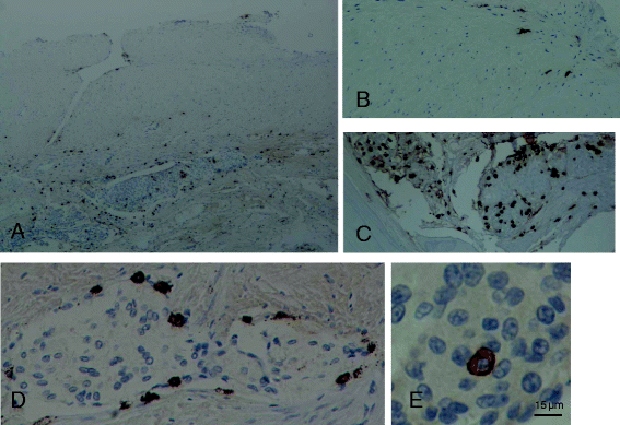
Photomicrographs of tissue samples of dura and bone infiltrated by meningioma of the meningothelial type grade I obtained from a brain lesion following a left-left frontotemporal craniectomy. Mast cells were stained immunohistochemically for the presence of tryptase (brown color). a Dura showing the upper unaffected part and the lower part infiltrated meningioma cells (blue) and accumulated mast cells (brown); magnification = ×40. b Unaffected dura; magnification = ×100. c Bone infiltrated by meningioma cells showing a cluster of mast cells; magnification = ×100. d Mast cells surrounding clusters of meningioma cells infiltrating the dura; magnification = ×400. e One mast cell surrounded by meningioma cells; this mast cell does not appear to be degranulated. Bar = 15 μm
Fig. 2
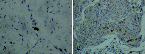
Photomicrographs of tissue samples of dura infiltrated by meningioma of the meningothelial type grade I obtained from a brain lesion following a left-left frontotemporal craniectomy. Mast cells were stained immunohistochemically for the presence of CD117 (brown color). a Unaffected dura showing two mast cells. b Dura infiltrated by meningioma cells showing a number of mast cells (brown color). Magnification = ×200
Similar articles
- Secretory meningioma: immunohistochemical findings and evaluation of mast cell infiltration.
Tirakotai W, Mennel HD, Celik I, Hellwig D, Bertalanffy H, Riegel T. Tirakotai W, et al. Neurosurg Rev. 2006 Jan;29(1):41-8. doi: 10.1007/s10143-005-0402-9. Epub 2005 Jul 12. Neurosurg Rev. 2006. PMID: 16010579 - Evaluation of mast cells and hypoxia inducible factor-1 expression in meningiomas of various grades in correlation with peritumoral brain edema.
Reszec J, Hermanowicz A, Rutkowski R, Bernaczyk P, Mariak Z, Chyczewski L. Reszec J, et al. J Neurooncol. 2013 Oct;115(1):119-25. doi: 10.1007/s11060-013-1208-1. Epub 2013 Jul 23. J Neurooncol. 2013. PMID: 23877362 Free PMC article. - Mast cells evaluation in meningioma of various grades.
Reszec J, Hermanowicz A, Kochanowicz J, Turek G, Mariak Z, Chyczewski L. Reszec J, et al. Folia Histochem Cytobiol. 2012;50(4):542-6. doi: 10.5603/14744. Folia Histochem Cytobiol. 2012. PMID: 23264217 - Tumor infiltrating immune cells in gliomas and meningiomas.
Domingues P, González-Tablas M, Otero Á, Pascual D, Miranda D, Ruiz L, Sousa P, Ciudad J, Gonçalves JM, Lopes MC, Orfao A, Tabernero MD. Domingues P, et al. Brain Behav Immun. 2016 Mar;53:1-15. doi: 10.1016/j.bbi.2015.07.019. Epub 2015 Jul 26. Brain Behav Immun. 2016. PMID: 26216710 Review. - The role of systemic inflammatory cells in meningiomas.
Haslund-Vinding J, Møller JR, Ziebell M, Vilhardt F, Mathiesen T. Haslund-Vinding J, et al. Neurosurg Rev. 2022 Apr;45(2):1205-1215. doi: 10.1007/s10143-021-01642-x. Epub 2021 Oct 30. Neurosurg Rev. 2022. PMID: 34716512 Review.
Cited by
- Ovarian ectopic pregnancy: the role of complex morphopathological assay. Review and case presentation.
Istrate-Ofiţeru AM, Ruican D, Niculescu M, Nagy RD, Roşu GC, Petrescu AM, Drăguşin RC, Iovan L, Zorilă GL, Iliescu DG. Istrate-Ofiţeru AM, et al. Rom J Morphol Embryol. 2020 Oct-Dec;61(4):985-997. doi: 10.47162/RJME.61.4.01. Rom J Morphol Embryol. 2020. PMID: 34171048 Free PMC article. - Clinical Potential of Immunotherapies in Subarachnoid Hemorrhage Treatment: Mechanistic Dissection of Innate and Adaptive Immune Responses.
Zhang A, Liu Y, Wang X, Xu H, Fang C, Yuan L, Wang K, Zheng J, Qi Y, Chen S, Zhang J, Shao A. Zhang A, et al. Aging Dis. 2023 Oct 1;14(5):1533-1554. doi: 10.14336/AD.2023.0126. Aging Dis. 2023. PMID: 37196120 Free PMC article. Review. - Mast cell-mediated neuroinflammation may have a role in attention deficit hyperactivity disorder (Review).
Song Y, Lu M, Yuan H, Chen T, Han X. Song Y, et al. Exp Ther Med. 2020 Aug;20(2):714-726. doi: 10.3892/etm.2020.8789. Epub 2020 May 25. Exp Ther Med. 2020. PMID: 32742317 Free PMC article. Review. - Mast cells in meningiomas.
D'Amati A, Tamma R, Annese T, Rizzi A, Ribatti D. D'Amati A, et al. Eur J Histochem. 2024 Apr 18;68(2):3973. doi: 10.4081/ejh.2024.3973. Eur J Histochem. 2024. PMID: 38634735 Free PMC article. - Angiomatous meningioma associated with rapidly aggravated peritumoral leptomeningitis: A case report.
Nakajima H, Tsuchiya T, Shimizu S, Murata T, Suzuki H. Nakajima H, et al. Surg Neurol Int. 2023 Apr 28;14:159. doi: 10.25259/SNI_54_2023. eCollection 2023. Surg Neurol Int. 2023. PMID: 37151464 Free PMC article.
References
- Ostrom QT, Gittleman H, Farah P, Ondracek A, Chen Y, Wolinsky Y, Stroup NE, Kruchko C, Barnholtz-Sloan JS. CBTRUS statistical report: primary brain and central nervous system tumors diagnosed in the United States in 2006–2010. Neuro Oncol. 2013;15(Suppl 2):ii1–56. doi: 10.1093/neuonc/not151. - DOI - PMC - PubMed
Publication types
MeSH terms
LinkOut - more resources
Full Text Sources
Other Literature Sources
Medical

