Topically applied metal chelator reduces thermal injury progression in a rat model of brass comb burn - PubMed (original) (raw)
Topically applied metal chelator reduces thermal injury progression in a rat model of brass comb burn
Cheng Z Wang et al. Burns. 2015 Dec.
Abstract
Oxidative stress may be involved in the cellular damage and tissue destruction as burn wounds continues to progress after abatement of the initial insult. Since iron and calcium ions play key roles in oxidative stress, this study tested whether topical application of Livionex formulation (LF) lotion, that contains disodium EDTA as a metal chelator and methyl sulfonyl methane (MSM) as a permeability enhancer, would prevent or reduce burns.
Methods: We used an established brass comb burn model with some modifications. Topical application of LF lotion was started 5 min post-burn, and repeated every 8 h for 3 consecutive days. Rats were euthanized and skin harvested for histochemistry and immunohistochemistry. Formation of protein adducts of 4-hydroxynonenal (HNE), malonadialdehyde (MDA) and acrolein (ACR) and expression of aldehyde dehydrogenase (ALDH) isozymes, ALDH1 and ALDH2 were assessed.
Results: LF lotion-treated burn sites and interspaces showed mild morphological improvement compared to untreated burn sites. Furthermore, the lotion significantly decreased the immunostaining of lipid aldehyde-protein adducts including protein -HNE, -MDA and -ACR adducts, and restored the expression of aldehyde dehydrogenase isozymes in the unburned interspaces.
Conclusion: This data, for the first time, demonstrates that a topically applied EDTA-containing lotion protects burns progression with a concomitant decrease in the accumulation of reactive lipid aldehydes and protection of aldehyde dehydrogenase isozymes. Present studies are suggestive of therapeutic intervention of burns by this novel lotion.
Keywords: Brass comb burn; Burn progression; Iron chelation; Oxidative stress; Reactive aldehydes; Thermal injury; Wound healing.
Copyright © 2015 Elsevier Ltd and ISBI. All rights reserved.
Figures
Fig 1
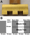
A, Brass comb probe consisting of three (3) 10-mm teeth separated by two (2) 10-mm notches modified from the previous Regas and Ehrlich model (1992). B, Diagram of the bottom view of the modified brass comb consisting of three (3)10×19 mm rectangles of burn sites separated by two (2)10×19 mm rectangles of interspaces. The second diagram is of the tissue sampling in each comb burn wound; two tissue blocks (9 × 30 mm each) were harvested.
Fig 2
Representative photographs of the burn wounds: A (5 min after injury) and B (72 hrs after injury). C (5 min after injury) and D (72 hrs after injury) with LF lotion treatment. Note that at 72 hrs the LF lotion-treated wound (D) showed a size of unburned interspace similar to that of the same wound 5 min after burn injury (C), while the untreated burn wound (B) had a significantly smaller interspace when compared to that of the same wound 5 min after burn (A).
Fig 3
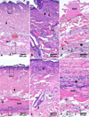
Representative H&E stained microphotographs of the burn sites: Middle of burn sites showing microscopic characteristics of burn wound without (A–C) or with (D–F) LF lotion treatment started 5 min post injury. B and C, higher power of the upper and lower insert boxes in A, respectively. E and F, higher power of the upper and lower insert boxes in D, respectively. SkM, skeletal muscle; SG, sebaceous gland; HF, hair follicles; △, dilation and congestion of capillaries, venules and arterioles; ▲, blocked vessels filled with denatured clots; *, inflammatory cell infiltration. ↑, Scale bar = 200 µm in A & D; 50 µm in B, C, E, F
Fig 4
Representative microphotographs of Masson’s trichrome staining of burn sites, 72 hrs post injury: A, Control, without burn; B, the middle of a burn site, burn alone; C, the middle of a burn site, burn plus LF lotion treatment post injury. SkM, skeletal muscle; SG, sebaceous gland; HF, hair follicles; ▲, blocked vessels filled with denatured clots; *, areas with inflammatory cell infiltration. Scale bar = 200 µm in A-C
Fig 5
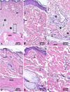
Representative H&E stained microphotographs of the interspaces: Middle of interspaces showing microscopic characteristics without (A–C) or with (D–F) LF lotion treatment. A–C, 72 hrs post injury; D–F, 72 hrs post injury plus LF lotion treatment. B and C, higher power of the upper and lower insert boxes in A, respectively. E and F, higher power of the upper and lower insert boxes in D, respectively. SkM, skeletal muscle; SG, sebaceous gland; HF, hair follicles; △, dilation and congestion of capillaries, venules or arterioles; *, inflammatory cell infiltration. Scale bar = 200 µm in A & D, 50 µm in B,C, E,F
Fig 6
Representative H&E microphotographs of survived interspace epidermis: A, control without burn; B, 72 hrs post injury; C, 72 hrs post injury plus LF lotion treatment. Yellow arrow line marks the length of survived interspace epidermis. SkM, skeletal muscle; SG, sebaceous gland; HF, hair follicles; ▲, blocked vessels filled with denatured clots; ↓, necrotic epidermis. Scale bar = 500 µm in A-C
Fig 7
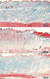
Representative Masson’s trichrome staining of the half interspace (4 mm from its middle line). A, control without burn; B, 72 hrs after burn; C, 72 hrs after burn plus LF lotion treatment post injury. SkM, skeletal muscle; SG, sebaceous gland; HF, hair follicles; ▲, blocked vessels filled with denatured clots; ↓, necrotic epidermis. Scale bar = 500 µm in A–C
Fig 8
Representative protein-HNE IHC microphotographs of interspaces : The methyl green counterstained IHC microphotographs show epidermis and dermis (A–C) and hypodermis (D–F). A & D, control, without burn; B & E, 72 hrs post burn; C & F, 72 hrs after a burn plus LF lotion treatment post injury; Ep, epidermis; SG, sebaceous gland; HF, hair follicles; ↓, patent vessels (D, F); △, dilated capillary in dermis (B); ▲, dilated and blocked (E) vessels in hypodermis. Scale bar = 100 µm (A–F)
Fig 9
Representative protein-MDA IHC microphotographs of interspaces: The methyl green counterstained IHC microphotographs show protein-MDA staining in the interspaces of A control, without burn; B, , 72 hrs after burn; C, 72 hrs after burn plus LF lotion treatment post injury. Ep, epidermis; SG, sebaceous gland; HF, hair follicle. Scale bar = 50 µm (A–C)
Fig 10
Representative ALDH1A1 and ALDH2 IHC microphotographs of interspaces: The methyl green counterstained IHC microphotographs show immunostaining of ALDH1A1 (A-C) and ALDH2 (D–F) in sebaceous gland (SG) and hair follicle (HF) epithelial cells. A & D, Control, without burn; B & E, 72 hrs after burn; C & F, 72 hrs after burn plus LF lotion treatment post injury. Ep, epidermis; SG, sebaceous gland; HF, hair follicle. Scale bar, 100 µm (A–F)
Similar articles
- Metal chelation attenuates oxidative stress, inflammation, and vertical burn progression in a porcine brass comb burn model.
El Ayadi A, Salsbury JR, Enkhbaatar P, Herndon DN, Ansari NH. El Ayadi A, et al. Redox Biol. 2021 Sep;45:102034. doi: 10.1016/j.redox.2021.102034. Epub 2021 Jun 8. Redox Biol. 2021. PMID: 34139550 Free PMC article. - Metal chelation reduces skin epithelial inflammation and rescues epithelial cells from toxicity due to thermal injury in a rat model.
El Ayadi A, Wang CZ, Zhang M, Wetzel M, Prasai A, Finnerty CC, Enkhbaatar P, Herndon DN, Ansari NH. El Ayadi A, et al. Burns Trauma. 2020 Oct 2;8:tkaa024. doi: 10.1093/burnst/tkaa024. eCollection 2020. Burns Trauma. 2020. PMID: 33033727 Free PMC article. - Metal chelator combined with permeability enhancer ameliorates oxidative stress-associated neurodegeneration in rat eyes with elevated intraocular pressure.
Liu P, Zhang M, Shoeb M, Hogan D, Tang L, Syed MF, Wang CZ, Campbell GA, Ansari NH. Liu P, et al. Free Radic Biol Med. 2014 Apr;69:289-99. doi: 10.1016/j.freeradbiomed.2014.01.039. Epub 2014 Feb 6. Free Radic Biol Med. 2014. PMID: 24509160 Free PMC article. - Aldehyde Dehydrogenase 2 as a Therapeutic Target in Oxidative Stress-Related Diseases: Post-Translational Modifications Deserve More Attention.
Gao J, Hao Y, Piao X, Gu X. Gao J, et al. Int J Mol Sci. 2022 Feb 28;23(5):2682. doi: 10.3390/ijms23052682. Int J Mol Sci. 2022. PMID: 35269824 Free PMC article. Review. - TRP Channels as Sensors of Aldehyde and Oxidative Stress.
Hellenthal KEM, Brabenec L, Gross ER, Wagner NM. Hellenthal KEM, et al. Biomolecules. 2021 Sep 24;11(10):1401. doi: 10.3390/biom11101401. Biomolecules. 2021. PMID: 34680034 Free PMC article. Review.
Cited by
- The P50 Research Center in Perioperative Sciences: How the investment by the National Institute of General Medical Sciences in team science has reduced postburn mortality.
Finnerty CC, Capek KD, Voigt C, Hundeshagen G, Cambiaso-Daniel J, Porter C, Sousse LE, El Ayadi A, Zapata-Sirvent R, Guillory AN, Suman OE, Herndon DN. Finnerty CC, et al. J Trauma Acute Care Surg. 2017 Sep;83(3):532-542. doi: 10.1097/TA.0000000000001644. J Trauma Acute Care Surg. 2017. PMID: 28697015 Free PMC article. - Thermal injury induces early blood vessel occlusion in a porcine model of brass comb burn.
Wang J, Wang CZ, Salsbury JR, Zhang J, Enkhbaatar P, Herndon DN, El Ayadi A, Ansari NH. Wang J, et al. Sci Rep. 2021 Jun 14;11(1):12457. doi: 10.1038/s41598-021-91874-0. Sci Rep. 2021. PMID: 34127701 Free PMC article. - Therapeutic Strategies to Reduce Burn Wound Conversion.
Palackic A, Jay JW, Duggan RP, Branski LK, Wolf SE, Ansari N, El Ayadi A. Palackic A, et al. Medicina (Kaunas). 2022 Jul 11;58(7):922. doi: 10.3390/medicina58070922. Medicina (Kaunas). 2022. PMID: 35888643 Free PMC article. Review. - The Bactericidal Tandem Drug, AB569: How to Eradicate Antibiotic-Resistant Biofilm Pseudomonas aeruginosa in Multiple Disease Settings Including Cystic Fibrosis, Burns/Wounds and Urinary Tract Infections.
Hassett DJ, Kovall RA, Schurr MJ, Kotagiri N, Kumari H, Satish L. Hassett DJ, et al. Front Microbiol. 2021 Jun 17;12:639362. doi: 10.3389/fmicb.2021.639362. eCollection 2021. Front Microbiol. 2021. PMID: 34220733 Free PMC article. Review. - Alda-1, an Aldehyde Dehydrogenase 2 Agonist, Improves Cutaneous Wound Healing by Activating Epidermal Keratinocytes via Akt/GSK-3β/β-Catenin Pathway.
Zhang S, Chen C, Ying J, Wei C, Wang L, Yang Z, Qi F. Zhang S, et al. Aesthetic Plast Surg. 2020 Jun;44(3):993-1005. doi: 10.1007/s00266-020-01614-4. Epub 2020 Jan 17. Aesthetic Plast Surg. 2020. PMID: 31953581
References
- Jackson DM. The diagnosis of the depth of burning. Br J Surg. 1953;40(164):588–596. - PubMed
- Singh V, et al. The pathogenesis of burn wound conversion. Ann Plast Surg. 2007;59(1):109–115. - PubMed
- Shupp JW, et al. A review of the local pathophysiologic bases of burn wound progression. J Burn Care Res. 2010;31(6):849–873. - PubMed
- Horton JW. Free radicals and lipid peroxidation mediated injury in burn trauma: the role of antioxidant therapy. Toxicology. 2003;189(1–2):75–88. - PubMed
Publication types
MeSH terms
Substances
LinkOut - more resources
Full Text Sources
Other Literature Sources
Medical
Miscellaneous


